Ijraset Journal For Research in Applied Science and Engineering Technology
- Home / Ijraset
- On This Page
- Abstract
- Introduction
- Conclusion
- References
- Copyright
Automated Anemia Screening via Nail Image Analysis Using Fast RCNN
Authors: Kadhunoori Aparna, Dr. O. Obulesu, M. Lalitha
DOI Link: https://doi.org/10.22214/ijraset.2024.64742
Certificate: View Certificate
Abstract
In the modern world, the health sector is quickly moving to digital platforms due to its practicality, ease of use, and affordability. According to the World Health Organization, anemia will continue to be a major global health issue in 2023, affecting 571 million women and 269 million young children. Anemia is still very common, especially in low- and middle-income countries. Photo plethysmography (PPG), a non-invasive technique for detecting anemia, has focused on selecting the appropriate light sources and wavelengths. Numerous methods, including data processing, photo detector design, and the use of lights with different spectra, can be used to detect anemia. Cost and excessive power use are the primary disadvantages. The proposed method includes a novel nail image-based anemia diagnosis approach to address the aforementioned issues. In order to detect leukonychia, which is characterized by the presence of white spots or streaks that are indicative of iron deficiency anemia, the primary goal is to suggest an improved method for analysing pictures of a person\'s nails. For example, this method makes use of state-of-the-art convolutional neural network architectures, such as Fast RCNN and RCNN. We show that Fast RCNN outperforms RCNN in terms of accuracy, reaching 92%. A significant step in the right direction toward enhancing anemia-related healthcare outcomes worldwide is the suggested strategy, which blends state-of-the-art technology with medical diagnostics. Additionally, the system creates opportunities for further improvements, such as adding real-time monitoring features and growing dataset which include a wider range of demographic traits.
Introduction
I. INTRODUCTION
Anemia, a disorder characterized by a lack of hemoglobin or red blood cells, affects the body's capacity to carry oxygen efficiently and causes symptoms like weakness, pallor, and exhaustion. The World Health Organization estimates that this global problem has large prevalence rates in vulnerable groups, including women and children, which is especially worrying. Early detection and treatment may be hampered by the cost, labour-intensive nature, and unpredictability of traditional diagnostic techniques for anemia, such as blood tests and conjunctiva pallor checks. This is particularly true in settings with minimal resources.
To tackle these issues, novel non-invasive approaches are being investigated, like employing image processing techniques to analyse the nail bed's color and texture. Particularly in underprivileged areas, this method presents a potentially accessible and affordable alternative for anemia screening. In order to improve overall healthcare accessibility and results, this approach may improve early detection and monitoring of anemia by utilizing advanced algorithms to assess nail features. Notwithstanding obstacles like picture variability and the requirement for thorough validation, this innovative method has the potential to completely transform the diagnosis and treatment of anemia.
The main objectives of proposed system listed below:
- To collect nails images and perform image annotation on each image.
- To effectively preprocess and apply data augmentation for increasing size of the dataset and reduce the noise.
- To train the model using deep learning techniques such as R-CNN, fast R-CNN and predict anemia using suitable algorithm.
II. RELATED WORKS
When it comes to the application of computerized algorithms, like machine learning, for the estimation of Hb and the diagnosis or detection of medical disorders, like anemia, the color of nail beds and the conjunctiva of the eyes are frequently linked to high accuracy. Many research using conjunctiva images of the eye have shown that the best algorithms for classification or detection rely on the problem domain to be solved in order to non-invasively diagnose anemia.
Table 1: Literature review
|
Author names |
Dataset |
Feature extraction method |
Algorithms |
Achieved Accuracy |
Limitations |
|
Tamir |
19 eye images |
Not indicated |
RGB thresholding algorithm |
With RGB thresholding techniques, 78.90% accuracy was achieved.
|
Less number of images (19) used to detect anemia |
|
Peksi |
20 nail and palm images |
Segmentation was used to calculate the average RGB value of images |
Naïve Bayes |
Achieved 90% accuracy |
Increase Database size and use more advanced techniques |
|
Yadav Singh |
400 retinal pictures |
Gray-level co-occurrence matrix (GLCM), Laws' texture energy (LTE)-based, Tamura's features, wavelet features, and HOG features |
SVM, k-NN, and decision tree |
Achieved 77.30% accuracy |
It is expensive as it is making use of hard ware tools |
|
Jain |
99 conjunctiva images |
Manual extraction of ROI of the palpable conjunctiva of the eyes |
Artificial neural networks (ANN) |
Attained 97% accuracy |
We may achieve improved accuracy by increasing dataset size |
It is clear that the evaluated related works which are non-expensive methods, such as machine learning techniques, are effective, economical, and give rapid outcomes when diagnosing medical conditions like anemia. This study examined the ability of the tangible palm, the color of nails, and the mucosa of the eyes in the identification of anemia, since the majority of investigations employed the conjunctiva of the eyes. This is due to the fact that children's conjunctiva might be shown to diseases or falling items, and finger is a key body parts for anemia identification. Thus, the suggested method uses pictures of fingernails to identify anemia.
III. METHODOLOGY
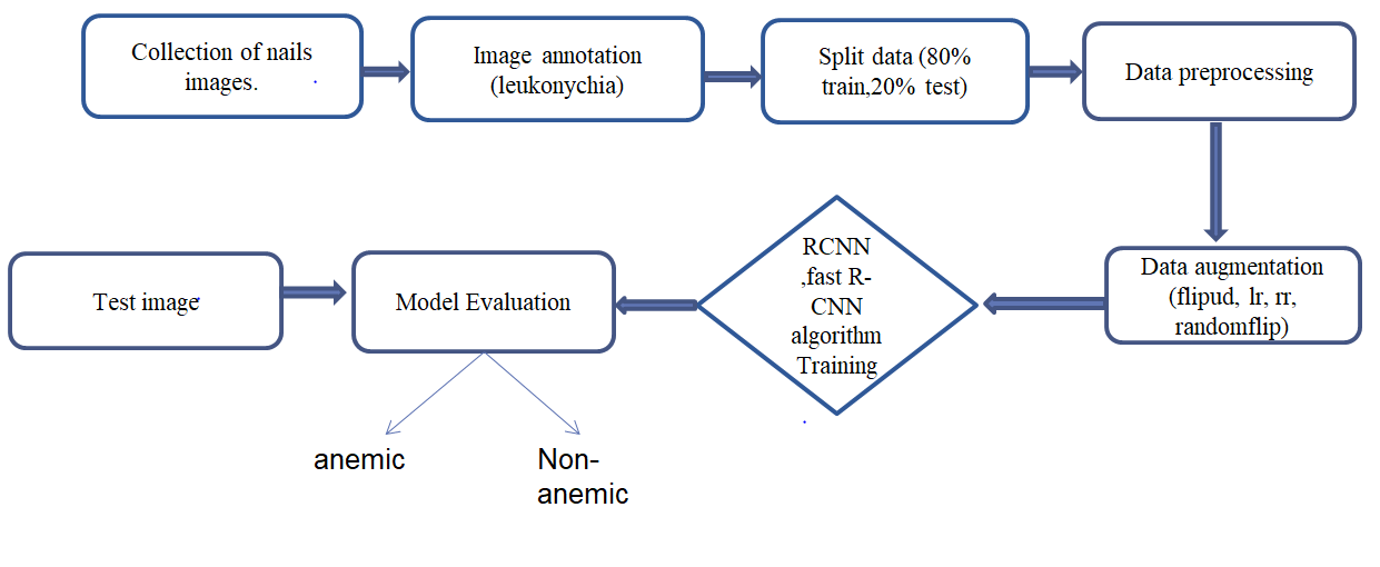
The architecture for anemia detection using RCNN and Fast R-CNN on finger nail images is a sophisticated framework that employs cutting-edge computer vision techniques to automatically identify potential indicators of anemia from images of finger nails shown in above Figure 1. The anemia detection system uses RCNN and Fast R-CNN to analyze fingernail images for signs of anemia. It involves collecting and annotating images with bounding boxes, preprocessing them through normalization and resizing, and expanding the dataset with data augmentation. The model is trained with both algorithms, and the most effective one is chosen for classifying images as anemic or non-anemic.

Figure 1: Flow Chart of the Proposed Methodology.
A. Datasets Used
The first step of the process is the entry of digital photographs of fingernails. These pictures come from a variety of places, including clinical research and medical databases. Every image captures possible visual clues associated with anemia by visualizing the fingernail area. The suggested method uses images of nails to forecast anemia. Images of nails with leukonychia are gathered from various websites and dataset resources to construct the dataset. An 80:20 ratio is used to split the entire dataset, with 80 used for training and 20 for testing. There are 140 images in the dataset.
B. Image Processing
Image annotation for anemia detection involves marking features like paleness, texture changes, and capillary density in fingernail images using color and texture labels, as well as bounding boxes. This process creates a dataset for training and validating machine learning models to detect anemia accurately and non-invasively. Ethical and privacy considerations are essential throughout to ensure responsible data handling.

Figure2: image annotation on finger nails
C. Data Preprocessing
Data preprocessing for anemia detection involves standardizing fingernail images by resizing, color correcting, and reducing noise to ensure uniformity and quality. This step also includes normalizing pixel values and cropping to focus on the fingernail region, which enhances the effectiveness of analysis and machine learning processes.
D. Data Augmentation
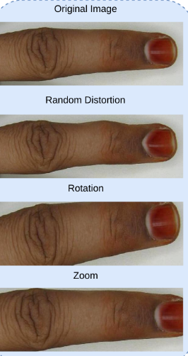
Data augmentation is essential for expanding and diversifying the dataset used in anemia detection from fingernail images. Techniques such as zooming, rotating, and flipping create new perspectives, improving the model's adaptability to various orientations and scales. This process enhances the model's ability to detect anemia-related patterns and increases the dataset size to 2,240 images, boosting overall performance and generalization.

Figure3: Augmented images of finger nails
IV. REGION BASED CONVOLUTIONAL NEURAL NETWORK (RCNN)
RCNN is a pivotal deep learning technique in computer vision planned to tackle image detection tasks. The RCNN model comprises several essential components. Region Proposal Network (RPN) plays a major role in generating potential regions within an image where objects might exist.
Working of RCNN in the proposed system is as follow:
The detection of anemia from fingernail images involves a series of steps starting with a dataset of anemic and non-anemic images, followed by pre-processing (resizing, normalization, and background removal) and augmentation (zooming, rotating, flipping) to enhance data quality. A Region-Based
Convolutional Neural Network (RCNN) approach is used, where a Region Proposal Network (RPN) identifies potential Regions of Interest (ROIs) in the image. Features from these ROIs are extracted using a pre-trained Convolutional Neural Network (CNN) and classified by fully connected layers to differentiate between anemic and non-anemic nails. The RCNN model then highlights and labels the detected regions accordingly.
V. FAST REGION BASED CONVOLUTIONAL NEURAL NETWORK(FAST-RCNN)
Fast R-CNN is a significant advancement over the original RCNN architecture, specifically designed for improving object detection and localization in the realm of computer vision.
Working of Fast-RCNN in proposed system is as follow:
The process for detecting anemia from fingernail images involves using a dataset of both anemic and non-anemic images, which are pre-processed and augmented to improve quality and performance. After annotating images with bounding boxes around areas of interest, a Convolutional Neural Network (CNN) extracts feature maps from these regions. Fixed-size feature vectors are then analyzed by fully connected layers to identify and classify the presence and severity of anemia, while also refining the bounding boxes around the affected areas.
VI. RESULTS
In this chapter, the outcomes of proposed system anemia detection with RCNN and Fast R-CNN applied on finger nail images are discussed. The RCNN and Fast R-CNN models were implemented using the PyTorch framework and trained on a high-performance computing cluster.

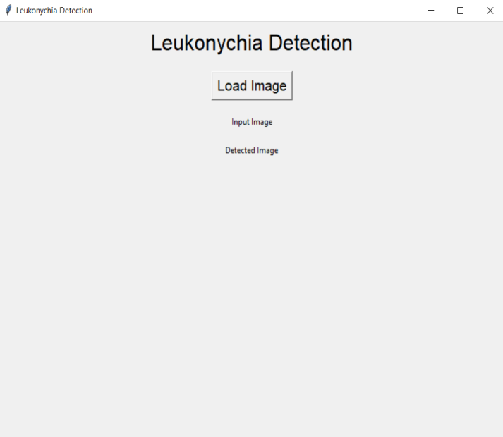
Figure 4: User interface Leukonychia based anemia detection
The user interface consists of “load image” button through which user can upload a finger nails image shown in figure 4. And the image run under the model which selected for prediction based on comparative performance analysis.

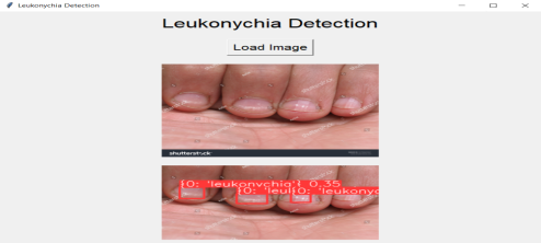
Figure5: Output of anemia detection
And the output consist of input image along with the predicted image i.e., probability of occurrence of anemia as show in the above figure 5. The input dataset has been divided into train and test in 80:20 ratio. The model has been trained for 50 epochs. The performance of both models was evaluated based on accuracy, precision and a comparative analysis was conducted to assess the superiority of Fast R-CNN over RCNN. The results of our experiments indicate a clear performance advantage of Fast R-CNN over RCNN in the task of anemia detection using finger nail images. The quantitative metrics are potted in the below table 2.
Table 2: Evaluation of evaluation metric
|
Model |
Accuracy |
Precision |
Recall |
F1-score |
|
RCNN |
87.2% |
0.873 |
0.892 |
0.882 |
|
Fast R-CNN |
92.8% |
0.932 |
0.938 |
0.935 |
As shown in the table, Fast R-CNN achieved an accuracy of 92.8%, outperforming RCNN by a notable margin of 5.6%. Similarly, Fast R-CNN demonstrated greater precision, recall, and F1-score outcomes compared to RCNN, indicating its superior ability to correctly identify anemia cases.
Conclusion
The proposed system is an early anemia detection with image processing methods which is implemented using two techniques known as RCNN and Fast RCNN. Through the experimentation and analysis, observed that Fast R-CNN consistently demonstrated higher accuracy 92.8% compared to the traditional RCNN method accuracy 87.2%. The application of Fast R-CNN yielded promising outcomes, showcasing its potential as an effective tool in anemia detection. The improved accuracy offered by Fast R-CNN indicates its capability to better identify anemic conditions from finger nail images. This could have significant implications for early detection and timely medical interventions, ultimately enhancing patient care and well-being. Future scope of this approach could involve acquiring larger and more diverse datasets of finger nail images, contributing to enhanced accuracy and broader applicability. Fine-tuning the model through transfer learning on medical-specific datasets could refine its performance and adaptability to specific patient groups.
References
[1] Acar E, Türk Ö, Ertu?rul ÖF, Aldemir E. Employing deep learning architectures for image-based automatic cataract diagnosis. Turk J Electr Eng Comput Sci. 2021; 29(SI-1): 2649-2662. [2] A. Halder et al., “Digital camera-based spectrometry for the development of Point-of-Care anemia detection on ultra-low volume whole blood sample,” IEEE Sensors J., vol. 17, no. 21, pp. 7149–7156, Nov. 2017. [3] A. Mitani et al., “Detection of anaemia from retinal fundus images via deep learning,” Nature Biomed. Eng., vol. 4, no. 1, pp. 18–27, Jan. 2020. [4] A. Tamir et al., “Detection of anemia from image of the anterior conjunctiva of the eye by image processing and thresholding,” in Proc. IEEE Region 10 Hum. Technol. Conf. (R10-HTC), Dec. 2017, pp. 697–701. [5] C. Adams. (2019). Ergonomic Lighting Levels by Room for Residential Spaces. [Online]. Available: https://www.thoughtco.com/lighting-levelsby-room-120664. [6] Chen Y, Zhong K, Zhu Y, Sun Q. Two-stage hemoglobin prediction based on prior causality. Front Public Health. 2022; 10:1079389. [7] Dimauro G, Caivano D, Girardi F. A new method and a non-invasive device to estimate anemia based on digital images of the conjunctiva. IEEE Access. 2018; 6: 46968-46975. [8] Delgado-Rivera G, Roman-Gonzalez A, Alva-Mantari A, et al. Method for the automatic segmentation of the palpebral conjunctiva using image processing. Proceedings of the 2018 IEEE International Conference on Automation/XXIII Congress of the Chilean Association of Automatic Control (ICA-ACCA); 2018:1-4. [9] G. Dimauro, L. Baldari, D. Caivano, G. Colucci, and F. Girardi, “Automatic segmentation of relevant sections of the conjunctiva for non-invasive anemia detection,” in 2018 3rd Int. Conf. On Smart and Sustain. Technol. (SpliTech), Dec. 2018, pp. 1–5. [10] Ghosh A, Mukherjee J, Chakravorty N. A low-cost test for anemia using an artificial neural network. Compute Methods Programs Biomed. 2023; 229:107251. [11] H. A. Elsalamony, “Anaemia cells detection based on shape signature using neural networks,” Measurement, vol. 104, pp. 50–59, Jul. 2017. [12] H. Von Schenck, M. Falkensson, and B. Lundberg, “Evaluation of ‘hemocue’ a new device for determining hemoglobin,” Clin. Chem., vol. 32, no. 3, pp. 526–529, 1986. [13] Irum A, Akram M, Ayub SM, Waseem S, Khan MJ. Anemia detection using image processing. Proceedings of the International Conference on Digital Information Processing, Electronics, and Wireless Communications; 2016. [14] Jain P, Bauskar S, Gyanchandani M. Neural network based non-invasive method to detect anemia from images of eye conjunctiva. Int J Imaging Syst Technol. 2019; 30(1): 112-125. [15] M. Sari et al., “Estimating the prevalence of anemia: A comparison of three methods,” Bulletin World Health Org., vol. 79, pp. 506–511, Dec. 2001. [16] O. Didzun et al., “Anaemia among men in India: A nationally representative cross-sectional study,” Lancet Global Health, vol. 7, no. 12, pp. 1685–1694, 2019. [17] P. Viola and M. J. Jones, “Robust real-time face detection,” Int. J. Comput. Vis., vol. 57, no. 2, pp. 137–154, May 2004. [18] Peksi NJ, Yuwono B, Florestiyanto MY. Classification of anemia with digital images of nails and palms using the naive Bayes method. Telematika. 2021; 18(1): 118. [19] Roychowdhury S, Sun D, Ren J, Bihis M, Hage P, Rahman H. Screening pallor site images for anemia-like pathologies. Proceedings of the IEEE International Conference on Biomedical Health Informatics; 2017:2017. [20] S. Collings, O. Thompson, E. Hirst, L. Goossens, A. George, and R. Weinkove, “Non-invasive detection of anaemia using digital photographs of the conjunctiva,” PLoS ONE, vol. 11, no. 4, Apr. 2016, Art. no. e0153286. [21] S. Filonov. (2019). Tracking Your Eyes with Python. [Online]. Available: https://medium.com/stepanfilonov/tracking-your-eyes-with-python3952e66194a6.
Copyright
Copyright © 2024 Kadhunoori Aparna, Dr. O. Obulesu, M. Lalitha. This is an open access article distributed under the Creative Commons Attribution License, which permits unrestricted use, distribution, and reproduction in any medium, provided the original work is properly cited.

Download Paper
Paper Id : IJRASET64742
Publish Date : 2024-10-22
ISSN : 2321-9653
Publisher Name : IJRASET
DOI Link : Click Here
 Submit Paper Online

