Ijraset Journal For Research in Applied Science and Engineering Technology
- Home / Ijraset
- On This Page
- Abstract
- Introduction
- Conclusion
- References
- Copyright
Automatic Detection of Genetic Diseases in Pediatric Age using Pupillometry
Authors: Ganji Deepthi , Dr. V. Uma Rani
DOI Link: https://doi.org/10.22214/ijraset.2023.55969
Certificate: View Certificate
Abstract
Inherited retinal diseases cause severe visual deficits in children. They are classified in outer and inner retina diseases, and often cause blindness in childhood. The diagnosis for this type of illness is challenging, given the wide range of clinical and genetic causes (with over 200 causative genes). It is routinely based on a complex pattern of clinical tests, including invasive ones, not always appropriate for infants or young children. A different approach is thus needed, that exploits Chromatic Pupillometry, a technique increasingly used to assess outer and inner retina functions. This paper presents a novel Clinical Decision Support System (CDSS), based on Machine Learning using Chromatic Pupillometry in order to support diagnosis of Inherited retinal diseases in pediatric subjects. An approach that combines hardware and software is proposed: a dedicated medical equipment (pupillometer) is used with a purposely designed custom machine learning decision support system. Two distinct Support Vector Machines (SVMs), one for each eye, classify the features extracted from the pupillometric data. The designed CDSS has been used for diagnosis of Retinitis Pigmentosa in pediatric subjects. The results, obtained by combining the two SVMs in an ensemble model, show satisfactory performance of the system, that achieved 0.846 accuracy, 0.937 sensitivity and 0.786 specificity. This is the first study that applies machine learning to pupillometric data in order to diagnose a genetic disease in pediatric age.
Introduction
I. INTRODUCTION
Inherited Retinal Diseases (IRDs) represent a significant cause of severe visual deficits in children [1]. They frequently are cause of blindness in childhood in Established Market Economies (1/3000 individuals). IRDs can be divided into diseases of the outer retina, namely photoreceptor degenerations (e.g., Leber Congenital Amaurosis, Retinitis Pigmentosa, Stargardt disease, Cone Dystrophy, Acromatopsia, Choroideremia, etc.), and diseases of the inner retina, mainly retinal ganglion cell degeneration (e.g. congenital glaucoma, dominant optic atrophy, Leber hereditary optic neuropathy). Both conditions are characterized by extremely high genetic heterogeneity with over 200 causative genes identified to date, which represent a remarkable obstacle to a rapid and effective diagnosis,also considering that the same gene could cause different and heterogeneous clinical phenotypes.
The clinical evaluation of IRDs is routinely based on a complex pattern of clinical tests, including invasive ones, that are not always appropriate for infants or young children. For example, electrophysiological testing, that represents the most informative clinical investigation for the diagnosis of inner and outer retinal diseases, often requires sedation of the children. Sedation affects the retinal response and requires a complex healthcare environment (e.g., operating room, pediatric, anesthesiologist, dedicated instrumentation, etc.)

A novel approach to support the diagnosis of IRDs would be useful. To this regard, chromatic pupillometry has been proposed as a highly sensitive and objective test to quantify the function of different light-sensitive retinal cells and, therefore, it has been shown helpful to detect the retinal dysfunction caused by IRDs as summarized in the following. Photoreceptor cells (rods and cones) exhibit fast temporal kinetics and cause a brisk pupillary constriction in response to light, whereas the inner retinal melanopsin containing intrinsic photosensitive Retinal Ganglion Cells (ipRGCs) exhibits slower temporal kinetics and elicits a sustained pupillary constriction to light stimuli, persisting after light cessation. The relative contributions of the three receptor types (rod, cone, and melanopsin photopigments) to the Pupillary Light Reflex (PLR) have been examined by manipulating the characteristics of large-field (90) flash stimuli and the adaptation conditions (light vs. dark adapted). For example, high-luminance, long-wavelength (red) flashes presented against a rod-suppressing adapting field elicit a PLR that is predominately cone-mediated whereas lowluminance, short-wavelength (blue) flashes presented to the dark-adapted eye elicits a PLR that is primarily rodmediated. For high-luminance, short-wavelength flashes presented to the dark-adapted eye, there is an initial transient pupil constriction (rod- and cone-mediated) that is followed by a melanopsin-mediated sustained constriction that can last for more than 30s after stimulus offset. The prolonged melanopsin-mediated constriction has been used in clinical protocols to assess inner-retina function. Thus, the use of chromatic pupil responses may be a novel way to diagnose and monitor diseases affecting either the outer or inner retina. This evidence suggested that a clinical decision support system (CDSS) based on chromatic pupillometry could be developed in order to support diagnosis of IRDs.
Our activity was performed within a research project, which main goal is defining effective protocols and systems for an early diagnosis and monitoring through chromatic pupillometry. The team that worked to this project is structured in three operative units: Department of Information Engineering of the University of Florence, Eye Clinics at the University of Campania Luigi Vanvitelli and at the University of Milan. This team designed a novel CDSS for diagnosis of Retinitis Pigmentosa (RP) in pediatric subjects.
II. LITERATURE REVIEW
- Genotype–Phenotype Correlation And Mutation Spectrum In A Large Cohort Of Patients With Inherited Retinal Dystrophy Revealed By Next-Generation Sequencing
Inherited retinal dystrophy (IRD) is a leading cause of blindness worldwide. Because of extreme genetic heterogeneity, the etiology and genotypic spectrum of IRD have not been clearly defined, and there is limited information on genotype-phenotype correlations. The purpose of this study was to elucidate the mutational spectrum and genotype-phenotype correlations of IRD. Methods: We developed a targeted panel of 164 known retinal disease genes, 88 candidate genes, and 32 retina-abundant microRNAs, used for exome sequencing. A total of 179 Chinese families with IRD were recruited. Results: In 99 unrelated patients, a total of 124 mutations in known retinal disease genes were identified, including 79 novel mutations (detection rate, 55.3%). Moreover, novel genotype-phenotype correlations were discovered, and phenotypic trends noted. Three cases are reported, including the identification of AHI1 as a novel candidate gene for nonsyndromic retinitis pigmentosa. Conclusion: This study revealed novel genotype-phenotype correlations, including a novel candidate gene, and identified 124 genetic defects within a cohort with IRD . The identification of novel genotype-phenotype correlations and the spectrum of mutations greatly enhance the current knowledge of IRD phenotypic and genotypic heterogeneity, which will assist both clinical diagnoses and personalized treatments of IRD patients.
2. Chromatic Pupil Responses. Preferential Activation Of The Melanopsin-Mediated Versus Outer Photoreceptor-Mediated Pupil Light Reflex
To weight the rod-, cone-, and melanopsin-mediated activation of the retinal ganglion cells, which drive the pupil light reflex by varying the light stimulus wavelength, intensity, and duration. Experimental study. Forty-three subjects with normal eyes and 3 patients with neuroretinal visual loss. A novel stimulus paradigm was developed using either a long wavelength (red) or short wavelength (blue) light given as a continuous Ganzfeld stimulus with stepwise increases over a 2 log-unit range. The pupillary movement before, during, and after the light stimulus was recorded in real time with an infrared illuminated video camera. The percent pupil contraction of the transient and sustained pupil response to a low- (1 cd/m(2)), medium- (10 cd/m(2)), and high-intensity (100 cd/m(2)) red- and blue-light stimulus was calculated for 1 eye of each subject. From the 43 normal eyes, median and 25th, 75th, 5th, and 95th percentile values were obtained for each stimulus condition. In normal eyes at lower intensities, blue light evoked much greater pupil responses compared with red light when matched for photopic luminance. The transient pupil contraction was generally greater than the sustained contraction, and this disparity was greatest at the lowest light intensity and least apparent with bright (100 cd/m(2)) blue light.
A patient with primarily rod dysfunction (nonrecordable scotopic electroretinogram) showed significantly reduced pupil responses to blue light at lower intensities. A patient with achromatopsia and an almost normal visual field showed selective reduction of the pupil response to red-light stimulation. A patient with ganglion cell dysfunction owing to anterior ischemic optic neuropathy demonstrated global loss of pupil responses to red and blue light in the affected eye. Pupil responses that differ as a function of light intensity and wavelength support the hypothesis that selected stimulus conditions can produce pupil responses that reflect phototransduction primarily mediated by rods, cones, or melanopsin. Use of chromatic pupil responses may be a novel way to diagnose and monitor diseases affecting either the outer or inner retina.
3. Pupil Responses Derived From Outer And Inner Retinal Photoreception Are Normal In Patients With Hereditary Optic Neuropathy
We compared the pupil responses originating from outer versus inner retinal photoreception between patients with isolated hereditary optic neuropathy (HON, n = 8) and healthy controls (n = 8). Three different testing protocols were used. For the first two protocols, a response function of the maximal pupil contraction versus stimulus light intensity was generated and the intensity at which half of the maximal pupil contraction, the half-max intensity, was determined. For the third protocol, the pupil size after light offset, the re-dilation rate and re-dilation amplitude were calculated to assess the post-light stimulus response. Patients with HON had bilateral, symmetric optic atrophy and significant reduction of visual acuity and visual field compared to controls. There were no significant mean differences in the response curve and pupil response parameters that reflect mainly rod, cone or melanopsin activity between patients and controls. In patients, there was a significant correlation between the half-max intensity of the red light sequence and visual field loss. In conclusion, pupil responses derived from outer or inner retinal photoreception in HON patients having mild-to moderate visual dysfunction are not quantitatively different from age-matched controls. However, an association between the degree of visual field loss and the half-max intensity of the cone response suggests that more advanced stages of disease may lead to impaired pupil light reflexes.
4. A Convolutional Neural Network Approach To Detect Congestive Heart Failure
Congestive Heart Failure (CHF) is a severe pathophysiological condition associated with high prevalence, high mortality rates, and sustained healthcare costs, therefore demanding efficient methods for its detection. Despite recent research has provided methods focused on advanced signal processing and machine learning, the potential of applying Convolutional Neural Network (CNN) approaches to the automatic detection of CHF has been largely overlooked thus far. This study addresses this important gap by presenting a CNN model that accurately identifies CHF on the basis of one raw electrocardiogram (ECG) heartbeat only, also juxtaposing existing methods typically grounded on Heart Rate Variability. We trained and tested the model on publicly available ECG datasets, comprising a total of 490,505 heartbeats, to achieve 100% CHF detection accuracy. Importantly, the model also identifies those heartbeat sequences and ECG’s morphological characteristics which are class-discriminative and thus prominent for CHF detection. Overall, our contribution substantially advances the current methodology for detecting CHF and caters to clinical practitioners’ needs by providing an accurate and fully transparent tool to support decisions concerning CHF detection.
5. Choriocapillaris Evaluation In Choroideremia Using Optical Coherence Tomography Angiography
The choriocapillaris plays an important role in supporting the metabolic demands of the retina. Studies of the choriocapillaris in disease states with optical coherence tomography angiography (OCTA) have proven insightful. However, image artifacts complicate the identification and quantification of the choriocapillaris in degenerative diseases such as choroideremia. Here, we demonstrate a supervised machine learning approach to detect intact choriocapillaris based on training with results from an expert grader. We trained a random forest classifier to evaluate en face structural OCT and OCTA information along with spatial image features. Evaluation of the trained classifier using previously unseen data showed good agreement with manual grading.
III. METHODOLOGY
The clinical evaluation of IRDs is routinely based on a complex pattern of clinical tests, including invasive ones, that are not always appropriate for infants or young children. For example, electrophysiological testing, that represents the most informative clinical investigation for the diagnosis of inner and outer retinal diseases, often requires sedation of the children. Sedation affects the retinal response and requires a complex healthcare environment (e.g., operating room, pediatric, anesthesiologist, dedicated instrumentation, etc.)with high costs for the health system.
Therefore, the clinical diagnosis is not easy and requires specialized centers. Consequently, it takes a long time for the young patients and their relatives to receive a correct and complete screening.
In many cases the electrophysiological responses are below the noise level (for example, extinguished scotopic electroretinogram response is the condition confirming the diagnosis).These responses are therefore not suitable for monitoring changes in visual functionality, that is relevant for evaluating disease progression and therapy efficacy.
A. Disadvantages
The clinical diagnosis is not easy and requires specialized centers. Consequently, it takes a long time for the young patients and their relatives to receive a correct and complete screening.
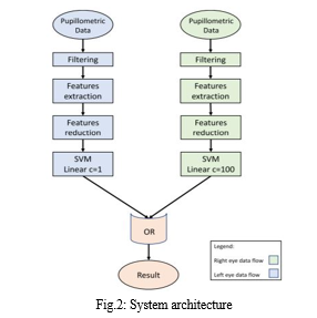
In this paper author is describing concept to detect eyes pediatric age genetic diseases using Pupillometry device data as this device is very accurate and it’s not require huge number of clinical test to detect disease. All existing techniques require huge number of clinical test to diagnose eye pupil disease in children’s and it’s not good for children’s health, so author using Pupillometry device which capture pupil diameters continuously and records that data in raw format in the file. Later we can analyse that data using Machine Learning SVM algorithm to detect presence of disease. Here author using two different SVM classifiers to train right and left eye pupil data and then performing OR operations between two classifier using ENSEMBLE VOTING classifier to get classifier with better accuracy. SVM will assign disease class label as 1 to train data if pupils size diameter is huge and if its size is normal then classifier will assign value 0.
B. Advantages
- The study that applies machine learning to pupillometric data in order to diagnose a genetic disease in pediatric age
- The ensemble system achieved 84.6% accuracy, 93.7% sensitivity and 78.6% specificity.

C. Modules
To implement this data author using Pupillometry raw data and perform below functions to diagnose pupils disease.
- Upload Pupillometry Data: Using this module we will upload raw data which contains continuous recording of pupils data.
- Filtering: Raw data contains huge number of buggy values and we will filter that raw data to extract only useful information such as pupil min and max diameter
- Features Extraction: Using this module all pupil min and max features extracted from raw data.
- Features Reduction: Using this module we will remove unnecessary features from raw data such as camera name, position etc to reduce features set. In this module we will extract features such as Patient ID, MAX, MIN, DELTA, CH etc. Extracted data can be used to split into train and test data
- Right SVM: Using this module we will train SVM with right pupil data
- Left SVM: using this module we will train SVM with left Pupil data and then apply SVM on test data to calculate prediction accuracy, sensitivity and specificity.
- Ensemble Algorithm (Left & Right SVM): Using this module we will combine both classifier to get classifier with high prediction accuracy.
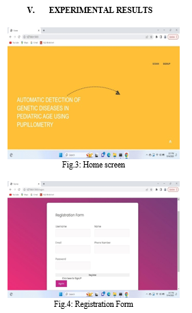
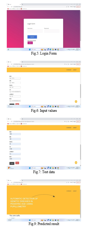
- Predict Disease: using this module we will upload test data and then apply SVM classifier to predict disease.
IV. IMPLEMENTATION
A. Algorithms
- Support Vector Machines: A support vector machine (SVM) is a type of deep learning algorithm that performs supervised learning for classification or regression of data groups. In AI and machine learning, supervised learning systems provide both input and desired output data, which are labeled for classification. Support vectors are data points that are closer to the hyperplane and influence the position and orientation of the hyperplane. Using these support vectors, we maximize the margin of the classifier. Deleting the support vectors will change the position of the hyperplane. These are the points that help us build our SVM.
- Clinical Decision Support System (CDSS): Our activity was performed within a research project, which main goal is defining effective protocols and systems for an early diagnosis and monitoring through chromatic pupillometry. The team that worked to this project is structured in three operative units: Department of Information Engineering of the University of Florence, Eye Clinics at the University of Campania Luigi Vanvitelli and at the University of Milan. This team designed a novel CDSS for diagnosis of Retinitis Pigmentosa (RP) in pediatric subjects.


Conclusion
This paper describes a new approach for supporting clinical decision for diagnosis of retinitis pigmentosa starting from analysis of pupil response to chromatic light stimuli in pediatric patients. The system was developed to clean artefacts, extract features and help the diagnosis of RP using a ML approach based on an ensemble model of two fine-tuned SVMs. Performances were evaluated with a leave-one-out cross-validation, also used to identify the best combination of internal parameters of the SVM, separately for both the left and right eyes. The class assigned to each eye were combined in the end with an OR-like approach so as to maximize the overall sensitivity of the CDSS; the ensemble system achieved 84.6% accuracy, 93.7% sensitivity and 78.6% specificity. The small amount of data available for this work, calls for further tests with a larger data pool for validating the performance of the system. Future scope includes testing the same approach with different devices. A problem that came out with great evidence, at the signal acquisition stage, is the frequent presence of movement artifacts. This is due to the particular shape of the device, together with the young age of the enrolled patients. Devices with different frame, including also systems based on smartphones, are going to be investigated. Moreover, considering the duration of the whole acquisition protocol, the procedure would benefit of some systems to capture the attention of the young patient (and his/her sight).
References
[1] X.-F. Huang, F. Huang, K.-C. Wu, J. Wu, J. Chen, C.-P. Pang, F. Lu, J. Qu, and Z.-B. Jin, ‘‘Genotype–phenotype correlation and mutation spectrum in a large cohort of patients with inherited retinal dystrophy revealed by next-generation sequencing,’’ Genet. Med., vol. 17, no. 4, pp. 271–278, Apr. 2015. [2] R. Kardon, S. C. Anderson, T. G. Damarjian, E. M. Grace, E. Stone, and A. Kawasaki, ‘‘Chromatic pupil responses. Preferential activation of the melanopsin-mediated versus outer photoreceptor-mediated pupil light reflex,’’ Ophthalmology, vol. 116, no. 8, pp. 1564–1573, 2009. [3] J. C. Park, A. L. Moura, A. S. Raza, D. W. Rhee, R. H. Kardon, and D. C. Hood, ‘‘Toward a clinical protocol for assessing rod, cone, and melanopsin contributions to the human pupil response,’’ Invest. Ophthalmol. Vis. Sci., vol. 52, no. 9, pp. 6624–6635, Aug. 2011. [4] A. Kawasaki, S. V. Crippa, R. Kardon, L. Leon, and C. Hamel, ‘‘Characterization of pupil responses to blue and red light stimuli in autosomal dominant retinitis pigmentosa due to NR2E3 mutation,’’ Investigative Ophthalmol. Vis. Sci., vol. 53, no. 9, pp. 5562–5569, 2012. [5] A. Kawasaki, F. L. Munier, L. Leon, and R. H. Kardon, ‘‘Pupillometric quantification of residual rod and cone activity in Leber congenital amaurosis,’’ Arch. Ophthalmol., vol. 130, no. 6, pp. 798–800, Jun. 2012. [6] A. Kawasaki, S. Collomb, L. Léon, and M. Münch, ‘‘Pupil responses derived from outer and inner retinal photoreception are normal in patients with hereditary optic neuropathy,’’ Exp. Eye Res., vol. 120, pp. 161–166, Mar. 2014. [7] P. Melillo, A. de Benedictis, E. Villani, M. C. Ferraro, E. Iadanza, M. Gherardelli, F. Testa, S. Banfi, P. Nucci, and F. Simonelli, ‘‘Toward a novel medical device based on chromatic pupillometry for screening and monitoring of inherited ocular disease: A pilot study,’’ in Proc. IFMBE, vol. 68, 2019, pp. 387–390. [8] E. Iadanza, R. Fabbri, A. Luschi, F. Gavazzi, P. Melillo, F. Simonelli, and M. Gherardelli, ‘‘ORÁO: RESTful cloud-based ophthalmologic medical record for chromatic pupillometry,’’ in Proc. IFMBE, vol. 73, 2020, pp. 713–720. [9] E. Iadanza, R. Fabbri, A. Luschi, P. Melillo, and F. Simonelli, ‘‘A collaborative RESTful cloud-based tool for management of chromatic pupillometry in a clinical trial,’’ Health Technol., pp. 1–14, Aug. 2019, doi: 10.1007/s12553-019-00362-z. [10] S. B. Kotsiantis, I. Zaharakis, and P. Pintelas, ‘‘Supervised machine learning: A review of classification techniques,’’ Emerg. Artif. Intell. Appl. Comput. Eng., vol. 160, pp. 3–24, Jun. 2007. [11] J. A. Alzubi, ‘‘Optimal classifier ensemble design based on cooperative game theory,’’ Res. J. Appl. Sci., Eng. Technol., vol. 11, no. 12, pp. 1336–1343, Jan. 2016 [12] J. Alzubi, A. Nayyar, and A. Kumar, ‘‘Machine learning from theory to algorithms: An overview,’’ J. Phys., Conf. Ser., vol. 1142, Nov. 2018, Art. no. 012012. [13] O. A. Alzubi, J. A. Alzubi, S. Tedmori, H. Rashaideh, and O. Almomani, ‘‘Consensus-based combining method for classifier ensembles,’’ Int. Arab J. Inf. Technol., vol. 15, no. 1, pp. 76–86, Jan. 2018. [14] P. Sajda, ‘‘Machine learning for detection and diagnosis of disease,’’ Annu. Rev. Biomed. Eng., vol. 8, no. 1, pp. 537–565, Aug. 2006. [15] J. A. ALzubi, B. Bharathikannan, S. Tanwar, R. Manikandan, A. Khanna, and C. Thaventhiran, ‘‘Boosted neural network ensemble classification for lung cancer disease diagnosis,’’ Appl. Soft Comput., vol. 80, pp. 579–591, Jul. 2019.
Copyright
Copyright © 2023 Ganji Deepthi , Dr. V. Uma Rani. This is an open access article distributed under the Creative Commons Attribution License, which permits unrestricted use, distribution, and reproduction in any medium, provided the original work is properly cited.

Download Paper
Paper Id : IJRASET55969
Publish Date : 2023-10-02
ISSN : 2321-9653
Publisher Name : IJRASET
DOI Link : Click Here
 Submit Paper Online

