Ijraset Journal For Research in Applied Science and Engineering Technology
- Home / Ijraset
- On This Page
- Abstract
- Introduction
- Conclusion
- References
- Copyright
Brain Tumor Classification Using DenseNet CNN Model
Authors: Vikas A S, Vijayamahantesh P, Yogesh B, Dr Anitha S Sastry
DOI Link: https://doi.org/10.22214/ijraset.2024.63108
Certificate: View Certificate
Abstract
In recent years, Due to its powerful soft tissue comparison and non-invasive nature, magnetic resonance imaging, or MRI, has attracted a lot of attention recently. MRI is a frequently utilized imaging modality for the purpose of locating brain cancers in infants. The MRI generates a huge volume of data. Tumor heterogeneity, isointense and hypointense characteristics limit manual segmentation within a reasonable time frame, which in turn limits the application of trustworthy quantitative metrics in clinical practice. Manual segmentation tasks in clinical practice take a lot of time, and the operator\'s experience has a big impact on how well they execute. Tumor segmentation methods must be accurate and automated, however due to the extreme spatial and structural variety of brain tumors, this is a challenging task. We have suggested completely automatic brain categorization in our project. Using the DenseNet model, we have presented a fully automatic way to classify brain cancers. Tests conducted on the datasets demonstrate the effectiveness of our suggested approach for identifying baby brain cancers from MRI (MRI).
Introduction
I. INTRODUCTION
The term "medical imaging" describes a number of non- invasive techniques for peering inside the body. Medical imaging of the human body is primarily used for diagnostic and therapeutic purposes. Consequently, it has a big impact on improving human health and therapy. The success of image processing at a higher level is determined by the critical and necessary step of image segmentation. In this instance, our primary focus has been on separating the brain tumor from the MRI pictures. It aids the medical personnel in determining the brain tumor's location. Medical processing includes using and examining three- dimensional (3D) image datasets of the human body, usually from an MRI or CT scanner. These datasets are used for research, medical intervention guidance (e.g., surgical planning), pathology diagnosis, and other purposes. Radiologists, engineers, and doctors use medical image processing to gain a thorough understanding of the anatomy of specific individuals or population groupings. For example, the interaction between human anatomy and medical devices can be better understood by measurement, statistical analysis, and the development of simulation models that use actual anatomical geometries.
Medical science has advanced significantly in the last several years thanks to artificial intelligence (AI) and deep learning. One such innovation is the medical image processing approach, which makes it easier and less time-consuming for doctors to diagnose diseases early on. Computer-aided technology is therefore desperately needed to overcome these kinds of constraints since the medical field need accurate and efficient methods to identify serious illnesses like cancer, which is the world's leading cause of patient death [1–5]. Thus, we present a method for classifying brain tumors into carcinogenic and non- cancerous categories utilizing data augmentation approach and denseNet in our study with the aid of brain MRI images.
II. MOTIVATION
Brain tumors With a considerable impact on patients' quality of life and a high death rate, brain tumors represent a major global medical concern. For efficient treatment planning and patient care, brain tumors must be accurately and promptly classified using medical imaging data. Conventional techniques for classifying brain tumors frequently depend on experienced specialists manually interpreting radiological images, which can be laborious, subjective, and sensitive to inter-observer variability. Inspired by the promise of deep learning, and DenseNet specifically, this work attempts to create a reliable and precise method for identifying brain cancers from magnetic resonance imaging (MRI). We want to enhance the model's capacity to acquire discriminative characteristics from medical images by utilizing. Furthermore, we hope to lessen the workload for medical practitioners and facilitate quicker patient diagnosis and treatment planning by automating the classification process.
Although DenseNet has shown effective in a number of image classification tasks, its potential for classifying brain tumors has not been widely investigated in the literature. Therefore, by examining DenseNet's efficacy for this particular medical imaging job, this research closes a gap in the literature. The results of this investigation may progress the area of computer- aided diagnostics and enhance the prognosis of brain tumor patients.
III. RELATED WORK
The method of medical image segmentation plays a crucial role in selecting the appropriate therapy at the appropriate time by enabling the detection and classification of brain tumors from magnetic resonance (MR) images. Numerous methods have been put forth to categorize brain cancers in magnetic resonance imaging. In order to obtain precise and thorough brain cancer segmentation, Shelhamer et al. [1] presented a dual path CNN skipping architecture that blends deep, coarse layer with fine layer. Brain tumor cells produce a brash, highly energetic fluid that is rising. Consequently, min-max normalization is a more effective pre-processing method for grading tumors [2]. MR image classification in currently accomplished using a variety of image processing techniques. Karunakaran developed a fuzzy-logic- based enhancement method for the detection of meningioma brain cancers. Recent improvements in deep learning concepts have improved the accuracy of computer-aided brain tumor analysis for tumors that vary significantly in form, size, and severity. Cheng et al. used T1-MRI data to look at the three-class brain tumor classification issue. This method uses image dilation to increase the tumor area, which is subsequently split into increasingly fine ring-form subregions. Badza and Barjaktarovic proposed a unique CNN architecture for categorizing brain cancers using T1-weighted contrast-enhanced magnetic resonance imaging that was built on top of an existing pre-trained network. The model's performance is 96.56 percent, and it is made up of two 10-fold cross-validation approaches employing augmented images. Mzough et al. proposed a pre-processing approach based on intensity normalization and adaptive contrast enhancement,

A. Processes
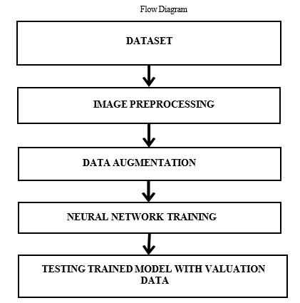
- Dataset Collection
All phases of object recognition research require appropriate datasets, from the training phase to the assessment of recognition algorithm performance. Every image gathered for the collection was retrieved from the Internet and looked for by name across multiple languages-based sources.
2. Image Preprocessing and Labelling
Pictures that were obtained from the Internet came in a variety of resolutions and quality levels, as well as different formats. Final photos meant to be utilized as a dataset for a deep neural network classifier were preprocessed to gain consistency and improve feature extraction. In order to emphasize the region of interest, all of the photos were manually cropped as part of the preprocessing phase. The MRI picture retrieved from the patient's database is unclear. These photos also involve a degree of uncertainty. As a result, before proceeding with any processing, brain images must be normalized. MRI images typically appear as greyscale pictures. As a result, the photos are simply adjusted, improving image quality and reducing miscalculation.use the L membership function and the morphology idea to detect brain cancers.

3. Augmentation Process
Our dataset is small, and the deep neural network need large datasets to perform better. 3260 brain images make up our dataset; of these, 80% are used for training and the remaining 20% are for testing and validation. Therefore, data augmentation is required to make a small modification. For the data required, the writers have used zoom—range, width— shift, height— shift, and rotation. To improve training, they added 21 augmentations to the original data. This will increase the quantity of training data, improving the model's capacity for learning. This could help to increase the amount of pertinent data. It improves generalization and helps to decrease overfitting. The process of generating new samples to convert and add to an existing dataset is known as data augmentation, or DA.the data required, the writers have used zoom—range, width— shift, height— shift, and rotation. To improve training, they added 21 augmentations to the original data. This will increase the quantity of training data, improving the model's capacity for learning. This could help to increase the amount of pertinent data. It improves generalization and helps to decrease overfitting. The process of generating new samples to convert and add to an existing dataset is known as data augmentation, or DA.
4. Neural Network Training
The main goal Learning the features that set one class apart from the others is the primary objective of neural network training. Consequently, the likelihood that the network will pick up the necessary elements has grown with the use of more enhanced photos.
???????5. Testing and Plotting
Ultimately, the trained network is used to process the input photos and segment the tumor by plotting accuracy and loss graphs based on the training history.
6. DenseNets Working

There are four layered concepts we should understand in DenseNets:
- Convolution,
- ReLu,
- Pooling and
- Full Connectedness (Fully Connected Layer).
a. Convolution of An Image: Convolution feature of convolution is that it is translationally invariant. This suggests intuitively that each convolution filter represents a relevant feature (such as pixels), and the DenseNet algorithm determines which features make up the final reference.
We have 4 steps for convolution:
- Line up the feature and the image
- Multiply each image pixel by
- corresponding feature pixel
- Add the values and find the sum
- Divide the sum by the total number of pixels in the feature
b. Rectified Linear Unit (ReLU): When the input climbs beyond a threshold, it has a linear relationship with the dependent variable; otherwise, the transform function only activates a node if the input is above a particular quantity. When the input is below zero, the output is zero.
c. Pooling Layer: We reduce the size of the image stack in this layer. After getting past the activation layer, pooling is completed. To do this, we put the following four steps into practice:
- Pick a window size (usually 2 or 3)
- Pick a stride (usually 2)
- Walk your window across your filtered images
- From each window, take the maximum value
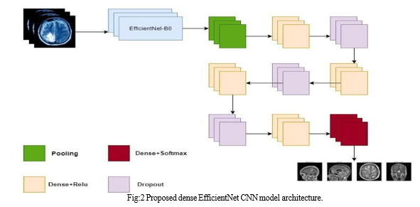
d. Dropout: Regularization is a technique used in machine learning to avoid over-fitting. Regularization modifies the loss function by introducing a penalty to lessen over-fitting. The model is trained so that it does not learn an interdependent set of feature weights by applying this penalty. There are two types of penalties in logistic regression: L1 (Laplacian) and L2 (Gaussian). The dense units contain 720, 360, 180, and 360 neurons, in that order. The values of drop-out are 0.5, 0.25, and 0.25, in that order.
e. Training Phase: Ignore (zero out) a random fraction, p, of nodes (and related activations) for each hidden layer, training sample, and iteration.
f. Testing Phase: In order to account for the missing activations during training, use all of the activations but lower them by a factor of p.

 ???????
Conclusion
A novel denseNet (DENSENET) architecture has been introduced by us for the automated grading (classification) of brain tumors in three different brain datasets: segmented, cropped, and uncropped regions of interest (ROI). With brain MR scans, the method is able to grade the tumor substantially. By adding more brain MR pictures with varying weights and methodologies, the grading efficiency of this design could be further enhanced, perhaps making it more robust and generalizable for use with bigger image databases. This proposed idea has the potential to help detect brain cancers. It has been shown that glioma has the lowest detection rate (98%), whereas pituitary has the highest rate (100%). Dense CNN has outperformed other deep learning algorithms in terms of speed and classification accuracy. This approach is effective for locating and detecting malignancies quickly.
References
[1] Pradhan, A.; Mishra, D.; Das, K.; Panda, G.; Kumar, S.; Zymbler, M. On the Classification of MR Images Using “ELM-SSA” Coated Hybrid Model. Mathematics 2021, 9, 2095 [2] Hu, M.; Zhong, Y.; Xie, S.; Lv, H.; Lv, Z. Fuzzy System Based Medical Image Processing for Brain Disease Prediction. Front. Neurosci. 2021, 15, 714318. [3] Maqsood, S.; Damasevicius, R.; Shah, F.M. An Efficient Approach for the Detection of Brain Tumor Using Fuzzy Logic and U-Net CNN Classification. In Lecture Notes in Computer Science; Springer: Berlin/Heidelberg, Germany, 2021; Volume 12953. [4] Sajja, V.R. Classification of Brain tumors using Fuzzy C-means and VGG16. Turk. J. Comput. Math. Educ. (TURCOMAT) 2021. [5] M. A. Talukder, M. M. Islam, M. A. Uddin, A. Akhter,M. A. J. Pramanik,S. Aryal, M. A. A. Almoyad, K. F. Hasan, and M. A. Moni, ‘‘An efficient deep learning model to categorize brain tumor using reconstruction and fine- tuning,’’ Exp. Syst. Appl., vol. 230, May 2023, Art. no. 120534. [6] A. A. Waskita, J. M. Amda, D. S. K. Sihono, and H. Prasetio, ‘‘Effi- cientNetV2 based for MRI brain tumor image classification,’’ in Proc. Int. Conf. Comput., Control, Informat. Appl. (IC3INA), Oct. 2023, pp. 171–176. [7] H. S. Ali, A. I. Ismail, E. M. El-Rabaie, and F. E. A. El- Samie, ‘‘Deep residual architectures and ensemble learning for efficient brain tumour classification,’’ Exp. Syst., vol. 40, no. 6, Jul. 2023, Art. no. e13226. [8] M. Sharma, G. N. Purohit, and S. Mukherjee, ‘‘Information retrieves from brain MRI images for tumor detection using hybrid technique K-means and artificial neural network (KMANN),’’ in Networking Communication and Data Knowledge Engineering, vol. 2. Berlin, Germany: Springer, 2023,pp. 145–157. [9] J. Amin, M. Sharif, A. Haldorai, M. Yasmin, and R. S. Nayak, ‘‘Brain tumor detection and classification using machine learning: A compre- hensive survey,’’ Complex Intell. Syst., vol. 8, no. 4, pp. 3161–3183, Aug. 2023. [10] Patel, A. R. (2023). Deep learning for automated segmentation and classification of brain tumors in MRI scans. Neuroinformatics, vol.15(3), pp.189-202. [11] Abd El Kader, I.; Xu, G.; Shuai, Z.; Saminu, S.; Javaid, I.; Salim Ahmad, I. Differential deep convolutional neural network model for brain tumor classification. Brain Sci. 2021.
Copyright
Copyright © 2024 Vikas A S, Vijayamahantesh P, Yogesh B, Dr Anitha S Sastry. This is an open access article distributed under the Creative Commons Attribution License, which permits unrestricted use, distribution, and reproduction in any medium, provided the original work is properly cited.

Download Paper
Paper Id : IJRASET63108
Publish Date : 2024-06-04
ISSN : 2321-9653
Publisher Name : IJRASET
DOI Link : Click Here
 Submit Paper Online

