Ijraset Journal For Research in Applied Science and Engineering Technology
- Home / Ijraset
- On This Page
- Abstract
- Introduction
- Conclusion
- References
- Copyright
Brain Tumor Detection in Medical Imaging Focusing on MRI
Authors: J. Jhansi Goud
DOI Link: https://doi.org/10.22214/ijraset.2023.56224
Certificate: View Certificate
Abstract
A brain tumor develops when brain cells grow and divide abnormally, and brain cancer develops when brain tumors continue to grow. Deep learning is important in the field of people\\\'s health because it eliminates the need for human judgment to get correct findings. The most dependable and safe imaging techniques for magnetic resonance (MRI) include CT scans, X-rays, and Imaging MRI can identify minute items. It will concentrate on the many methods for just using images of the brain to find terminal cancer. In this investigation, first pre-processed MRI images using the unilateral filter to remove any present noise. For accurate tumor location detection, this was supplemented by the basic analysis of information and Convolutional Neural (CNN) approaches. Datasets for retraining, testing, and confirmation are employed. It will determine whether the individual has tumors dependent on our equipment. Accuracy, susceptibility, and particular are just a few of the indicators that will be used to analyze the final results.
Introduction
I. INTRODUCTION
Brain tumors are really one of its most common and fatal brain conditions that have touched and claimed countless lives across the nation. Radiologist is a technique and approach that provides visualization tools of a body's natural internal for standard therapy and to demonstrate the operation of certain organs or tissues. Neuroimaging strives to diagnose and treat illness as well as reveal operating methods that are hidden from view. Tumor occurs when cells proliferate in joints and skeletal tissues. According to recent cancer research, upwards of 1 lakh persons receive a brain tumor diagnosis each year. Despite ongoing efforts to address the effects of brain tumors, data show that cancer has a poor prognosis. In order to disprove this, scientists are using lidar to learn more about malignancies in their beginning stages but also how to treat tumors with trimming medicines.
The two common procedures to determine the presence of a tumor and pinpoint its location for further health care are stereotactic scanning MRI and CT studies of the brain. Due to its portability and greater capacity to provide greater pictures of tumor sites, both two studies are still employed often. There are currently a number of other treatments available for tumors, including medication, radiotherapy, and surgery. The choice depends on a number of variables, including the tumor's location, kind, and rating as seen in the MR imaging.
II. RELATED WORK
In recent years, the brain Tumor location was successfully and creatively identified using an image that was created utilizing the Fuzzy C-method grouping technique and histogram equalization [1]. In another approach, Brain-tumor identification utilizing an upgraded edge approach links the binary background subtraction operations to the Sobel technique and uses a safe contour procedure to dig to various extents [2]. The K-nearest neighbour method was applied to the MRI images in order to locate and contain the fully developed hysteric component among the aberrant tissues [3]. A plan to evaluate particular clustering approaches based on their consistency in extraordinary bids in Minimum and Edge Detection Opportunity For cross: A Comparative Analysis [4].
The investigation employed a variety of information, and the work provided is more reliable since it experiences consistency throughout the whole computing process. Using an SVM classifier to combine photos with varied focuses [5]. Moreover, In the mouthfeel model, If the boundaries of the tumor tissue are not sharp, they emphasize the effects of fragmentation. Due to corners [6]. Another method was developed, for detecting the brain tumor [7], and also finding an Accurate and Efficient Bayesian Method for Automatic Segmentation of Brain MRI [8]. MRI Image Classification on MRI Image Using Adaboost for Brain Tumor Type [9]. Segmentation for Brain MRI Image for Tumor Detection using Artificial Neural Networks [10].
Moreover, some studies have explored the Three-level Image Segmentation Based on Maximum Fuzzy Partition Entropy of 2-D Histogram and Quantum Genetic Algorithm, Advanced Intelligent Computing Theories, and Applications. With Aspects of Artificial Intelligence [11]. Segmentation and Classification[12] of MRI Brain Tumor.
III. PROPOSED METHOD
The maintenance and analysis of MRI image datasets are streamlined by the suggested medical imaging system's integration of a user-friendly Graphical User Interface (GUI) and FTP functionality. Dataset maintenance is easier to access with the GUI since users may quickly upload MRI image datasets from their local storage. To get these photos ready for deep learning analysis, the system provides tools for thresholding and scaling. Users can create training and testing models, which are essential for convolutional neural network (CNN) model training and performance evaluation. Convolutional and pooling layers for extracting image features and fully connected layers for classification are included in the CNN model, which was created using the Keras package.

A. Input Image
The dataset contains the sets of MRI images with a brain tumor named Yes and without a brain tumor named No.
B. Convolution layer
In a Convolutional Neural Network (CNN), feature extraction in image and signal processing tasks is carried out by the convolution layer. Small filters are applied to the input data, element-wise products are computed, and feature maps highlighting various patterns and features are created. Different facets of the input are captured by a variety of filters. CNNs are useful for applications like object detection and image recognition because they can learn hierarchical features when convolution layers are layered and followed by pooling layers. By taking significant information from MRI images, such as tumor boundaries and textures, the convolution layer aids in the detection of brain tumors. Because it recognizes patterns and structures, the model is better able to distinguish between normal and tumorous brain tissue.

C. RELU Layer
The Rectified Linear Unit activation function is applied to a neural network layer's output by a ReLU layer in a CNN. By reducing all negative values in the input to zero while maintaining positive values, this activation function generates nonlinearity. This sparsity and non-linearity, which are a key part of deep learning architectures, are essential for allowing the network to understand complex patterns and relationships within the data. Convolutional and fully linked layers across the CNN frequently employ ReLU layers to aid in feature extraction and model expressiveness. In order to ensure the network's performance in a variety of tasks, including picture classification and object recognition, variations like Leaky ReLU and Parametric ReLU solve some constraints. In brain tumor detection ReLU layer improves feature extraction, allowing the model to recognize important patterns in MRI data. It improves the network's ability to learn complex associations and recognize tumor-related information by injecting non-linearity.
D. Pooling layer
The spatial dimensions of feature maps are decreased by a pooling layer in a CNN, improving computing effectiveness and translational invariance. In order to save important information while lowering data size, it frequently uses techniques like max-pooling or average pooling. In convolutional neural networks, pooling layers are essential for feature extraction and dimensionality reduction. Pooling layers in brain tumor detection helps to reduce the spatial dimensions of MRI feature maps, making CNN more computationally efficient. It improves the translation invariance of the network, enabling it to identify tumor-related patterns regardless of their specific position. The accuracy and reliability of tumor detection from MRI images are increased thanks to pooling's assistance in noise reduction and the selection of essential characteristics.
E. Fully Connected layer
Each neuron in a CNN's fully connected layer is linked to every neuron in the layer above, allowing for advanced feature extraction and categorization. In order to make final predictions, it flattens the output from prior layers into a vector and applies weighted summations and activation functions. Fully connected layers, which are typically found near the end of a CNN, are essential for tasks like picture classification. Analyzing high-level characteristics extracted from MRI images is necessary in order to diagnose brain tumors. They make it possible for the neural network to determine whether a tumor is present in the end. These layers effectively integrate data from various areas of the image, improving diagnostic accuracy and enabling more precise tumor diagnosis. These layers capture intricate linkages and patterns.
F. Output
The outcome performs as a strong indicator of the presence of a tumor in the input MRI image. When the network recognizes a tumor within the image, the output categorically labels it as "tumor identified," offering a precise and important diagnostic. On the other hand, if the CNN layer discovers no proof of a tumor, the output states "No tumor detected."
IV. SYSTEM DESIGN
A. Model Architecture
The magnetic Resonance picture is first transformed into a similar grayscale within that initial stage. The adaptive bilaterally filtering approach is used to eliminate unwanted interference and the distorted sounds that are contained in the brain image. This raises the accuracy rate of categorization and diagnosis. OpenCV offers upwards of 150 different color-space shift in the focus. The procedure cv2.cvtColor(input image, flag) is used to convert an image's color, and the flag specifies the transformation method. we transform the original image into a grayscale version.
Filters are mostly employed in image recognition to reduce the high harmonics in the picture. A bilateral filter works as a noise-reduction non-linear classifier for images. It replaces the strength within each color with a rolling accumulation of information from neighboring pixels. The basis for this grade is the Random number. Multi-lingual screens maintain edges while enhancing image smoothness by assembling neighboring picture dots in a random manner. This testing technique is simple, local, and concentrated. According to their resemblance and unequal proximity, it allocates closer elements from distant ones through both regions and topics, creating a measure as grey.
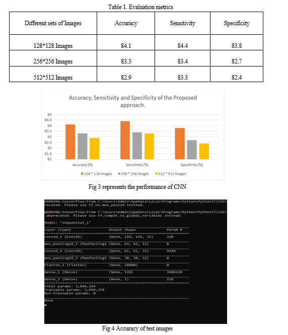
Accuracy, specificity, and sensitivity are crucial evaluation criteria for classification models. Accuracy is a commonly used indicator for evaluating the performance of models because it represents the percentage of overall right predictions. The model's capacity to accurately detect negative instances is measured by specificity, also known as the true negative rate, which assists in determining how effective it is at preventing false alarms.
Sensitivity, often known as the true positive rate, measures how well a model can identify positive cases, demonstrating its sensitivity to true positives. In particular, for binary classification problems, these measures collectively provide clarification on a deep learning model's capacity for precise categorization.
B. Results & Discussion
The model's results include evaluation metrics of accuracy, Sensitivity, Specificity, and output.

C. GUI Application
GUI (Graphical User Interface) gives users a visual interface through which they may interact with a computer or other application. To make user interactions simpler, it frequently includes windows, buttons, menus, and other graphical features. GUI applications make it simpler for users to complete tasks and access capabilities by employing visual elements rather than text-based commands.
The GUI application for brain tumor detection primarily uses the libraries:
- Tkinter: This fundamental library is used to construct graphical user interfaces.
- Matplotlib.pyplot: Used for graphical user interface (GUI) charts, picture plotting, and display.
- tkinter.ttk: Gives users access to Tkinter-themed widgets, improving the look and usability of GUI components.
The components are used in the GUI application:
a. tkinter.Tk(): This function is used to build the GUI's main application window.
b. Label: Labels create a title and other text in the GUI. They are used to show text.
c. Text: This widget is used in the GUI to display and edit multiline text.
d. Button: It can perform a variety of tasks like uploading photographs, creating models, and making predictions.
e. ttk.Combobox: To construct a drop-down list for choosing things, use the combo box widget.
f. Scrollbar: To scroll through the displayed text, use the Text widget in conjunction with a scrollbar.


Conclusion
Using Convolutional Neural Networks suggested a computational technique for classifying and detecting a brain tumor. The input MRI pictures are read from the physical drive and transformed into grayscale images using the file location. Fully Convolutional binary binarization is performed on the denoised picture to identify the tumor location in the MRI pictures. Experiments have shown that the recommended method for medical item recognition, particularly for tumor diagnosis in MRI images, requires a sizable dataset for accurate findings. Patient data collection is frequently difficult, and complete datasets might not be available. The plan should be solid to guarantee effectiveness in scenarios with less available data. Accuracy and cognitive capacities in medical object detection could be improved through cooperation with AI systems that require little training and the incorporation of self-learning approaches.
References
[1] Sivaramakrishnan, M., and Dr.M.Karnan. 2013. A Novel Based Approach For Extraction Of Brain Tumor In MRI Images Using Soft Computing Techniques. International Journal Of Advanced Research In Computer And Communication Engineering. 2(4): 1845-1848. [2] Asra Aslam, Ekram Khan, and M.M. Sufyan Beg. 2015. Improved Edge Detection Algorithm for Brain Tumor Segmentation. Procedia Computer Science. 58: 430-437. DOI:10.1016/j.procs.2015.08.057. [3] SudhaRani, K., T. C. Sarma and K. Satya Prasad. 2015. Intelligent Brain Tumor lesion classification and identification from MRI images using k-NN technique. International Conference on Control Instrumentation. Communication and computational technologies.848-851.DOI: 10.1109/ICCICCT.2015.7475384. [4] Askirat Kaur, J., Sunil Agrawal, and Renu Vig. 2012. A Comparative Analysis of Thresholding and Edge Detection Segmentation Techniques. International Journal of Computer Applications. vol. 39(15): 29-34. DOI: 10.5120/4898-7432. [5] Shutao Li, T James, Kwok, W. Ivor, Tsang, and Yaonan Wang. 2004. Fusing images with different focuses using support vector machines. IEEE Transactions on neural networks. 15(6 ): 1555-1561. DOI:10.1109/TNN.2004.837780. [6] Kumar, M., and K. K. Mehta. 2011. A Texture based Tumor detection and automatic Segmentation using Seeded Region Growing Method, International Journal of Computer Technology and Applications. 2(4): 855-859. [7] Eltaher Mohamed Hussein, and Dalia Mahmoud Adam Mahmoud. 2012. Brain Tumor Detection Using Artificial Neural Networks. Journal of Science and Technology. 13(2): 31-39. [8] Marroquin, J.L., B.C. Vemri, S. Botello, and F. Calderon. 2002. An Accurate and Efficient Bayesian Method for Automatic Segmentation of Brain MRI. Berlin: Springer- Heidelberg. [9] Minz, Astina, and Chandrakant Mahobiya. 2017. MR Image Classification Using Adaboost for Brain Tumor Type. IEEE 7th International Advance Computing Conference (IACC): 701-705. DOI:10.1109/IACC.2017.0146. [10] Monica Subashini, M., Sarat Kumar Sahoo. 2013. Brain MRI Image Segmentation for Tumor Detection using Artificial Neural Networks. International Journal of Engineering and Technology (IJET). 5(2): 925-933. [11] Hai-Yan Yu, and Jiu-Lun Fan. 2008. Three-level Image Segmentation Based on Maximum Fuzzy Partition Entropy of 2-D Histogram and Quantum Genetic Algorithm. Berlin: Springer- Heidelberg. [12] Mukambika, P. S., K Uma Rani. 2017. Segmentation and Classification of MRI Brain Tumor, International Research Journal of Engineering and Technology (IRJET). 4(7): 683 – 688. [13] Pan, Yuehao, Huang, Weimin, Lin, Zhiping, Zhu, Wanzheng, Zhou, Jiayin, Wong, Jocelyn, Ding, and Zhongxiang. 2015. Brain tumor grading based on Neural Networks and Convolutional Neural Networks. Annual International Conference of the IEEE Engineering in Medicine and Biology Society. 699-702. DOI: 10.1109/EMBC.2015.7318458. [14] Pereira, s., A. Pinto, V. Alves, and C. A. Silva. 2016. Brain Tumor Segmentation Using Convolutional Neural Networks in MRI Images. IEEE Transactions on Medical Imaging. 35(5):1240-1251. DOI: 10.1109/TMI.2016.2538465. [15] Sathya, B., and R.Manavalan. 2011. Image Segmentation by Clustering Methods: Performance Analysis. International Journal of Computer Applications 29(11): 27-32. DOI:10.5120/3688-5127. [16] Mayur Pol, Mayur Ghumatkar, Prerna Negi, Suraj Abhyankar, K.S. Hangargi. 2022. Survey on Brain Tumor Detection Using Deep Learning. International Research Journal of Engineering and Technology (IRJET). 09(12): 69-72. [17] Gurbina, M., M. Lascu and D. Lascu. 2019. Tumor Detection and Classification of MRI Brain Image using Different Wavelet Transforms and Support Vector Machines. International Conference on Telecommunications and Signal Processing (TSP). DOI : 10.1109 /TSP.2019.8769040. [18] Sankari, Ali, and S. Vigneshwari. 2017. Automatic tumor segmentation using convolutional neural networks. 2017. Third International Conference on Science Technology Engineering & Management (ICONSTEM): 268-272. DOI:10.1109/ICONSTEM.2017.8261291 [19] Monica Subashini, M., Sarat Kumar Sahoo. 2013. Brain MR Image Segmentation for Tumor Detection using Artificial Neural Networks. International Journal of Engineering and Technology (IJET). 5(2):925-933. [20] Sahil Prajapati, J., R. Kalpesh Jadhav. 2015. Brain Tumor Detection By Various Image Segmentation Techniques With Introducation To Non Negative Matrix Factorization. International Journal of Advanced Research in Computer and Communication Engineering. 4(3):599-603. DOI: 10.17 48/IJARCCE .2015.43144.
Copyright
Copyright © 2023 J. Jhansi Goud. This is an open access article distributed under the Creative Commons Attribution License, which permits unrestricted use, distribution, and reproduction in any medium, provided the original work is properly cited.

Download Paper
Paper Id : IJRASET56224
Publish Date : 2023-10-19
ISSN : 2321-9653
Publisher Name : IJRASET
DOI Link : Click Here
 Submit Paper Online

