Ijraset Journal For Research in Applied Science and Engineering Technology
- Home / Ijraset
- On This Page
- Abstract
- Introduction
- Conclusion
- References
- Copyright
Brain Tumor Detection Using Deep Learning
Authors: Abhishek Sawle, Shubham Bhosale, Shraddha Abhang, Pranay Ghagare, Prof. Dakshata Argade
DOI Link: https://doi.org/10.22214/ijraset.2024.59360
Certificate: View Certificate
Abstract
This paper explores the application of deep learning in detecting brain tumors through the comparison of ResNet50 and VGG19 classification models. The study also includes the comparison of Res-U-Net and U-Net models for tumor segmentation. The process involves feeding an input of Brain MRI scan into the classification model, which provides two outputs: detection of the tumor or confirmation of the patient\'s health. If a tumor is detected, the segmentation model locates it at a pixel level. The proposed approach has the potential to improve the accuracy and speed of brain tumor diagnosis, which could lead to more effective treatment.
Introduction
I. INTRODUCTION
Brain tumors pose a significant health risk, and early diagnosis is crucial for effective treatment. However, the classification of these tumors can be a challenging task for physicians and radiologists. Recent advances in artificial intelligence, particularly deep learning algorithms, offer promising solutions for the rapid and accurate detection of brain tumors.
Deep learning algorithms leverage neural networks to extract and encode features, enabling data abstraction and facilitating the localization and classification of tumors. Convolutional neural networks (CNNs), such as ResNet50 and VGG19, are particularly effective in image classification tasks. Additionally, segmentation of brain tumors is a crucial step in the detection and diagnosis process. This paper compares two segmentation models, Res-U-Net and U-Net, to determine their effectiveness in detecting brain tumors. The use of deep learning algorithms in medical image analysis has the potential to revolutionize the field, especially in areas where access to expert physicians and radiologists is limited. The paper's comparison of ResNet50, VGG19 for classification and Res-U-Net, U-Net for segmentation of brain tumors provides insights into the optimal models for detecting and diagnosing these tumors accurately and efficiently. Ultimately, the integration of deep learning algorithms into medical imaging has the potential to improve patient outcomes by enabling earlier diagnoses and more effective treatment options.
II. DATASET
Magnetic resonance imaging (MRI) is a powerful radiologic imaging technology used to produce high-quality two-dimensional or three-dimensional images of the brain and brainstem. These images can provide valuable insights into the structure and function of the brain, as well as help diagnose various disorders, including brain tumors.
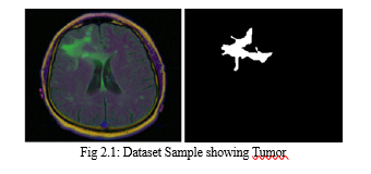
In this study, a dataset consisting of 7860 images and manual FLAIR abnormality segmentation masks was utilized. The images were obtained from The Cancer Imaging Archive (TCIA), and correspond to 110 patients in the lower-grade glioma collection of The Cancer Genome Atlas (TCGA) who had at least fluid-attenuated inversion recovery (FLAIR) sequencing and genomic cluster data available. The utilization of this dataset allows for the development and testing of machine learning algorithms for the automatic detection and segmentation of brain tumors. By utilizing these images and manual segmentation masks, researchers can train and test deep learning models to improve the accuracy and efficiency of brain tumor diagnosis. The integration of deep learning and medical imaging has the potential to revolutionize the diagnosis and treatment of brain tumors, ultimately improving patient outcomes.

III. PROPOSED SYSTEM
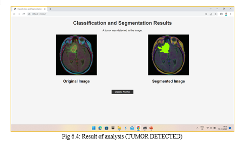
The proposed system for detecting brain tumors involves the gathering of patient information in the form of Magnetic Resonance Imaging (MRI) scans, which are then fed as input into the classification model. The main objective of the Classifier Model is to categorize the MRI scans into two categories: those with tumors and those without tumors. If the tumor is not detected, the patient is considered to have "no tumor." On the other hand, if a tumor is detected, the image is then sent to the segmentation model, which determines the precise location of the tumor at the pixel level.
The classification model uses deep learning algorithms to analyze the MRI scan data and identify the presence or absence of a tumor. This step is critical as it determines whether further analysis and diagnosis are necessary. In the case of no tumor detection, the patient can be classified as healthy and moved forward for other routine check-ups.
If the classification model detects a tumor, the MRI scan is then passed on to the segmentation model. This model further processes the image data and determines the exact location of the tumor at the pixel level. By doing so, it provides a detailed understanding of the tumor's size and shape, which can assist doctors in planning the most effective course of treatment for the patient.

Overall, the proposed system can significantly improve the accuracy and speed of brain tumor diagnosis, leading to better patient outcomes. The automated approach using deep learning algorithms reduces the likelihood of errors associated with manual diagnosis and is more efficient. This system has the potential to become a crucial tool in the diagnosis and treatment of brain tumors.
IV. ALGORITHMS
In this study, we used four deep learning algorithms for the tasks of brain tumor detection and segmentation. The algorithms we considered were ResNet50, VGG19, Res-U-Net, and U-Net.
A. VGG19
VGG19 is a deep convolutional neural network that is widely used for image recognition and classification tasks. It was developed by researchers at the University of Oxford in 2014 and is based on the VGG-Net architecture. VGG19 consists of 19 layers, including multiple convolutional and fully connected layers, making it one of the more complex neural networks used in computer vision. One of the key benefits of VGG19 is its ability to achieve high accuracy in image recognition tasks. It has been used in a wide range of applications, including facial recognition, object detection, and medical imaging. The network achieves high accuracy by extracting features from the raw image data at multiple levels, allowing it to capture both low-level and high-level information about the image. Another key benefit of VGG19 is its simplicity and ease of use. The network architecture is easy to understand and implement, making it accessible to a wide range of users, including those without a background in machine learning. This simplicity also makes it easy to modify the network architecture to suit specific needs or applications, such as adding or removing layers to improve performance. In conclusion, VGG19 is a powerful convolutional neural network that has become a popular tool in the field of computer vision. Its ability to achieve high accuracy in image recognition tasks, along with its simplicity and ease of use, has made it a valuable tool for a wide range of applications. As the field of computer vision continues to evolve, it is likely that VGG19 and other deep learning models will continue to play an increasingly important role in helping us to better understand and interact with visual data.
B. ResNet50
ResNet50 is a powerful deep neural network that has been widely used for image recognition and classification tasks. It is a variant of the ResNet architecture, which was introduced by researchers at Microsoft in 2015. The key innovation of ResNet50 is the use of residual connections, which allow the network to learn features from raw image data more effectively than traditional neural networks. ResNet50 consists of 50 layers, which are organized into a series of building blocks. Each building block contains multiple convolutional layers, along with a shortcut connection that allows the network to bypass certain layers. This shortcut connection is what gives ResNet50 its ability to learn features from raw image data more effectively than traditional neural networks. One of the key benefits of ResNet50 is its ability to handle complex image recognition tasks with high accuracy. In fact, ResNet50 has achieved state-of-the-art performance on a wide range of image recognition benchmarks, including the ImageNet dataset. This makes it a valuable tool for a wide range of applications, from facial recognition and object detection to medical imaging and autonomous vehicles.
In conclusion, ResNet50 is a powerful deep neural network that has revolutionized the field of image recognition and classification. Its use of residual connections and shortcut connections allows it to learn features from raw image data more effectively than traditional neural networks, making it a valuable tool for a wide range of applications
C. U-Net
U-Net is a deep convolutional neural network architecture that was developed for biomedical image segmentation. It was introduced by researchers at the University of Freiburg in 2015 and has since been widely adopted in a range of applications, from medical imaging to satellite imagery. The U-Net architecture is named for its U-shaped structure, which allows it to capture both local and global features of the input image.
One of the key innovations of U-Net is the use of skip connections, which allow the network to preserve fine-grained information about the input image. These skip connections connect the layers in the encoder and decoder parts of the network, allowing information to flow both downwards and upwards. This structure enables the network to achieve high accuracy in segmentation tasks while also maintaining a high degree of spatial resolution.
Another key benefit of U-Net is its ability to handle datasets with a small number of labeled images. This is achieved through the use of data augmentation techniques, such as flipping and rotating the input images, which allows the network to learn more robust and generalizable features. This makes U-Net particularly useful in medical imaging applications, where it can be difficult to obtain large datasets with a high degree of label accuracy.
In conclusion, U-Net is a powerful convolutional neural network architecture that has become a popular tool in the field of biomedical image segmentation.
D. Res-U-Net
Res-U-Net is a hybrid deep neural network architecture that combines the benefits of ResNet and U-Net. It was introduced in 2018 by researchers at the University of Trento and is specifically designed for medical image segmentation tasks. The Res-U-Net architecture consists of multiple residual blocks, skip connections, and a U-shaped structure, making it a powerful tool for high-accuracy medical image segmentation.
One of the key innovations of Res-U-Net is the use of residual connections, which allow the network to learn more robust and generalizable features from the input image. These residual connections enable the network to bypass certain layers and capture the fine-grained features of the image more effectively. Additionally, Res-U-Net also includes skip connections, which connect the encoder and decoder parts of the network, allowing the network to maintain a high degree of spatial resolution throughout the segmentation process.
In many medical imaging applications, such as tumor segmentation, the number of pixels belonging to the target class is significantly smaller than the number of pixels belonging to the background class. Res-U-Net addresses this issue by incorporating a weighted loss function, which assigns higher weights to pixels belonging to the target class. This enables the network to focus more on accurately segmenting the target class, improving the overall accuracy of the segmentation results.
In conclusion, Res-U-Net is a powerful deep neural network architecture that combines the benefits of ResNet and U-Net for high-accuracy medical image segmentation. Its use of residual connections and skip connections allows it to capture fine-grained features of the input image while maintaining spatial resolution.
V. ALGORITHM ANALYSIS
In this technical paper, we compare the performance of four deep learning algorithms for brain tumor detection and segmentation. The algorithms considered are ResNet50, VGG19, ResUNet, and U-Net.
Deep learning has become an increasingly popular technique for computer vision and image classification in recent years. ResNet50 and VGG19 are two widely used deep learning models for image classification. ResNet50 is a 50-layer convolutional neural network that uses skip connections to alleviate the vanishing gradient problem. VGG19, on the other hand, is a 19-layer CNN that uses small 3x3 filters throughout the network.


Conclusion
Deep learning models have shown great potential in the field of medical image analysis, and this analysis highlights the effectiveness of ResNet50 and Res-U-Net in particular for brain tumor detection and segmentation, respectively. The advantages of ResNet50\'s skip connections and Res-U-Net\'s combination of U-Net and ResNet architectures contribute to their superior performance in these tasks. The results showed that ResNet50 outperformed VGG19 for brain tumor detection, while ResUNet outperformed U-Net for brain tumor segmentation. Specifically, ResNet50 achieved an accuracy of 97.95% for brain tumor detection, while ResUNet achieved a Dice coefficient of 0.88 for brain tumor segmentation. The potential benefits of deep learning models in medical imaging are immense, including the potential for faster and more accurate diagnosis and treatment planning.
References
[1] Suhib Irsheidat, Rehab Duwairi, Brain Tumor Detection Using Artificial Convolutional Neural Networks, 2020 11th International Conference on Information and Communication Systems (ICICS) [2] Prof. Sujata Bhairnallykar, Aniket Prajapati, Anurag Rajbhar, Sahil Mujawar, Convolutional Neural Network (CNN) for Image Detection, 2020, International Research Journal of Engineering and Technology (IRJET) [3] Jianxin Zhang; Zongkang Jiang; Jing Dong; Yaqing Hou; Bin Liu, Attention Gate ResU-Net for Automatic MRI Brain Tumor Segmentation, 2019, IEEE Access (Volume: 8) [4] Rajat Mehrotra, M. A. Ansari, Rajeev Agrawal and R. S. Anand, \"A Transfer Learning approach for Al-based classification of brain tumors\", Elsevier Journal on Machine Learning with Applications, Dec 2020. [5] Palash Ghosal, Lokesh Nandanwar, Swati Kanchan, Ashok Bhadra, Jayasree Chakraborty and Debashis Nandi, \"Brain Tumor Classification Using ResNet-101 Based Squeeze and Excitation Deep Neural Network\", /EEE International Conference on Advanced Computational and Communication Paradigms (ICACCP), Oct 2019. [6] Yann LeCun, Yoshua Bengio, Geoffery Hinton, “Deep Learning”, Nature, Volume 521, pp. 436-444, Macmillan Publishers, May 2015. [7] Abir T.A., Siraji J.A., Ahmed E., Khulna B., Analysis of a novel MRI based brain tumour classification using probabilistic neural network (PNN), International Journal of Science and Research Science Engineering Technology, 4 (8) (2018) [8] Chelghoum R., Ikhlef A., Hameurlaine A., Jacquir S., Transfer learning using convolutional neural network architectures for brain tumor classification from MRI images, (2020) [9] Jalab H.A., Hasan A., Magnetic resonance imaging segmentation techniques of brain tumors: A review, Archive Neuroscience, 6 (2019)
Copyright
Copyright © 2024 Abhishek Sawle, Shubham Bhosale, Shraddha Abhang, Pranay Ghagare, Prof. Dakshata Argade. This is an open access article distributed under the Creative Commons Attribution License, which permits unrestricted use, distribution, and reproduction in any medium, provided the original work is properly cited.

Download Paper
Paper Id : IJRASET59360
Publish Date : 2024-03-24
ISSN : 2321-9653
Publisher Name : IJRASET
DOI Link : Click Here
 Submit Paper Online

