Ijraset Journal For Research in Applied Science and Engineering Technology
- Home / Ijraset
- On This Page
- Abstract
- Introduction
- Conclusion
- References
- Copyright
Brain Tumor Detection Using Deep Learning Technique
Authors: Raksha Nayak, Sanjana Rao U S, Shreeta Jayakar Shetty, Vinaya S, Shashikala
DOI Link: https://doi.org/10.22214/ijraset.2024.61745
Certificate: View Certificate
Abstract
One of the most aggressive disorders affecting both children and adults is brain tumors. Brain tumors develop very quickly, and if not treated at the proper time, they decrease the patient\'s chances of survival. It is crucial to find brain tumors at an early stage. To increase patients’ life expectancy, proper treatment planning and precise diagnostics are most important. Magnetic Resonance Imaging (MRI) is the most effective method for finding brain tumors. Therefore, to find the types of tumors, an automated brain tumor detection system is needed [1]. This work uses Deep Learning (DL) architectures like Convolutional Neural Network (CNN) and EfficientNetB0 for Transfer Learning to detect the brain tumor. This model is used to predict the types of brain tumors
Introduction
I. INTRODUCTION
Millions of cells make up the human body, and the brain is a crucial component. The brain's tissue contains abnormal cells that can develop into brain tumors. Due to its escalating effects and high mortality rate across all age groups, it is regarded as one of the world's deadliest diseases. It is the second-leading cancer-causing factor in India [3]. There are currently 120 different types of tumors known, as they all come in different sizes and shapes, making diagnosis more difficult. For a long time, brain abnormalities have been identified using medical imaging techniques like Positron Emission Tomography (PET), Computed Tomography (CT), MRI and Magnetoencephalography (MEG) [4]. Due to its ability to discern between structure and tissue based on contrast levels, the MRI multimodality imaging approach is the most widely used and effective method of diagnosis for brain tumors [5].
II. LITERATURE REVIEW
In [1], CNN was used to categorize tumors. There are a total of 253 MRI scan images, 155 of them were tumors, while the remaining 98 are not. In this instance, training uses 80% of the data, whereas testing uses 20%. Depth wise Use of a Mobilenet Architecture was used to implement separable convolutions. The accuracy detection rate for this work is 92%. This procedure can predict the presence or absence of a tumor.
In [2], There were 253 brain MRI images included in this data set, 155 of which are reported to have brain tumors, and 98 of which have no brain tumors. Radiation therapy, chemotherapy, and surgery are all possible methods of treating brain tumors. Here, CNN, support vector machines (SVM), and artificial neural networks (ANN) were employed for the detection of brain tumors. This study consists of 80% of the total data, 10% of validation data, and the remaining 10% of testing data. In the future, 3D brain scans can be added to this work.
In [3], DL based methods for segmenting brain tumors include Fully Automatic Heterogeneous Segmentation using Support Vector Machine (FAHS-SVM). An algorithm for learning called the Extreme Learning Machine (ELM) consists of one or more layers of hidden nodes. Regression and classification were just two applications for such networks. In this study, a method for segmenting and classifying brain tumors was described. This work achieves 98.51% accuracy. Here many datasets were required and the procedure takes a very long time.
In [4], to get enough data for DL brain MRI images were enhanced. After that, the images were pre-processed to reduce noise and prepare them for subsequent processes. To create an image that is free of unnecessary features and to segment the tumor zone, autoencoders were utilized. KMeans is an unsupervised learning technique. Image augmentation is the process of enlarging the dataset by creating copies of the original images using various processing techniques, such as random rotation, shifts, shears and flips. 95.55% of the test data were accurate.
In [5], a new automatic detection and categorization system were suggested. The dataset used in this study contains 2870 MRI images from the training set and 394 testing images. This model seeks to categorize brain tumors with a minimum of complexity and a maximum of accuracy. This model has 95.17% accuracy rate.
In [6], Deep Convolutional Neural Networks (DCNN) was employed DL networks. The accuracy and computational complexity of the suggested strategy was analyzed.
The suggested approach was compared with the traditional CNN approach and improvement of accuracy was about 3% was made.
In [7], this work uses CNN to segment brain tumors from 2D MRI images, followed by conventional classifiers and DL techniques. “TensorFlow” and “Keras” were used in the proposed strategy since Python is an effective programming language for quick work. The accuracy achieved in this model was 99.74%. Here 273 images used for testing and 2473 images for training in a 9:1 ratio using an 11-epoch procedure. This model has a 14-stage, 9-layer CNN model.
In the work proposed in [8], an automatic feature extractor, a modified hidden layer architecture and an activation function. Compared to other methods like Fourier CNN, Mask Region-Based CNN (R-CNN) and Adjacent Features Propagation networks (AFPNet) better in detecting brain tumors. This model was to identify brain tumors. The dataset consists of 30,000 images, that was used to train the DL model. There are 15,000 healthy brain images in the data set. It has a 93% accuracy rate. The suggested model has outperformed previous methods like AFPNet, mask RCNN and Fourier CNN (FCNN) in identifying brain tumors.
In [9], in order to distinguish between binary (normal and abnormal) and multiclass (meningioma, glioma, and pituitary) brain tumors, this model includes two DL models. They made use of two freely accessible datasets that each contains 152 and 3064 MRI images. They started by using a 23-layer CNN. In the future, it will be decided whether to employe more layers or different regularization methods to cope with a tiny image dataset. This model achieved 97.8% accuracy rate.
In [10], Deep Neural Network (DNN) and SVM approaches were used it helps to enhance the image for detection. The method involves processing the MRI image, preprocessing it in grayscale, scaling the image and then transforming it into a two-dimensional spatial image with a flexible wavelet transform. For the detection of a brain tumor, the spatial features were extracted using an AlexNet model and presented to an SVM and K-Nearest Neighbor (KNN). This would be the most accurate location of the tumor, making subsequent medical operations to remove the tumor easier. 92% accuracy was attained by this model.
In [11], the model suggests a novel technique for identifying brain tumors from distinct brain images by using various image processing techniques. Edema, which is a common in brain tumors, can alter the pixel intensities around the tumor and distort neighboring structures. The collection includes 2297 images and a subset of 2262 tumor images. The accuracy obtained through training on histogram equalized images was 98% on the train set and 80% on the test set. The overall accuracy of this approach is 97.94%.
In the work [12], DNNs like CNN and Visual Geometry Group (VGG-16) were examined using brain MRI images. Both models have produced useful results, although VGG-16 performs better than CNN despite requiring more memory and processing time. The dataset consists of scanned MRI images from 253 patients, 155 who have tumors and 98 of who does not. The accuracy through CNN was 93.36% and the accuracy through VGG-16 was 97.16%.
In [13], the goal of this work was to demonstrate a system for classifying and identifying brain tumors from MRI images
utilizing the CNN algorithm and DL techniques. The input layer comes first, followed by the convolution layer, the normalization layer, the pooling layer and activation function, which was Rectified Linear Units (ReLU). There were 3064 images in the dataset. The proposed method had testing success rate of 98.029% and a training success rate of 98.29%.
In [14], in order to assist healthcare systems, this study proposed model to automatically detect and classify brain tumor in MRI images that focuses on high accuracy in image segmentation, low noise sensitivity, more processing speed based on DL technology and CNN classifier. The suggested algorithm was based on CNN architecture for brain tumor detection and classification. This model's accuracy score was 99%.
In [15], this work tends to improve the level and effectiveness of MRI equipment in classifying brain tumors and determining their type. There were 4480 images in the dataset. Afterwards, the images are divided into three categories: training, validation, and testing. This model's accuracy score was 98.75%.
In [16], for 3D images, this model used the Efficient Net Architecture. For classifying brain tumor, this work mostly used EfficientNetB0. BraTs 2021 was the dataset utilized in this model and it contains 585 images for training. It incorporates structural multi-parametric MRI (mpMRI) scans in each instance. In this study, the input image is 256*256*64 in size and the loss function was binary cross entropy. With the tiny training dataset, this strategy will eventually achieve a higher score and lessen the overfitting issue.
In [17], to identify and categorize there are three types of tumors namely glioma, meningioma and pituitary transfer learning is applied. The dataset consists of 3264, 2-D MRI scans and 4 classes namely no tumor, glioma, meningioma, and pituitary that make up the dataset used in this study for the best accuracy, the EfficientNetB0 architecture was employed in this model. This project's main goal was to distinguish between normal and abnormal pixels. The collection includes 500 no tumor MRI images, 901 pituitary tumor images and 937 glioma tumor images. It was discovered, after comparison with other architectures like VGG 16 and Mobilenet, that EfficientNetB0 produced a good accuracy of 97.61%. Since VGG 16 does not use any skip connection, there may be significant information loss while moving deeper into the network.
III. METHODOLOGY
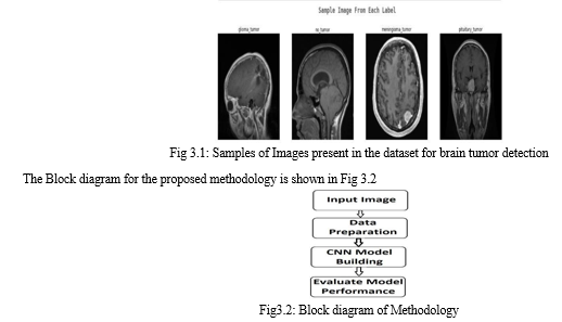
The dataset consists of four classes: Glioma tumor, No tumor, Meningioma tumor and Pituitary tumor respectively. The testing set contains 101 images of Glioma tumor, 105 images of No tumor, 128 images of Meningioma tumor and 99 images of Pituitary tumor. In the training set, there are 826 images of Glioma tumor, 397 images of No tumor, 825 images of Meningioma tumor and 827 images of Pituitary tumor.
The sample images include classes of Glioma tumor, no tumor, Meningioma tumor and Pituitary tumor is shown in the below Fig3.1

- Input Image: The image for this work is obtained through “Brain MRI Images for Brain tumor detection” dataset from Kaggle website.
- Data Preparation: The process of data preparation entails cleaning and transforming raw data before processing and analysis. It is an important step that typically involves reformatting, correcting, and combining datasets to improve data before processing. In this model, four different types of brain tumors were detected, so a variable called labels like glioma, meningioma, pituitary and no tumor were declared.
- Convolution Neural Network (CNN) Model Building: In this case, the CNN model is built using transfer learning and the EfficientNetB0 architecture.
- Evaluate Model Performance: The performance of the model is evaluated by using the confusion matrix and classification report obtained from the model.
The CNN algorithm used here uses EfficientNetB0 architecture to classify the given input image
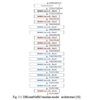
The architecture of the EfficientNetB0 model is shown in Fig 3.3. The EfficientNetB0 baseline model is employed as the entry point in this suggested technique, which accepts an input image with a dimension of 224x224x3. The mobile inverted bottleneck convolution (MBConv) and numerous convolutional (Conv) layers with a 3x3 receptive field are then used by the model to extract features from all of the layers. The primary rationale for using the EfficientNetB0 in this situation is that it has balanced depth, width, and resolution, which can result in a model that is scalable, accurate, and simple to install.

EfficientNetB0 uses a fixed set of scaling coefficients to scale each dimension in comparison to other Deep Convolutional Neural Networks (DCNNs). This method outperformed previous cutting-edge algorithms developed using the ImageNet dataset. EfficientNet generated remarkable performance despite transfer learning, demonstrating its effectiveness beyond that of ImageNetNetB0 as a whole. The model had scales of 0 to 7 when it was first released, indicating an improvement in parameter size and even precision. Users and developers can now access and provide enhanced ubiquitous computing enhanced with DL capabilities across several platforms for a variety of applications thanks to the current EfficientNet. It is important to note that the focus of this work is the empirical investigation of contemporary DCNNs and EfficientNetB0 for the categorization of tumors kinds.
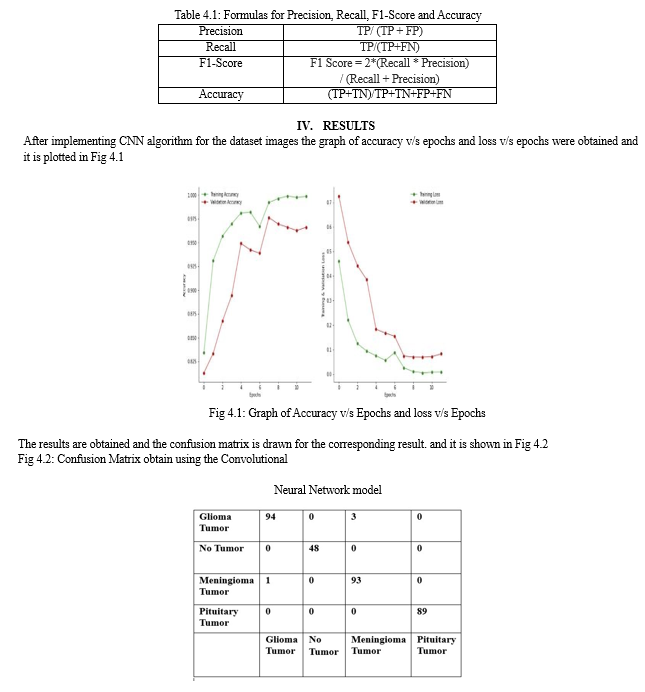
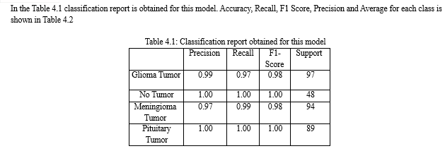
The classification report obtained for the model gives the values of precision, recall and f1-score, it can be calculated from the confusion matrix using True Positive (TP), True Negative (TN), False Positive (FP) and False Negative (FN). There are four ways to determine if the forecasts were accurate or not
- TN / True Negative: Both the case and the prediction were negative.
- TP / True Positive: The case was favorable and was anticipated to be favorable.
- FN/False Negative: The case was affirmative but the result was projected to be negative.
- FP/False Positive: the result was negative but was expected to be positive
a. Precision: Precision is the ability of a classifier to avoid identifying something that is actually negative as positive. The ratio of true positives to the sum of true positives and false positives is how it is defined for each class. Accuracy of optimistic prognoses is precision.
b. Recall: The ability of a classifier to locate each case that qualifies as positive is known as recall. The ratio of true positives to the sum of true positives and false negatives for each class is how it is defined. Recall: - The proportion of positives that were correctly identified as positive.
c. F1 Score: The weighted average is the F1 score, which varies from 0.0 to 1.0 depending recall and precision. If F1 scores are not accurate, accuracy is determined by precision and recall, which are factors in the computation of the scores.
d. Support: Support is defined as the proportion of actual occurrences of the class in the dataset. Unbalanced support in the training data, which may imply structural issues with the classifier's reported scores, may suggest the need for stratified sampling or rebalancing. instead of diagnosing the evaluation process, support that is consistent across models.
e. Accuracy: It gives you information on the overall accuracy of the model, which is the proportion of all samples that the classifier correctly detected.


And it was found to be effective in identifying brain tumors with high accuracy. However, the identification of tumors remains challenging due to variations in location, shape, and structure, which make segmentation as difficult task. The dataset used in the model contains 927 Glioma images, 952 Meningioma images, 926 Pituitary images and finally 502 no tumor images. From this count, it can be inferred that the dataset is biased. The number of no tumor images that are considered for this study is less compared to the other classes of tumor images. This is the one drawback of the dataset that is being used in this work. This will affect the performance of the model. Despite these challenges, the proposed method using EfficientNetB0 can provide a scalable yet accurate and easily deployable model for brain tumor detection in MRI.
Conclusion
Brain tumor diagnosis is a critical task that requires high accuracy to avoid complications. MRI is one of the best techniques to detect brain tumors, but its complexity makes it challenging to identify the tumor\'s size and location variability. This work proposed a DL model for detecting brain tumors, the methodology used EfficientNetB0 architecture with transfer learning to build the CNN model. The proposed method achieved excellent results, indicating its effectiveness beyond the usual ImageNet dataset. The model\'s performance was evaluated using a confusion matrix and a classification report obtained from the model. The study\'s findings contribute to the automatic medical image analysis field and can provide an accurate and reliable method for recognizing brain tumors in MRI. However, the study\'s results are limited to the dataset used, here more research is needed to verify the model\'s effectiveness on different datasets.
References
[1] Bathe, Kavita, et al. \"Brain Tumor Detection Using Deep Learning Techniques.\" Available at SSRN 3867216 (2021). [2] Siddique, Md Abu Bakr, et al. \"Deep convolutional neural networks model-based brain tumor detection in brain MRI images.\" 2020 Fourth International Conference on I-SMAC (IoT in Social, Mobile, Analytics and Cloud) (I-SMAC). IEEE, 2020. [3] Analysis of a fine-tuned deep convolutional model in classifying and detecting malaria parasites from blood smears.\" KSII Transactions on Internet and Information Systems (TIIS) 15.1 (2021): 147-165. [4] Raut, Gajendra, et al. \"Deep Learning Approach for Brain Tumor Detection and Segmentation.\" 2020 International Conference on Convergence to Digital World-Quo Vadis (ICCDW). IEEE, 2020. [5] Vankdothu, Ramdas, and Mohd Abdul Hameed. \"Brain tumor MRI images identification and classification based on the recurrent convolutional neural network.\" Measurement: Sensors 24 (2022): 100412. [6] Sathe, Samson, R. Arun, and Vishnu Raj. \"A Modified Deep Convolutional Neural Network for Brian Abnormalities Detection.\" International Journal of Engineering Research & Technology (IJERT) ISSN (2020): 2278-0181. [7] Chattopadhyay, Arkapravo, and Mausumi Maitra. \"MRI-based Brain Tumor Image Detection Using CNN based Deep Learning Method.\" Neuroscience Informatics (2022): 100060. [8] Nayan, Al-Akhir, et al. \"A deep learning approach for brain tumor detection using magnetic resonance imaging.\" arXiv preprint arXiv:2210.13882 (2022). [9] Khan, Md Saikat Islam, et al. \"Accurate brain tumor detection using deep convolutional neural network.\" Computational and Structural Biotechnology Journal 20 (2022): 4733-4745. [10] Razzaq, Saqlain, et al. \"Brain Tumor Detection from MRI Images Using Bag of Features And Deep Neural Network.\" 2020 International Symposium on Recent Advances in Electrical Engineering & Computer Sciences (RAEE & CS). Vol. 5. IEEE, 2020 [11] Methil, Aryan Sagar. \"Brain Tumor Detection using Deep Learning and Image Processing.\" 2021 International Conference on Artificial Intelligence and Smart Systems (ICAIS). IEEE, 2021. [12] Grampurohit, Sneha, et al. \"Brain tumor detection using deep learning models.\" 2020 IEEE India Council International Subsections Conference (INDISCON). IEEE, 2020. [13] Fayyadh, Sultan B., and Abdullahi A. Ibrahim. \"Brain Tumor Detection and Classifiaction Using CNN Algorithm and Deep Learning Techniques.\" 2020 International Conference on Advanced Science and Engineering (ICOASE). IEEE, 2020 [14] Ramtekkar, Praveen Kumar, Anjana Pandey, and Mahesh Kumar Pawar. \"A proposed model for automation of detection and classification of brain tumor by deep learning.\" 2nd International Conference on Data, Engineering and Applications (IDEA). IEEE, 2020. [15] Saleh, Ahmad, Rozana Sukaik, and Samy S. Abu-Naser. \"Brain tumor classification using deep learning.\" 2020 International Conference on Assistive and Rehabilitation Technologies (iCareTech). IEEE, 2020. [16] Trinh, Quoc-Huy, et al. \"EfficientNet for Brain-Lesion Classification.\" Brainlesion: Glioma, Multiple Sclerosis, Stroke and Traumatic Brain Injuries: 7th International Workshop, BrainLes 2021, Held in Conjunction with MICCAI 2021, Virtual Event, September 27, 2021, Revised Selected Papers, Part I. Cham: Springer International Publishing, 2022 [17] Balaji, Gopinath, Ranit Senour Harsh Kirty. \"Detection and Classification of Brain tumors Using Deep Convolutional Neural Networks.\" arXiv preprint arXiv:2208.13264 (2022). [18] Montalbo, Francis Jesmar P., and Alvin S. Alon. \"Empirical
Copyright
Copyright © 2024 Raksha Nayak, Sanjana Rao U S, Shreeta Jayakar Shetty, Vinaya S, Shashikala . This is an open access article distributed under the Creative Commons Attribution License, which permits unrestricted use, distribution, and reproduction in any medium, provided the original work is properly cited.

Download Paper
Paper Id : IJRASET61745
Publish Date : 2024-05-07
ISSN : 2321-9653
Publisher Name : IJRASET
DOI Link : Click Here
 Submit Paper Online

