Ijraset Journal For Research in Applied Science and Engineering Technology
- Home / Ijraset
- On This Page
- Abstract
- Introduction
- Conclusion
- References
- Copyright
Brain Tumor Growth Prediction Based on Segmentation Using Resolution Convolution Network
Authors: Roshni Srivastava, VIvek Kumar Sharma, Yashitaa Anil, Prof. Er. Somya Kumari
DOI Link: https://doi.org/10.22214/ijraset.2024.61244
Certificate: View Certificate
Abstract
This study presents a novel approach to brain tumor growth prediction using Resolution Convolution Network (RCN) segmentation. The method involves precise segmentation of brain tumor regions from magnetic resonance imaging (MRI) scans followed by application of RCN to predict growth. The RCN architecture integrates high-resolution convolutional layers, which facilitates the extraction of detailed features that are crucial for accurate segmentation and subsequent prediction. Performance evaluation is performed on a comprehensive dataset of MRI brain scans. The results show the effectiveness of the proposed method in accurate segmentation of tumor regions and prediction of tumor growth progression. The findings suggest that this approach holds promise for improving clinical decision support systems and facilitating personalized treatment strategies for patients with brain tumors.
Introduction
I. INTRODUCTION
The brain, vital for decision-making must be protected from injury and diseases. Brain tumors either near or within the brain tissue pose a serious risk due to the limited space of the skull. Their growth increases pressure in the skull, which can cause edema, reduced blood flow, and displacement of healthy tissue. Brain tumors, whether malignant or benign, are inherently serious because they tend to invade confined spaces within the skull. The threat they pose depends on factors such as tumor type, location, size, and stage of development. Diagnosis often occurs at an advanced stage because the protection of the brain by the skull prevents early identification unless special diagnostic tools are used. Early detection of symptoms is important for timely intervention. Brain tumors pose significant challenges in the field of medical diagnosis and treatment due to their complex growth patterns and potential for rapid progression. This article explores the applications of RCNs in predicting brain development and highlights their potential to improve decision-making in neuro-oncology learning have led to the development of new methods to predict tumor growth.
One such method is segmentation-based prediction using network connectivity (RCN), which uses high-resolution data and deep learning techniques to indicate tumor progression and predict growth trajectories. By using RCN to segment brain tumor images, researchers aim to extract key features and patterns to reveal predictive patterns, ultimately facilitating early detection, accommodating eight self-healings, and improving patient outcomes. This article explores the applications of RCNs in predicting brain development and highlights their potential to improve decision-making in neuro-oncology.
II. RELATED WORK
Several recent studies have focused on advances in brain tumor segmentation and prediction techniques with the aim of improving the accuracy and efficiency of medical imaging diagnostic processes. Roy and Bandyopadhyay [1] introduced a method for proportional measurement of tumor affected area using a multi-step adaptive approach for image filtering and segmentation. Their algorithm showed promising results in brain tumor diagnosis and staging, providing a fast alternative to manual segmentation. Pan et al. [4] used Convolutional Neural Networks (CNN) to develop a brain tumor detection system based on brain MRI pixel statistics. Their CNN-based approach outperformed traditional methods and showed improved sensitivity and specificity parameters. Rastogi, Johari and Tiwari [24] proposed the use of a 2D-VNet model for brain segmentation and prediction for effective discrimination between tumor and healthy tissue. However, the lack of detailed information about model limitations poses challenges for further improvement. Gupta and Gupta [25] introduced a new deep neural network training strategy that combines data distillation and augmentation to improve model performance. Although this approach is promising, concerns regarding representative sample selection and noise removal in data augmentation must be addressed to achieve optimal results.
The purpose of the proposed project is to address these inherent limitations such as sensitivity to noise, open lines, and difficulty in choosing the initial starting point. To overcome these challenges, research aimed at developing more robust segmentation models is ongoing and improve predictive capabilities for detecting tumor growth at early stages.
This study aims to improve efficiency and accuracy by adopting a full-resolution CNN architecture and integrating Modified Sunflower Optimization (MSFO) with the Inverse Boltzmann Machine (IBM) technique.
III. DATA SOURCE
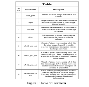
BraTS stands as a pivotal platform dedicated to the advancement of brain tumor segmentation techniques using state-of-the-art methods applied to multimodal MRI scans. In the BraTS 2020 initiative, the spotlight remains on the intricate segmentation of brain tumors, particularly gliomas, within pre-operative MRI scans sourced from a multitude of institutions.
Alongside this, BraTS 2020 extends its scope to encompass the clinical implications of segmentation tasks, delving into patient survival prediction, discerning pseudo progression from true tumor recurrence, and exploring algorithmic uncertainties in tumor segmentation.
A. Tasks and Evaluation Framework
Within the framework of BraTS 2020, four distinct tasks are delineated, each backed by meticulous evaluation criteria:
- Manual segmentation labels of tumor sub-regions.
- Clinical data reflecting overall survival.
- Clinical evaluation of tumor progression status.
- Estimation of uncertainty surrounding predicted tumor sub-regions.
B. Description of Imaging Data
The BraTS dataset comprises NIfTI files (.nii.gz) encapsulating a comprehensive array of imaging modalities:
- Native (T1) and post-contrast T1-weighted (T1Gd) volumes.
- T2-weighted (T2) volumes.
- T2 Fluid Attenuated Inversion Recovery (T2-FLAIR) volumes.
Originating from diverse clinical protocols and scanner platforms across nineteen contributing institutions, these scans undergo meticulous manual segmentation. Annotations, meticulously crafted and vetted by neuro-radiologists, encompass the GD-enhancing tumor (ET), peritumoral edema (ED), and the necrotic and non-enhancing tumor core (NCR/NET).
All datasets are subjected to rigorous pre-processing, including co-registration to a unified anatomical template, interpolation to a standardized resolution (1 mm^3), and skull-stripping.

IV. METHODOLOGY
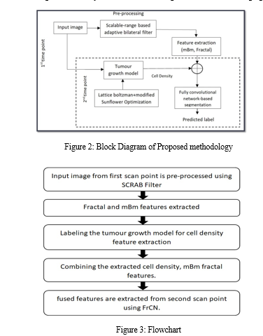
The research initiative aims to address the daunting challenges of dealing with brain tumors and recognizes their significant impact on neurological health. The methodology comprises four interconnected modules carefully designed to enhance different aspects of image analysis, tumor growth simulation and semantic segmentation.
The start of the process is marked by the deployment of the Scalable Range-Based Adaptive Bilateral Filter. This module optimizes image quality by dynamically adjusting the range parameter, effectively reducing noise, smoothing imperfections and preserving important edge details.
Following this, attention shifts to feature extraction, a core component of a methodology aimed at distilling the essence of data by extracting only the most relevant characteristics. This strategic simplification not only streamlines subsequent processing steps, but also ensures the preservation of vital information necessary for accurate analysis.
A core aspect of the methodology is the introduction of a tumor growth model that uses mathematical equations and the Lattice Boltzmann method to facilitate accurate simulation of tumor growth. In addition, the integration of the Modified Sunflower Optimization algorithm refines the models, improving their predictive capabilities and enabling more informed decision-making in medical contexts. Subsequently, the methodology dives into semantic segmentation, which is a critical aspect of image analysis. Using full-resolution convolutional networks (FCNs), the team performs pixel-by-pixel classification to achieve granular image segmentation. This careful approach not only improves prediction accuracy, but also allows semantic classes to be distinguished with unprecedented accuracy. By seamlessly integrating these modules, the methodology presents a comprehensive framework for image processing, tumor growth simulation, and semantic segmentation. Each module builds on the previous one, culminating in a robust pipeline capable of delivering accurate analysis and invaluable insights across medical imaging and related domains.
V. RESULTS AND DISCUSSION
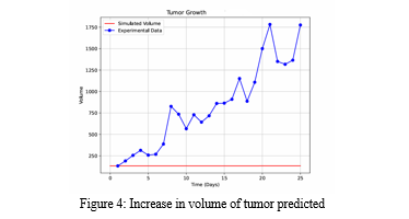
The results of the project demonstrate the effectiveness of the proposed methodology in the analysis and prediction of brain tumor growth trajectories. Through the integration of Resolution Convolutional Networks (RCN) with advanced segmentation techniques, accurate segmentation of tumor regions was achieved, enabling precise delineation of tumor boundaries and identification of tumor characteristics.
Additionally, a predictive modelling approach used longitudinal imaging data and machine learning algorithms to predict tumor growth trajectories and predict treatment response. The results indicate promising performance metrics, including high segmentation accuracy and robust predictive capabilities, underscoring the potential of the proposed methodology to improve diagnosis, treatment planning, and patient outcomes in neuro-oncology.
The discussion also addresses potential limitations of the methodology, such as issues related to data variability and model generalizability, and suggests avenues for future research and improvement. Overall, the results and discussion underscore the transformative potential of the proposed methodology in advancing the field of neuro-oncology and improving patient care.

Conclusion
Research seeks to address the formidable challenges posed by brain tumors and recognizes their significant impact on neurological function and patient well-being. By contributing to the advancement of medical imaging methodologies and predictive modeling within neuro-oncology, the research marks significant progress in the pursuit of more effective strategies to address brain tumor pathologies. The applied methodology includes four interconnected modules that form a comprehensive framework adapted to extend various aspects of image analysis, tumor growth simulation and semantic segmentation. This approach, starting with image quality optimization through a range-based adaptive bilateral filter, seeks to minimize noise while preserving essential edge detail, creating a robust foundation for subsequent processing stages. The core of the methodology is an innovative tumor growth model that uses mathematical equations and optimization algorithms to accurately simulate tumor progression. Through the integration of advanced methodologies such as semantic segmentation using full-resolution convolutional networks, this methodology facilitates careful analysis and prediction of tumor growth trajectories, facilitating early detection and informed decision-making in neuro-oncology. Through the seamless integration of these modules, the methodology offers a comprehensive approach to brain tumor analysis with the potential to significantly impact diagnosis, treatment planning, and patient outcomes. The research, which contributes to the development of medical imaging and predictive modeling methodologies in neuro-oncology, represents a significant step forward in the effort to design more effective strategies to fight brain tumors and preserve neurological function.
References
[1] S. Roy And S. K. Bandyopadhyay, \"Detection and Qualification Of Brain Tumor From MRI Of Brain And Symmetric Analysis,\" International Journal Of Information And Communication Technology Research, Volume 2 No.6, June 2012, Pp584-588 [2] Tiwari A, Srivastava S, Pant M. Brain tumor segmentation and classification from magnetic resonance images: Review of selected methods from 2014 to 2019. Pattern Recognition Letters. 2020;131:244-260 [3] Vaishali et al. (2015) Wavelet-based feature extraction for brain tumor diagnosis-a survey.Int J Res Appl Sci Eng Technol (IJRASET) 3(V), ISSN:2321-9653 [4] Pan, Yuehao & Huang, Weimin & Lin, Zhiping & Zhu, Wanzheng & Zhou, Jiayin & Wong, Jocelyn & Ding, Zhongxiang. (2015). Brain tumor grading based on Neural Networks and Convolutional Neural Networks.Conference proceedings: Annual International Conference of the IEEE Engineering in Medicine and Biology Society. IEEE Engineering in Medicine and Biology Society. Conference. 2015. 699-702. 10.1109/EMBC.2015.7318458. [5] K. Simonyan and A. Zisserman, “Very deep convolutional networks for large-scale image recognition.” September, 2015. Available online at: https://arxiv.org/abs/1409.1556 [6] K. Sudharani, T. C. Sarma and K. Satya Rasad, \"Intelligent Brain Tumor lesion classification and identification from MRI images using a K-NN technique,\"2015 International Conference on Control, Instrumentation, Communication and Computational Technologies (ICCICCT), Kumaracoil, 2015, pp. 777-780.DOI: 10.1109/ICCICCT.2015.7475384 [7] S. Pereira, A. Pinto, V. Alves, and C. A. Silva, \"Brain Tumor Segmentation Using Convolutional Neural Networks in MRI Images,\" in IEEE Transactions on Medical Imaging, vol. 35, no. 5, pp. 1240-1251, May 2016. [8] Pavel Dvo?ák & Bjoern Menze, “Local Structure Prediction with Convolutional Neural Networks for Multimodal Brain Tumor Segmentation” Conference Paper, International MICCAI Workshop on Medical, July,2016. [9] Minz, Astina, and Chandrakant Mahobiya. \"MR Image Classification Using Adaboost for Brain Tumor Type.\" 2017 IEEE 7th International Advance Computing Conference (IACC) (2017): 701-705. [10] P.S. Mukambika, K Uma Rani, \"Segmentation and Classification of MRI Brain Tumor,\" International Research Journal of Engineering and Technology (IRJET), Vol.4, Issue 7, 2017, pp. 683 - 688, ISSN: 2395-0056 [11] Sankari Ali, and S. Vigneshwari. \"Automatic tumor segmentation using convolutional neural networks.\" 2017 Third International Conference on Science Technology Engineering & Management (ICONSTEM) (2017): 268-272. [12] Varuna Shree, N., Kumar, T.N.R. Identification and classification of brain tumor MRI images with feature extraction using DWT and probabilistic neural network. Brain Inf. 5, 23-30 (2018) DOI:10.1007/s40708-017-00755 [13] Devkota, B. & Alsadoon, Abeer & Prasad, P.W.C. & Singh, A.K. & Elchouemi, A. (2018). Image Segmentation for Early Stage Brain Tumor Detection using Mathematical Morphological Reconstruction. Procedia Computer Science.125. 115-123. 10.1016/j.procs.2017.12.017. [14] A.K. Anaraki, M. Ayati, F. Kazemi, “Magnetic resonance imaging baser brain tumour grades classification and grading via convolutional neural networks and genetic algorithms.” Biocybernetics and Biomedical Engineering, Vol. 39, No.1, pp.63- 74, January 2019. [15] R. Thillaikkarasi, S. Saravanan, “An enhancement of deep learning algorithm for brain Tumor segmentation using kernel based CNN with M SVM”. Journal of Medical System, Vol. 43, No. 4, February 2019. [16] Z. N. K. Swati, Q. Zhao, M. Kabir, F. Ali, Z. Ali, S. Ahmed, and J. Lu, “Brain tumor classification for MR images using transfer learning and fine-tuning.” Computerized Medical Imaging and Graphics, July 2019. [17] S. M. Kamrul Hasan and C. A. Linte, \"U-NetPlus: A Modified Encoder-Decoder U-Net Architecture for Semantic and Instance Segmentation of Surgical Instruments from Laparoscopic Images,\" 41st Annual International Conference of the IEEE Engineering in Medicine and Biology Society (EMBC), Berlin, Germany, pp. 7205-7211, 2019. [18] Kumar A, Ramachandran M, Gandomi AH, Patan R, Lukasik S, Soundarapandian RK. A deep neural network based classifier for brain tumor diagnosis. Applied Soft Computing. 2019;82:105528 [19] Yin B, Wang C, Abza F. New brain tumor classification method based on an improved version of whale optimization algorithm. Biomedical Signal Processing and Control. 2020;56:101728 [20] Kaplan K, Kaya Y, Kuncan M, Ertunç HM. Brain tumor classification using modified local binary patterns (LBP) feature extraction methods. Medical Hypotheses. 2020;139:109696 [21] A. Cinar and M. Yildirim, “Detection of tumors on brain MRI images using the hybrid convolutional neural network architecture.” Medical Hypotheses, March 2020. [22] D. Lu, N. Potomac, I. Gracheva, E. Hattingen, and J. Triesch, “Human-expert-level brain tumor detection using deep learning with data distillation and augmentation.” June, 2020. Available online at: https://arxiv.org/abs/2006.12285 [23] Z. Yuan, W. Wang, H. Wang, N. Razmjooy, “A new technique for optimal estimation of the circuit-based PEMFCs using developed Sunflower Optimization Algorithm,” Research paper on Energy Reports, Vol.6, pp.662-671, November 2020 [24] D. Rastogi, P. Johri and V. Tiwari, \"Brain Tumor Segmentation and Tumor Prediction Using 2D-VNet Deep Learning Architecture,\" 10th International Conference on System Modelling & Advancement in Research Trends (SMART), MORADABAD, India, pp. 723-732, 2021. [25] S. Gupta and M. Gupta, \"Deep Learning for Brain Tumor Segmentation using Magnetic Resonance Images,\" IEEE Conference on Computational Intelligence in Bioinformatics and Computational Biology (CIBCB), Melbourne, Australia, pp. 1-6, 2021. [26] V.V.S. Sasank, S. Venkateswarlu,“An automatic tumour growth prediction based segmentation using full resolution convolutional network for brain tumour”. Biomedical Signal Processing and Control, Vol.71, Part A, January 2022.
Copyright
Copyright © 2024 Roshni Srivastava, VIvek Kumar Sharma, Yashitaa Anil, Prof. Er. Somya Kumari. This is an open access article distributed under the Creative Commons Attribution License, which permits unrestricted use, distribution, and reproduction in any medium, provided the original work is properly cited.

Download Paper
Paper Id : IJRASET61244
Publish Date : 2024-04-29
ISSN : 2321-9653
Publisher Name : IJRASET
DOI Link : Click Here
 Submit Paper Online


