

Ijraset Journal For Research in Applied Science and Engineering Technology
Brain Tumor Segmentation Using Deep Learning
Authors: S. Krithika, S. Naga Kalyan, S. Praneeth, S. Shashank , Dr. K. Rajeshwar Rao
DOI Link: https://doi.org/10.22214/ijraset.2023.54585
Certificate: View Certificate
Abstract
Now a day’s tumor is second leading cause of cancer. Due to cancer large no of patients are in danger. The medical field needs fast, automated, efficient, and reliable technique to detect tumor like brain tumor. Detection plays very important role in treatment. If proper detection of tumor is possible then doctors keep a patient out of danger. Various image processing techniques are used in this application. Using this application doctors provide proper treatment and save tumor patients. A tumor is nothing but excess cells growing in an uncontrolled manner. Brain tumor cells grow in a way that they eventually take up all the nutrients meant for the healthy cells and tissues, which results in brain failure. Currently, doctors locate the position and the area of brain tumor by looking at the MR Images of the brain of the patient manually. This results in inaccurate detection of the tumor and is considered very time consuming.
Introduction
I. INTRODUCTION
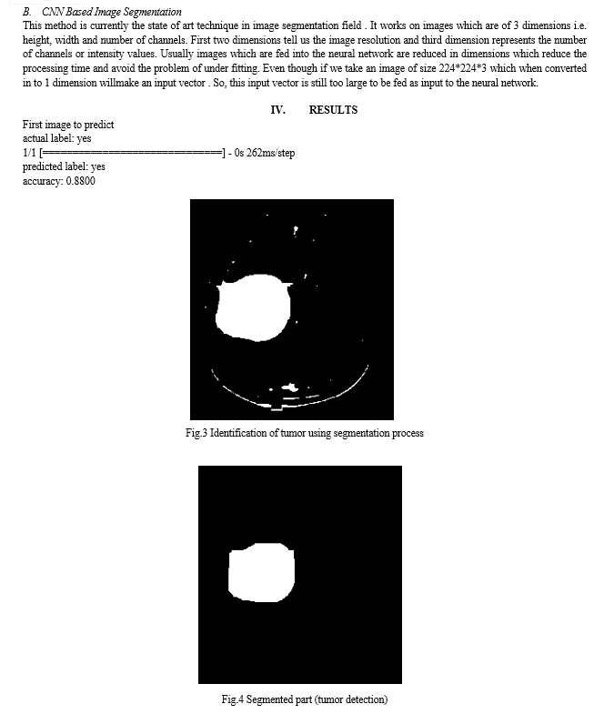
The human body is made up of many organs and brain is the most critical and vital organ of them all. One of the common reasons for disfunction of brain is brain tumor. A tumor is nothing but excess cells growing in an uncontrolled manner. Brain tumor cells grow in a way that they eventually take up all the nutrients meant for the healthy cells and tissues, which results in brain failure. A Brain Cancer is very critical disease which causes deaths of many individuals. The brain tumor segmentation and detection system is used so that it can be diagnosed at early stages. Cancer classification is the most challenging tasks in clinical diagnosis. This project deals with such a system, which uses computer, based procedures to detect tumor blocks and classify the type of tumor using Convolution Neural Network Algorithm for MRI images of different patients. Detecting Brain tumor using Image Processing techniques its involves the four stages is Image Pre-Processing, Image segmentation, Feature Extraction, and Classification. Image processing and neural network techniques are used for improve the performance of detecting and segmentation of brain tumor in MRI images. A tumor is a mass of tissue it grows out of control. We can use a Deep Learning architectures CNN (Convolution Neural Network) generally known as NN (Neural Network to detect the brain tumor. The performance of model is to predict image tumor is present or not in image. If the tumor is present, it return yes otherwise return no.
A. Objective Of Project
To provide doctors good software to identify tumor and their causes. Save patient’s time. Provide a solution appropriately at early stages. Get timely consultation.
B. Limitations
- Lack of Annotated Data: Deep learning models require a large amount of annotated data to achieve optimal performance. However, collecting and annotating medical images for brain tumor segmentation is a time-consuming and expensive process, which can limit the availability of such datasets.
- Computational Requirements: Training and evaluating deep learning models for brain tumor segmentation can require significant computational resources, such as powerful GPUs and large amounts of memory. This can limit the accessibility of deep learning-based methods to smaller research labs and clinical settings with limited resources
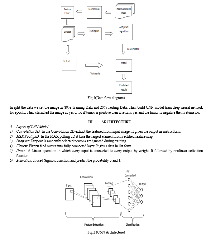
II. IMPLEMENTATION
The proposed system has mainly five modules. Dataset, Preprocessing, segmentation, Split the data, Build CNN model train Deep Neural network for epochs, and classification. In dataset we can take multiple MRI images and take one as input image. In pre-processing image to encoded the label and resize the image.
Conclusion
A fully automatic and accurate process is shown in this project, using a deep convolution neural network with well-known medical image architecture for the detection of entire brain tumors and intra-tumor regions. The CNN model built has been trained with the BRATS dataset to detect the tumors. BraTS\'2018 training and challenge validation data sets have been tested and quantitatively trained. The different tests have shown that the detection results have been quite successful with higher accuracy with CNN models and evaluation actions have confirmed that our results are very comparable ,however the proposed approach can be further enhanced. We present a CNN architecture which differs from those traditionally used in computer vision. Our CNN exploits both local features as well as more global contextual features simultaneously. Also, different from most traditional uses of CNNs, our networks use a final layer that is a convolutional implementation of a fully connected layer which allows a 40 fold speed up.
References
[1] L.Guo,L.Zhao,Y.Wu,Y.Li,G.Xu,andQ.Yan,“Tumordetection in MR images using oneclass immune feature weighted SVMs,” IEEE Transactions on Magnetics, vol. 47, no. 10, pp. 3849–3852,2011. [2] R.Kumari,“SVMclassificationanapproachondetectingabnormalityinbrainMRIimages,”Inter nationalJournalofEngineeringResearchandApplications,vol.3,pp.1686–1690,2013. [3] DICOM Samples Image Sets, http://www.osirix-viewer.com/. [4] “Brainweb:SimulatedBrainDatabase” http://brainweb.bic.mni.mcgill.ca/cgi/brainweb1. [5] ObtainableOnline: www.cancer.ca/~/media/CCE 10/08/2015. [6] J. C. Buckner, P. D. Brown, B. P. O’Neill, F. B. Meyer , C. J. Wetmore,J. H Uhm, \"Central nervous system tumors.\" In Mayo Clinic Proceedings,Vol. 82, No. 10, pp. 1271- 1286, October 2007. [7] Deepa , Singh Akansha. (2016). - Review of Brain Tumor Detection from tomography. International Conference on Computing for Sustainable Global Development (INDIACom) [8] R. A. Novellines, M. D. - Squire\'s fundamentals of radiology; Six Edition; UPR, 2004. [9] Preston, D. c. (2006). Magnetic Resonance Imaging (MRI) of the Brain and Spine from Basics. casemed.case.edu . [10] Hendrik RE. (2005) Glossary of MR Terms from American College of Radiology . [11] A. Demirhan, M. Toru, and I. Guler, “Segmentation of tumor and edema along with healthy tissues of brain using wavelets and neural networks,” IEEE Journal of Biomedical and Health Informatics, vol. 19, no. 4, pp. 1451–1458, 2015. 18CP813 BRAIN TUMOR DETECTION 25 [12] Nilesh Bhaskarrao Bahadure, A.K. (2017, March 6). Retrieved from https://www.hindawi.com/journals/ijbi/2017/9749108/.
Copyright
Copyright © 2023 S. Krithika, S. Naga Kalyan, S. Praneeth, S. Shashank , Dr. K. Rajeshwar Rao. This is an open access article distributed under the Creative Commons Attribution License, which permits unrestricted use, distribution, and reproduction in any medium, provided the original work is properly cited.

Download Paper
Paper Id : IJRASET54585
Publish Date : 2023-07-03
ISSN : 2321-9653
Publisher Name : IJRASET
DOI Link : Click Here
 Submit Paper Online