Ijraset Journal For Research in Applied Science and Engineering Technology
- Home / Ijraset
- On This Page
- Abstract
- Introduction
- Conclusion
- References
- Copyright
Classification of Melanoma Skin Cancer using Transfer Learning
Authors: Foram J. Parekh, Rauki Yadav
DOI Link: https://doi.org/10.22214/ijraset.2024.61334
Certificate: View Certificate
Abstract
Melanoma, a fatal variety of skin cancer, is a significant public health issue on a global scale. For patients to have better results, melanoma must be detected accurately and early. In this study, transfer learning techniques are used in deep learning to provide a novel method of melanoma cancer detection. By using pre-trained convolutional neural networks (CNNs), such as VGG, MobileNet, and EfficientNet, as feature extractors, the research takes advantage of the capability of transfer learning. These networks, which were first created for tasks involving generic image identification, are refined using a large dataset of dermoscopic images of skin lesions in order to adapt them to the particular needs of melanoma detection. Essential preprocessing techniques, including image resizing, image normalization, and lesion segmentation using the Otsu segmentation method, are used to improve the dataset\'s quality and consistency. Image normalization has been used to reduce the complexity and processing time of the image. Skin lesions have been segmented using Otsu segmentation. These procedures increase the robustness of the melanoma detection model and help to prepare the images for efficient training. The findings of this study highlight the possibility for an earlier and more precise diagnosis and provide encouraging insights into the viability of transfer learning as a formidable tool for melanoma detection. The overall objective of this work is to improve the diagnosis and patient care for melanoma cancer. This work contributes to ongoing efforts to provide practical and effective tools for dermatologists and healthcare professionals.
Introduction
I. INTRODUCTION
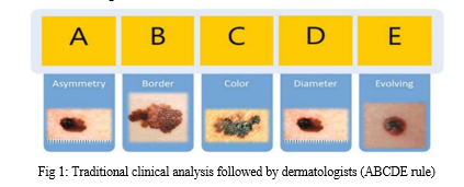
High occurrence of skin cancer compared to other cancer types is a dominant factor in making it one of the most severe health issues in the world. Historically, melanoma is a rare cancer, but in the past five decades, the worldwide occurrence of melanoma has drastically risen. In fact, it is one of the prominent cancers in average years of life lost per death. Adding to the strain, the financial burden of melanoma treatment is also expensive. Squamous Cell Carcinoma and Basal Cell Carcinoma, are eminently curable if diagnosed and treated on in the early stages. The five-year survival rate of patients with early stage diagnosis of melanoma is around 99%. Therefore, timely detection of skin cancer is the key factor in reducing the mortality rate. Melanoma originates from skin melanocytes that have undergone malignant transformation. Melanocytes produce dark pigments on the skin, hair, eyes, and spots of the body. Therefore, melanoma tumors are mostly brown or black. But in a few cases, melanomas do not produce pigment and appear pink, red or purple. A common methodology to detect melanomas is the use of the ABCDE rule: asymmetry, borders, color, diameter, and evolving. These are the warning signs that are monitored in order to diagnose melanoma. High levels of asymmetry or border irregularities are the first alert sign, as well as a strange color of the mole and more than 6mm diameter. All these signs are monitored to analyze their evolution along the time.

II. LITERATURE SURVEY
This section includes information related to different implementation method for classification of skin cancer.
- Melanoma Skin Cancer Detection using Image Processing and Machine Learning
Types of skin diseases skin cancer is found to be the deadliest kind of disease found in humans. This is found most commonly among the fair skin. Skin cancer is found to be 2 types Malignant Melanoma and Non- Melanoma. Malignant Melanoma is one of the deadly and dangerous type cancers, even though it’s found that only 4% of the population is affected with this, it holds for 75% of the death caused due to skin cancer. Melanoma can be cured if its identified or diagnosed in early stages and the treatment can be provided early, but if melanoma is identified in the last stages, it is possible that Melanoma can spread across deeper into skin and also can affect other parts of the body, then it becomes very difficult to treat. Melanoma is caused due to presence of Melanocytes which are present with in the body. This project is to determine the accurate prediction of skin cancer and also to classify the skin cancer as malignant or non-malignant melanoma. To do so, some pre-processing steps were carried out which followed Hair removal, shadow removal, glare removal and also segmentation. SVM and Deep Neural networks will be used to classify. classifier will be trained to learn the features and finally used to classify. The novelty of the present methodology is that it should do the detection in very quick time hence aiding the technicians to perfect their diagnostic skills.
2. A Convolutional Neural Network Framework for Accurate Skin Cancer Detection
Skin alterations are caused due to multiple factors, like allergies, infections, exposition to the sun, etc. The last one is a common practice of most people, who looks for a tan of their skin. However, this search for beauty can have a negative effect on the appearance of skin lesions. This is a typical example of one of the reasons for skin cancer. Melanoma and non-melanoma skin cancer are highly present in Caucasians. The most common nonmelanoma affections are basal cell carcinoma and squamous cell carcinoma. There were more than one million cases in 2018, being the 5th most common cancer. On the other side, melanoma is a less occurring cancer (in the 19th position), with around three hundred thousand new cases last year. Despite the lower number of detections, melanoma causes most of the mortality cases within the skin cancer area. Melanoma is caused by an abnormal multiplication of melanocytes, the cells that produce pigment and give color to the skin. The sooner the melanoma is detected, the greater the chances of cure. Nevertheless, it could spread to other parts of the body if it is not detected early, causing an irremediable effect. The problem resides in the capacity of the detection of melanomas, as they are similar in characteristics to other benign nevi. Dermatologists find hard to distinguish between a benign and a malign mole, being a challenge to find an appropriate rule to classify them. Further works will be focused on the testing of more deep networks and other hierarchies, including preprocessing steps in order to distinguish better between the six non-nevi classes. A specific analysis of the features of each class is a necessary task to generate an adequate classifier. The inclusion of probabilistic techniques to make an accurate prediction of different classifiers is another research line.
3. Skin Cancer Diagnosis Based on Optimized Convolutional Neural Network
A new optimized technique was proposed for skin cancer diagnosis from the input images. The method was based on Convolutional neural network. An improved version of the whale optimization algorithm was adopted for optimizing the efficiency result of CNN. The utilized optimization algorithm is adopted for the optimal selection of weights and biases in the network to minimize the error of the network output and the desired output. For performance analysis of the proposed method, it is tested on two different benchmarks including Dermquest and DermIS and the results were compared with 10 different methods including semi-supervised method, Spot-moletool, AlexNet, Ordinary CNN, VGG-16, LIN, Inception-v3, and ResNet. The performance indexes here are specificity, accuracy, sensitivity, NPV, and PPV. Final results showed that using the proposed method gives the best achievement for the skin cancer diagnosis.
4. Deep Learning Solutions for Skin Cancer Detection and Diagnosis
High occurrence of skin cancer compared to other cancer types is a dominant factor in making it one of the most severe health issues in the world. Historically, melanoma is a rare cancer, but in the past five decades, the worldwide occurrence of melanoma has drastically risen. In fact, it is one of the prominent cancers in average years of life lost per death. Adding to the strain, the financial burden of melanoma treatment is also expensive. Out of the $8.1 Billion in all skin cancer treatment costs in the USA, $3.3 Billion are spent only on Melanoma. Squamous Cell Carcinoma and Basal Cell Carcinoma, are eminently curable if diagnosed and treated on in the early stages. The five-year survival rate of patients with early stage diagnosis of melanoma is around 99%.
Therefore, timely detection of skin cancer is the key factor in reducing the mortality rate. To restate, this project was conducted with the aim of developing convolutional neural network model to diagnose and detect skin cancer from lesion images. It also explored the data augmentation technique as a preprocessing step to strengthen the classification robustness of the CNN model. The best model, namely InceptionResnet achieved an average accuracy of 91%.
TABLE 1: Comparisons among Different classification papers
|
Key Features |
[1] |
[2] |
[3] |
[4] |
|
Title |
Melanoma Skin Cancer Detection using Image Processing and Machine Learning |
A Convolutional Neural Network Framework for Accurate Skin Cancer Detection |
Skin Cancer Diagnosis Based on Optimized Convolutional Neural Network |
Deep Learning Solutions for Skin Cancer Detection and Diagnosis |
|
Image Segmentation |
Shade removal, Glare removal, Hair removal, Otsu Segmentation |
melanoma class (mel), benign keratosis (bkl), Bascal cell carcinoma, Vascular skin |
Asymmetry, Border, Color, Diameter(ABCD rule) |
Melanoma, Melanocytic Nevus, Basal Cell Carcinoma, Actinic Keratosis, Benign Keratosis, Dermatofibroma, Vascular Lesion |
|
Feature Extraction |
Extract image using Neural Networks |
- |
By delaunay triangulation. |
- |
|
Dataset |
ISIC (International Skin Image Collaboration) Dataset |
DenseNet201, Inception -ResNetV2, InceptionV3, HAM10000 |
Dermquest Dataset, DermIS Digital Dataset |
ISIC (International Skin Image Collaboration) Dataset, InceptionResnet Dataset |
|
Classification |
Support Vector Machine (SVM) |
Deep Convolutional Neural Networks (DCNNs) |
Deep Convolutional Neural Networks (DCNNs) |
Convolutional Neural Network (CNN) |
III. PROPOSED SYSTEM
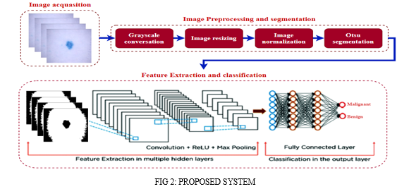
The images for melanoma detection are in the form of RGB. They are converted into grayscale to reduce the time complexity of the model. Following that images are resized in (128,128). Following the images are normalized for faster convergence of the model. Moreover, the images contain extra information other than the skin lesion part. Which may hamper the performance of the model. Hence, to preserve the metadata of the image, the Otsu segmentation is applied to segment the skin lesion from the image. Then the segmented images are given for the feature extraction and classification step.

IV. METHODOLOGY
Our proposed work used mainly three classifiers like VGG16, MobileNet and EfficientNetB2.
- VGG16: A convolutional neural network is also known as a ConvNet, which is a kind of artificial neural network. A convolutional neural network has an input layer, an output layer, and various hidden layers. VGG16 is a type of CNN (Convolutional Neural Network) that is considered to be one of the best computer vision models to date. The creators ofthis modelevaluated the networks and increased the depth using an architecture with very small (3 ×3) convolution filters, which showed a significant improvement on the prior-art configurations. They pushed the depth to 16–19 weight layers making it approx — 138 trainable parameters. VGG16 is object detection and classification algorithm which is able to classify 1000 images of 1000 different categories with 92.7% accuracy. It is one of the popular algorithmsfor image classification and is easy to use with transfer learning.
- MobileNet: A lightweight convolutional neural network (CNN) architecture, MobileNetV2, is specifically designed for mobile and embedded vision applications. Google researchers developed it as an enhancement over the original MobileNet model. Another remarkable aspect of this model is its ability to strike a good balance between model size and accuracy, rendering it ideal for resource-constrained devices. MobileNetV2 incorporates several key features that contribute to its efficiency and effectiveness in image classification tasks. o These features include depthwise separable convolution, inverted residuals, bottleneck design, linear bottlenecks, and squeeze-andexcitation (SE) blocks. o Each of these features plays a crucial role in reducing the computational complexity of the model while maintaining high accuracy.
- EfficientNetB2: EfficientNet uses a technique called compound coefficient to scale up models in a simple but effective manner. Instead of randomly scaling up width, depth or resolution, compound scaling uniformly scales each dimension with a certain fixed set of scaling coefficients. Using the scaling method and AutoML. The authors of efficient developed seven models of various dimensions, which surpassed the stateof-the-art accuracy of most convolutional neural networks, and with much better efficiency.The intuition for the networks is, if the input image is bigger, then the network needs more layers to increase the receptive field and more channels to capture more fine-grained patterns on the bigger image.
V. FUTURE ENHANCEMENTS
We aim to apply various pre-trained CNN architecture on melanoma images to experiment and analyze the proposed approach in different experimental scenarios.
Conclusion
In summary, this study initially focused on melanoma detection, a critical issue in healthcare. To enhance image quality, we employed a robust preprocessing pipeline, including image resizing, and normalization. Moreover, to remove the unnecessary metadata of images, segmentation of skin lesions has been achieved using Otsu segmentation. We will use pre-trained CNNs like EfficientNetB2, VGG16, and MobileNet to build an effective melanoma detection model to get accuracy for efficiently classifies the image into benign or malignant. This approach offers a promising solution for early diagnosis and underscores the potential of transfer learning in medical imaging
References
[1] abrham debasu mengistu , dagnachew melesew alemayehu “computer vision for skin cancer diagnosis and recognition using rbf and som “ international journal of image processing (ijip), volume (9) : issue (6) 2015. [2] poornima m s, dr. Shailaja k “detection of skin cancer using svm” , international research journal of engineering and technology (irjet) volume: 04 issue: 07| july - 2017. [3] salome kazeminia, christoph baur, arjan kuijper, bram van Ginneken, nassir navab, shadi albarqouni, anirban mukhopadhyay “gans for medical image analysis “, arxiv:1809.06222v2 [cs.cv] 21 dec 2018 [4] prashant b. Yadav, mrs. S.s. Patil “ recognition of dermatological disease area for identification of disease” ijsdr may 2016 volume 1, issue 5 [5] m.yuvaraju, d.divya, a.poornima, “segmentation of skin lesion from digital images using morphological filter”, international research journal of engineering and technology (irjet) volume: 03 issue: 05 | may-2016 [6] Asha Gnana Priya H, Anitha J, Poonima Jacinth J (2018) Identification of melanoma in dermoscopy images using image processing algorithms. In: 2018 international conference on control, power, communication and computing technologies, ICCPCCT 2018, pp 553–557 [7] Gao Z et al (2019) Privileged modality distillation for vessel border detection in intracoronary imaging.IEEE Trans Med Imaging 39(5):1524–1534 [8] Hussain Z, Gimenez F, Yi D, Rubin D (2017) Differential data augmentation techniques for medical imaging classification tasks. In: AMIA annual symposium proceedings, vol 2017. American Medical Informatics Association, p 979 [9] Nachbar F, Stolz W, Merkle T, Cognetta AB, Vogt T, Landthaler M, Bilek P, B-Falco O, Plewig G (1994) The ABCD rule of dermatoscopy: high prospective value in the diagnosis of doubtful melanocytic skin lesions. [10] Pereira dos Santos F, Antonelli Ponti M (2018) Robust feature spaces from pre-trained deep network layers for skin lesion classification. In: 2018 31st SIBGRAPI conference on graphics, patterns and images. [11] https://www.mdpi.com/1424-8220/21/23/8142
Copyright
Copyright © 2024 Foram J. Parekh, Rauki Yadav. This is an open access article distributed under the Creative Commons Attribution License, which permits unrestricted use, distribution, and reproduction in any medium, provided the original work is properly cited.

Download Paper
Paper Id : IJRASET61334
Publish Date : 2024-04-30
ISSN : 2321-9653
Publisher Name : IJRASET
DOI Link : Click Here
 Submit Paper Online

