Ijraset Journal For Research in Applied Science and Engineering Technology
- Home / Ijraset
- On This Page
- Abstract
- Introduction
- Conclusion
- References
- Copyright
Colon Cancer Detection of Colonoscopy Images Using CNN
Authors: Chaitali Vishal Sheth, Rauki Yadav
DOI Link: https://doi.org/10.22214/ijraset.2024.61465
Certificate: View Certificate
Abstract
Colon cancer requires an early and precise diagnosis in order to be effectively treated because it is a common and potentially fatal condition. This study proposes a reliable framework for colon cancer detection through image preprocessing and transfer learning approaches. The research compares the performance of three popular convolutional neural networks (CNNs) for this task: VGG-15, ResNet-50, and AlexNet.The image preprocessing phase entails a number of critical operations, including image conversion, resizing, and contrast enhancement using histogram equalization. In order to guarantee that the neural networks receive high-quality inputs, these preprocessing techniques strive to improve the quality and consistency of the colon cancer dataset. Pre-trained models extract and classify features from big datasets using transfer learning. The preprocessed colon cancer dataset is used to fine-tune the chosen CNN architectures VGG-15, ResNet-50, and AlexNet and adapt them for this particular medical imaging purpose.The results of this study show that transfer learning has the potential for colon cancer detection and provides helpful direction for researchers and practitioners looking for the best deep-learning architectures for early cancer diagnosis. This study adds to continuing efforts to improve the precision and effectiveness of colon cancer detection techniques, ultimately leading to better patient outcomes.
Introduction
I. INTRODUCTION
Colorectal cancer is a well-known tumour that affects both men and women across the world and is quite common. According to a study published by the World Health Organization in 2018, colon cancer ranked third, with 1.80 million people afflicted. To be more specific, it is the cancer that comes after it that is the second most frequent cause of cancer in women and the third most common cause of cancer in men. Colorectal cancer is thought to be caused by a lack of control over the integrity of epidermal cells, which may occur in the intestine or during a malignancy. A reliable method of detecting colon cancer at an early stage, followed by intensive treatment, has the potential to significantly lower the mortality rates that result. [10]
Common symptoms include diarrhoea, constipation, blood in the stool, abdominal pain, unexplained weight loss, fatigue, and low iron levels. Many people will not have symptoms in the early stages of the disease. The risk of colorectal cancer can be reduced by eating a healthy diet, staying physically active, not smoking tobacco and limiting alcohol. Regular screenings are crucial for early detection. Colon cancer is the second leading cause of cancer-related deaths worldwide. In 2020, more than 1.9 million new cases of colorectal cancer and more than 930 000 deaths due to colorectal cancer were estimated to have occurred worldwide. Large geographical variations in incidence and mortality rates were observed.
The incidence rates were highest in Europe and Australia and New Zealand, and the mortality rates were highest in Eastern Europe. By 2040 the burden of colorectal cancer will increase to 3.2 million new cases per year (an increase of 63%) and 1.6 million deaths per year (an increase of 73%).
FIGURE 1: ANATOMY OF LARGE INTENSTINE
A. Diagnosis For Colon Cancer
- Test for occult blood in faeces: Sample of faeces collected from the patient. Furthermore, to test for occult blood to suspect colorectal cancer, but which takes a long process.
- A DNA test on faeces: To test the presence of pre-cancerous polyps, discarded in the faeces, DNA mutations are examined and analyzed. Un- like an occult blood test, this gives results accurately, which distinguishes from cancer from polyps but fails to indicate a particular tumour is present.
- Flexible Sigmoidoscopy: Screening is done through a sigmoidoscope, which is a thin tube with a light attached in the end to provide light inside the colon and rectum to observe a patient’s colon. If some polyps recognized, then they are removed through colonoscopy after microscopic examination.
- X-ray with Barium enema: The patient’s trails are induced some amount of barium in the form of an enema, and X-ray is performed, which produces double contrast. Barium dye, used in the trails sticks to the inner lining of the bowel, producing more precise images of X-ray. Small polyps missed by the barium enema X-ray, recognized by flexible Sigmoidoscopy.
- Colonoscopy: As represented in Figure 1.3, a colonoscope is similar to a sigmoidoscope rather have more length, and it is a slim tube with an electronic camera affixed at one end, to capture an inward view of the colon, and then displayed. During the test, if any polyp seems to be abnormal, biopsies, or tissue samples are taken. A colonoscopy is painless, but sometimes mild sedative is given to some patients not to feel pain while performing the colonoscopy. Before the colonoscopy, laxative fluid is given to the patients to clean the colon.
- CT Colonography: Although colonoscopy is the best way for diagnosis of polyps and other abnormalities, in which some risks already analyzed. Perforation of inward tissues may result in bleeding and other complications. Hence, CT Colonoscopy is introduced. The patient put into the C.T. scanner and required observations made out using ultra-violet radiation passing through the body.[4]
- Scan and Imaging Techniques: Ultrasound (Magnetic Resonance Imagining) scans can show the diffusion of cancer in the body. The waves can be penetrated inside the body and do not harm the internal tissues. Even though we may not get the best results, but some can deduce.
B. Motivation:
Every year lots of people suffer from Colon Cancer and passes away. Due to late diagnosis only, few people can survive it. Early diagnosis may reduce the number of death and increase the survival rate. However, with the help of image processing and deep learning technique can help to identify the early stage of Colon Cancer. This will be helpful for taking early measures to recover from Colon Cancer.
II. LITERATURE SURVEY
This section includes information related to different implementation methods for Colon Cancer Detection .
A. An Efficient Deep Learning Approach for Colon Cancer Detection:
The main contribution of this research is to propose a new lightweight deep learningapproach based on CNN for efficient colon cancer detection. The efficiency of the proposed system is analyzed with histopathological images database and is compared with the existing approaches in this field. Results show that our method outperformed most of the previous deep learning approaches for colon cancer detection. The best accuracy, precision, recall, and F1-score of our model are 99.50%, 99%, 100%, and 99.49%, respectively. The proposed method is more robust and more efficient than other previous deep models for the detection of colon cancer. Our application can be useful in some cases where the pathologist who checked the colon images needs to be insured, so our system will help them to reach the right diagnosis. In the future, we will test our deep model on different datasets to see how it performs. In addition, we can employ and combine one of the optimization techniques such as a genetic algorithm with our deep model to select the best.
FIGURE 2: HISTOPATHOLOGICAL IMAGES FROM THE USED DATASET
B. Colonoscopy Stage Wise Detection Using Deep Learning Techniques:
An effort has been made to offer numerous study results that have been produced by a number of writers and researchers in order to detect Colorectal Cancer, with the goal of increasing awareness. Based on the past research, it is clear that it is always preferable to detect cancer at an earlier stage in order to have the best chance of recovering from it. Following a thorough investigation of these conventional techniques for diagnosing Colorectal Cancer, it was discovered that the accuracy of identification and the time required to diagnose the illness were both increased. It has also been found in prior research that the outcomes obtained via the use of deep learning processes are not very promising.
C. Two Stage Classification with CNN for Colorectal Cancer Detection:
A two-stage classification is presented to detect colorectal cancer from colonoscopy videos. In the first stage, frames of colonoscopy video are extracted and are rated as significant if it contains a polyp, and these results are then aggregated in a second stage to come to an overall decision concerning the final classification of that frame to be neoplastic and non- neoplastic. We investigated the applicability of deep learning to perform this two-stage classification and the CNN models, namely VGG16, VGG19, Inception V3, Xception, GoogLeNet, ResNet50, ResNet100, DenseNet, NASNetMobile, MobilenetV2, InceptionResNetV2 and fine-tuned version of each model is evaluated. It is observed that the two fine-tuned version of four models: VGG16, VGG19, ResNet100, and DenseNet121, achieved more than 90% of accuracy in both the stages, and the best result was achieved by fine-tuned VGG19 with a test accuracy of 95.75%. Transfer learning from the ImageNet dataset is one of the reasons that VGG19 outperforms some of the previous results mentioned in the literature where training CNN is done on raw data. Thus, we can expect performance gain if the transfer learning is made from the same domain dataset. After the categorization of neo-plastic and non-neoplastic polyps, the automated system will be more useful to the doctors if it can provide a precise 3D location.
D. Automated Detection and Characterization of Colon Cancer with Deep Convolutional Neural Networks:
The suggested DCNN model outperforms previous transfer learning models capable of classifying benign and adenocarcinoma colon tissues by replacing the sigmoid function for binary classification in the output activation layer. We have also proposed a training and evaluation technique for the training of the CNN architecture so that these textured images are high resolution without transforming them into low-resolution images. In addition, the method proposed was evaluated on a dataset, in which we gained a superior level of training and testing accuracy to other models of transfer learning. To the best ofour knowledge, we know of a previous work carried out in categorizing the benign colon tissue with adenocarcinoma on the same dataset. We get 100% precision, 99.80% recall, 99.87% f1-score, and 99.80% accuracy, which is greater than that. Based on the 7ndings of this investigation and previouslydescribed observations, we have a precision of greater than 6%. )e development of computer-supported technology for diagnosing malignant tumors will give pathologists a substantial amount of support.
FIGURE 3:PROPOSED METHODLOGY WORKFLOW
III. PROPOSED SYSTEM
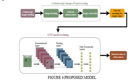
The colonoscopy images are first resized in one format of the (128,128) to reduce the time complexity of the pre-trained convolution neural network. The colonoscopy images in the dataset are in lower contrast. This artifact can hamper the model's performance. Thus, the contrast of the colonoscopy images is enhanced using histogram equalization. Moreover, the colonoscopy images are converted into RGB color space to have proper visualization. The labels of the colonoscopy images are in textual form, which needs to be converted into numerical form. Therefore, the one hot encoding is applied to convert labels from text to numerical labeling.

IV. METHODOLOGY
Our proposed work used mainly three classifiers like VGG16,Resnet50 and InceptionV3.
A. VGG16:
A convolutional neural network is also known as a ConvNet, which is a kind of artificialneural network. A convolutional neural network has an input layer, an output layer, andvarious hidden layers. VGG16 is a type of CNN (Convolutional Neural Network) that isconsideredtobeoneofthebestcomputervisionmodelstodate.Thecreatorsofthismodelevaluated the networks and increased the depth using an architecture with very small (3 ×3)convolution filters,which showed a significant improvement on the prior-art configurations. They pushed the depth to 16–19 weight layers making it approx — 138 trainable parameters. VGG16 is object detection and classification algorithm which is able to classify 1000 images of 1000 different categories with 92.7% accuracy. It is one of the popular algorithms for image classification and is easy to use with transfer learning.
B. RESNET50:
ResNet stands for Residual Network. It is an innovative neural network that was firstintroduced by Kaiming He, Xiangyu Zhang, Shaoqing Ren, and Jian Sun in their 2015computer vision research paper titled ‘DeepResidual Learning for Image Recognition.This model was immensely successful,as can be ascertained from the factthat it sensemblewon the top position at the ILSVRC 2015 classification competition with an error of only3.57%. Additionally, it also came first in the ImageNet detection, ImageNet localization,COCO detection, and COCO segmentation in the ILSVRC & COCO competitions of2015.ResNet has many variants that run on the same concept but have different numbers of layers.ResNet-50 isused to denote the variant that can work with 50 neural network layers.
C. INCEPTIONV3:
The inception v3 model was released in the year 2015, it has a total of 42 layers and a lower error rate than its predecessors. The different optimizations that make the inception V3 model better.
The major modifications done on the Inception V3 model are:
- Factorization into Smaller Convolutions
- Spatial Factorization into Asymmetric Convolutions
- Utility of Auxiliary Classifiers
- Efficient Grid Size Reduction
V. EXPERIMENTAL RESULTS
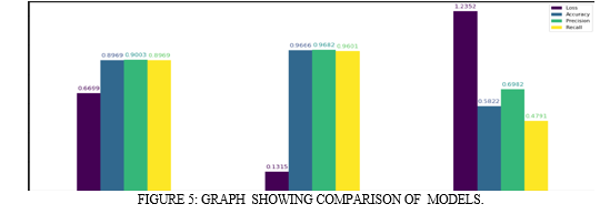
It is implemented on Dataset 128*128 Colonoscopy images of cancer as a raw data as an input and then we do preprocessing of raw data by histogram equalization. Loading the images and preprocess the images. Preprocessing includes grayscale to RGB conversion, resizing, contrast enhancement. One hot encoding for labeling is applied as it is multiclass classification.Among the processed images approx. 7000 images used in training dataset and 70% images used in testing dataset that belonging to seven classes. Then we train validate and test the data.Define VGG16 ,Resnet50 and InceptionV3 model as pretrained CNN with ReLu activation function apply upto 128 layer and at dense layer apply Sigmoid function and uses adam optimizer.VGG16 gives 89.69% accuracy,Resnet50 gives 96.66% accuracy and InceptionV3 gives 58.22% accuracy.We conclude that ResnetNet50 give more accurate result as compare to other model. Figure represents graphical representation of training and testing score of VGGNet16,Resnet50 and InceptionV3.

VI. FUTURE WORK
We can further extend the work for classification of Colon Cancer using the explainable AI, which provides a set of processes and methods that allows human users to comprehend and trust the results and output created by machine learning algorithms.
Conclusion
This study presents an effective approach for early and precise Colon Cancer Detection. By combining Image Preprocessing and Transfer Learning ,we employed preprocessing steps of image resizing, contrast enhancement and image conversion. Also, One Hot Encoding has been used to convert categorical target variable into numerical value. We are going to use various CNN Architectures like VGG-16, ResNet-50, AlexNet etc. and compare the result using modifying hyper parameter to get better accuracy for classifying the colonoscopy image into one of the seven classes. This research underscores the potential of Transfer Learning in improving Colon Cancer Diagnosis, offering valuable insights of healthcare professionals and researchers working toward enhanced patient outcomes.
References
[1] Hasan, M. I., Ali, M. S., Rahman, M. H., & Islam, M. K. (2022, August 24). Automated Detection and Characterization of Colon Cancer with Deep Convolutional Neural Networks. Journal of Healthcare Engineering, 2022, 1–12. https://doi.org/10.1155/2022/5269913 [2] Sakr, A. S., Soliman, N. F., Al-Gaashani, M. S., P?awiak, P., Ateya, A. A., & Hammad, M. (2022, August 24). An Efficient Deep Learning Approach for Colon Cancer Detection. Applied Sciences, 12(17), 8450. https://doi.org/10.3390/app12178450 [3] Murugesan, M., Madonna Arieth, R., Balraj, S., & Nirmala, R. (2023, February). Colon cancer stage detection in colonoscopy images using YOLOv3 MSF deep learning architecture. Biomedical Signal Processing and Control, 80, 104283. https://doi.org/10.1016/j.bspc.2022.104283 [4] Sharma, P., Bora, K., Kasugai, K., & Kumar Balabantaray, B. (2020). Two Stage Classification with CNN for Colorectal Cancer Detection. Oncologie, 22(3), 129–145. https://doi.org/10.32604/oncologie.2020.013870 [5] https://www.ncbi.nlm.nih.gov/pmc/articles/PMC9367621/ [6] https://www.hindawi.com/journals/jhe/2022/5269913/ [7] https://www.mdpi.com/2076-3417/12/17/8450 [8] https://www.geeksforgeeks.org/introduction-convolution-neural-network/ [9] https://www.analyticsvidhya.com/blog/2020/10/what-is-the-convolutional-neural-network- architecture/ [10] https://insightsimaging.springeropen.com/articles/10.1007/s13244-018-0639-9 [11] https://towardsdatascience.com/loss-functions-and-their-use-in-neural-networks-a470e703f1e9
Copyright
Copyright © 2024 Chaitali Vishal Sheth, Rauki Yadav. This is an open access article distributed under the Creative Commons Attribution License, which permits unrestricted use, distribution, and reproduction in any medium, provided the original work is properly cited.

Download Paper
Paper Id : IJRASET61465
Publish Date : 2024-05-02
ISSN : 2321-9653
Publisher Name : IJRASET
DOI Link : Click Here
 Submit Paper Online

