Ijraset Journal For Research in Applied Science and Engineering Technology
- Home / Ijraset
- On This Page
- Abstract
- Introduction
- Conclusion
- References
- Copyright
Dermatological Malignancy Detection Using CNN
Authors: Ms. Komal Parashar, M. V. Devendranathreddy, M. Deepak Reddy, S. Jashwitha
DOI Link: https://doi.org/10.22214/ijraset.2024.59154
Certificate: View Certificate
Abstract
Skin cancer is a prevalent and potentially life-threatening disease that continues to pose a significant public health concern. Starting stage detection Plays a crucial part in enhancing treatment efficacy. Furthermore, the advancement of deep learning has enabled the creation of automated frameworks for detecting maligant cancer. This proposed system utilizes a varied dataset encompassing various types of skin cancer, such as malignant melanomas and benign nevi, to educate a learning model. Utilizing CNN, pertinent features are automatically extracted from skin cancer images, facilitating precise classification. Assessment of the suggested system showcases encouraging outcomes concerning sensitivity, specificity, and overall precision. Comparative assessments against conventional techniques underscore the superior efficacy and effectiveness of the devised model in identifying potential skin cancer instances. Moreover, the system\'s interpretive capacity is heightened through the integration integration of attention mechanisms offering insights into the areas of focus within the skin cancer images
Introduction
I. INTRODUCTION
Skin cancer can be dangerous, and its severity relies on the hinge of skin cancer, its stage at the time of diagnosis, and how quickly it is treated. basal cell, carcinoma , squamous cell carcinoma , and melanoma[3] etc are top skin diseases.personal Detecting skin maligant using CNN is a common and effective approach with in the realm of randiomics. CNNs are well-suited in the objective like classification, making them suitable for identifying patterns and features in medical images. Here's a general outline of the steps you might follow to create a maligancy identity system using CNN. Improve the chances of successful treatment and reduce the risk of complications associated with advanced stages. Enhance accessibility to skin cancer detection, especially region featuring limited access to dermatologists or specialized healthcare services. Contribute to public health efforts by facilitating large-scale screenings and early interventions., which are self-executing contracts with predefined conditions, By achieving these objectives, the use of CNNs in detection aims to make a positive impact on patient outcomes, healthcare efficiency, and public health initiatives related to skin cancer awareness and prevention.
II. related work
Skin cancer detection is an active area of research, with extensive investigations aimed at devising precise and efficient approaches for early identification . Below are some notable works and approaches in the realm of cancer detection. pioneering groundbreaking advancements in skin randiomics technologies and methodologies has provided a dataset that researchers use for benchmarking their algorithms. This dataset includes a Multifariousnessskin lesion images with corresponding diagnoses. It's essential to check for the latest research publications and developments in the field, as advancements in technology and methodologies continue to shape the landscape of maligent detection[5].
A. Skin Cancer Classification using Resnet
Skin cancer detection using ResNet involves employing a learning model, specifically ResNet (Residual Neural Network), to analyze and classify skin lesions as either benign or malignant network Evaluate the data model on the test the dataset set to assess its generalization performance. Metrics like exactitude, exactness, retrieval rate , and F1 score for the proficiency the model's It's significant name of the success of dermatological malignancy detection using ResNet reliable on quality and diversity of the dataset, appropriate hyperparameter tuning, and magnitude of the model. Continuous monitoring and updates may be necessary to adapt to new data and enhance the model's performance over time[1].
B. Skin Cancer Classification using Vggnet
Obtain a dataset of dermatological cancer images. A ubicutious dataset for this task is reducing the structure of system. and fine-tuning it on a dataset of skin cancer images Resize images to the input size required by the VGGNet model (typically 224x224 pixels). Load the pre-trained VGGNet model.
You can use a pre-trained VGG16 or VGG19 model from a learning library like Keras or PyTorch. Train the modified VGGNet on your skin cancer dataset. Use the training for instruction and the validation set to monitor the model's performance and avoid overfitting. Fine-tune the model by adjusting turning parameters like lsteprate, grouping magnitude, and the number of epochs., Utilize metrics such as exactitude, exactness, retrieval rate to evaluate the model's effectiveness in skin cancer classification.[2]
C. Skin Cancer Classification using SVM and Transfer Learning
Dermatological cancer classification using svm and transfer learning: Detecting skin cancer using Support Vector Machines (SVM) involves training a classification learning data to classify skin cancer disease as either good or bad based on certain forward representation from images. Here's a simplified guide on how you can approach keeps a copy of the database, Collect a dataset of skin lesion images labeled as benign or malignant. Ensure that your dataset is diverse, representative, Extract relevant features from the images to feed into the SVM model. Easily used for radiomics include color histograms, texture features, and shape descriptors. partitioning the dataset into training and validation sets. The training set is employed for model training, while the validation set is utilized to assess its performance on unseen data., Evaluate the performance of. Adjust Once satisfied with the model's performance, deploy it to a system where it should be employed for cancer detection on Remember that this is a simplified guide, and Additional aspects to take into account are parameter adjustmen, cross-validation, and fine tuning the feature representation provides that can be explored to improve the model's performance.[4]
III. METHODS AND EXPERIMENTAL DETAILS
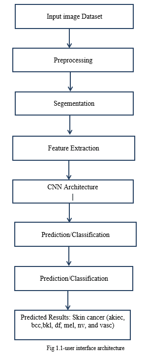
Building a malignancy identification system using CNN is a widely embraced learning implementations within healthcare In the preexisting frameworks three diseases it can manage but in the proposed system it can handle approxiametly 7 diseases like basel cell,dermatofibroma,actinickeratoses,,beingkeratosis,.melonoma.melanocyticnevi.To implement this Gather a large dataset of skin diseases images of ham10000 Each image should be Resize and normalize the images to ensure consistency in the source feed Augment the dataset by applying transformations like rotation, flipping, and zooming to bolster the model's adaptability.. A typical CNN architecture consists of Filter layers, Down sampling layers, and Dense layers ,Convolutional layers use filters to detect patterns and features in the input images. Pooling layers reduce the spatial dimensions, preserving the essential information. connected layers make decisions based on the features. Split the dataset into training, validation, and test sets privacy Train the CNN using the training set, adjusting the weights hinged upon the error (loss) calculated during each iteration.
Assess the todays efficacy on the evaluation dataset set using Evaluate the model's efficacy on the test set utilizing metrics like exactitude, exactness, retrieval rate, and measure-score Continuously monitor the proficency in the real-world setting. By following these steps, a CNN capable of discerning e patterns indicative of skin cancer from images, contributing to early detection and improved patient outcomes.
A. CNN Algorithm
CNNs, or convolutional neural networks, are a subset of A CNN's convolutional phase is its basic construction phase. With the aim of extract Extracting distinct characteristics from the input data at a local level, it applies a group of filters, also known as kernels. These filters create a responce map by swiping upon the randicoms and multiplying each element by the local region.
- Convolutional Layer: The input volume's spatial dimensions are down sampled through the utilization for pooling layers. By taking the maximum value from a collection of values in a region, a typical approach called "max pooling" reduces the spatial size while preserving the most crucial information. Pooling Enhances the network further resilient and reduces processing.
- Pooling Layer: The input volume's spatial dimensions are down sampled through the application of pooling layers. By taking the maximum value from a group of values in a region, a typical approach called "max pooling" reduces the spatial size while preserving the most crucial information. Pooling Augments the network's capabilities further resilient and reduces processing.
- Activation Function: A mathematical operation that is applied to a neural network neuron's (or node's) output is known as an activation function. It provides the network non-linearities, which enable a. The An activation function uses its input, or group of inputs, to determine a neuron's output.
- Training: With the intent of a (CNN) to generate accurate predictions on new, unseen data, the model's parameters (weights and biases) are optimized using a labeled dataset.
- Output Layer: Reliant on the task at hand, the output layer's activation function (softmax is frequently utilized for multi-class
B. Dataset
The HAM10000 dataset contains images of various skin lesions, and it includes multiple classes representing different skin diseases and conditions. The dataset includes the following disease classes:
- mel: Melanoma
- nv: Melanocytic nevus (common mole)
- bkl: Benign keratosis-like lesions (seborrheic keratosis, solar lentigines, and lichen-planus-like keratosis)
- bcc: Basal cell carcinoma
- akiec: Actinic keratosis and intraepithelial carcinoma / Bowen's disease
- vasc: Vascular lesions (angiomas, angiokeratomas, and pyogenic granulomas)
- df: Dermatofibroma
Each class represents a specific skin disease or category. Researchers and practitioners use this dataset to develop and Assess the performance of learning models for the automated classification and diagnosis of these skin lesions. It's noteworthy that... that accurate diagnosis of skin diseases often requires a trained dermatologist, and Machine learning models are crafted to assist in The diagnostic process instead of supplanting professional medical judgment.
IV. RESULTS AND DISCUSSION
In skin cancer detection using CNNs, The model's efficacy is typically evaluated using various metrics to assess its accuracy and effectiveness. Below are metrics and results that are often considered Precission Top of Form
among all the samples predicted as positive. It is computed as the proportion of correctly identified positive predictions relative to the aggregate of true positives and false positives. Recall gauges the model's proficiency in accurately pinpointing positive samples amidst all genuine positive instances.
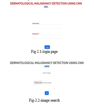
A. Login
Creating a login page in a dermatological malignancy detection system typically involves integrating user authentication and access control to ensure secure access to the application. Here's a simplified overview of how you might implement a login page for a cancer detection system
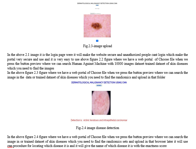
B. Preview
In the preview we should select image from the dataset and upload in the portal and we can get the output of which disease is it and we have the graph in which it shows how much accuracy it is there

 ???????
Conclusion
In conclusion, the application of CNN for dermatological malignancy detection represents a significant advancement in the field of skin and medical imaging. The use of learning techniques, particularly CNNs, Has demonstrated extraordinary efficacy in precise identification and classifying dermatological lesions, including potentially cancerous ones. This technology brings several key advantages
References
[1] S. Prasher, L. Nelson and S. Gomathi, \"Resnet 50 Based Classification Model for Skin Cancer Detection Using Dermatoscopic Images,\" 2023 3rd Asian Conference on Innovation in Technology (ASIANCON), Ravet IN, India, 2023,pp. 1-5, doi: 10.1109/ASIANCON58793.2023.10270158. [2] Nourabuared, “Skin Cancer Based on VGG19 and Transfer Learning”, In 3rd International Conference on Signal Processing and Information Security IEEE Conference, pp 1-4, DOI [3] N. C. Lynn and Z. M. Kyu, , Taipei, Taiwan, 2017, pp. 117-122, DOI: 10.1109/PDCAT.2017.00028. [4] Harikrishna, “Skin Cancer Classification using Transfer Learning IEEE International Conference on Advent Trends in Multidisciplinary Research and Innovation (ICATMRI-2020) [5] N. C. Lynn and Z. M. Kyu, \"Segmentation and Classification of Skin Cancer Melanoma from Skin Lesion Images,\" 2017 18th International Conference on Parallel and Distributed Computing, Applications and Technologies, Taipei, Taiwan, 2017, pp. 117-122, DOI: 10.1109/PDCAT.2017.00028.
Copyright
Copyright © 2024 Ms. Komal Parashar, M. V. Devendranathreddy, M. Deepak Reddy, S. Jashwitha. This is an open access article distributed under the Creative Commons Attribution License, which permits unrestricted use, distribution, and reproduction in any medium, provided the original work is properly cited.

Download Paper
Paper Id : IJRASET59154
Publish Date : 2024-03-19
ISSN : 2321-9653
Publisher Name : IJRASET
DOI Link : Click Here
 Submit Paper Online


