Ijraset Journal For Research in Applied Science and Engineering Technology
- Home / Ijraset
- On This Page
- Abstract
- Introduction
- Conclusion
- References
- Copyright
Detection of Diabetic Retinopathy (DR) using Fundus Images
Authors: Ritesh Sansare, Tejaswi Ganesh Mangave
DOI Link: https://doi.org/10.22214/ijraset.2024.65554
Certificate: View Certificate
Abstract
Diabetic retinopathy (DR) is a leading cause of vision impairment and blindness among adults, resulting from prolonged diabetes. Early detection and timely intervention are crucial to prevent severe visual outcomes. Manual examinations of retinal images, are often subjective, time-consuming, and require expert ophthalmologists. Deep learning has shown remarkable success in image analysis across various medical fields. Its application in diagnosing diabetic retinopathy can process and evaluate images rapidly, facilitating quicker diagnoses and enabling timely treatment decisions. The proposed work highlights the use of CNN, a class of deep learning algorithms to identify relevant features from retinal images, such as micro aneurysms, hemorrhages, and exudates, which are crucial for diagnosing diabetic retinopathy. The proposed system detects the type of DR (like No DR, Mild, Moderate, Severe and Proliferative DR) based on CNN classification with accuracy of 94.35%. The large dataset of the fundus image from Kaggle is used for experimentation. The system is proven effective in detecting diabetic retinopathy.
Introduction
I. INTRODUCTION
Individuals with diabetes are at risk for diabetic retinopathy (DR), a degenerative eye disease. It results from persistent high blood sugar levels, which can harm the small blood vessels in the retina, the light-sensitive tissue at the back of the eye. If left untreated, DR can lead to vision impairment or even blindness, making it one of the leading causes of blindness globally, especially among working-age adults [1][2][5][6].
Because of its link to diabetes, which is affecting a growing number of individuals globally, diabetic retinopathy (DR) is a serious global health concern. The World Health Organisation (WHO) estimates that 420 million persons between the ages of 20 and 79 had diabetes in 2021, and by 2030, that figure is expected to increase to 578 million. Over 100 million people worldwide are currently impacted by diabetes-related retinopathy (DR), with approximately 44 million facing vision-threatening consequence [2][3]. It has been observed that, Over 90% of people suffering from type 2 diabetes, which is driven by socio-economic, demographic, environmental, and genetic factor [3]. Moreover, according to statista report, India has the second-largest number of diabetics worldwide. An estimated 74 million Indians were diagnosed with diabetes in 2021, and by 2045, that number is predicted to increase to nearly 124 million, making diabetes one of the century's worst global health emergencies [4]. These figures emphasize the importance of early diagnosis, preventive measures, and advancements in technologies like AI and telemedicine for managing DR and reducing its impact.
A. Types of DR
There are two types of DR Non-Proliferative DR (NP-DR) and Proliferative DR (P-DR) [5]. The NP-DR is of milder form and usually it is symptomless. Occurs when blood vessels in the retina swell and leak. Figure 1 shows condition of eye in Non-Proliferative (NPDR) and Proliferative Diabetic Retinopathy. On the other hand, PDR refers to the generation of abnormal blood vessel in the retina’s surface and it is the progressive (advance) stage of diabetic retinopathy. PDR is characterized by the development of new but abnormal blood vessels in the retina. There are 4 stages of DR such as mild, moderate, Severe and PDR.
1) Mild DR (Mild non-proliferative retinopathy): This is the earliest stage of the disease in which small new blood vessels (neovascularization) start forming in the retina due to ischemia.
2) Moderate non-proliferative Retinopathy: The condition becomes worse in this case; the retina's supplying blood vessels may enlarge and deform. They might also lose their ability to carry blood as the illness worsens. Both situations result in distinctive alterations to the retina's presence.
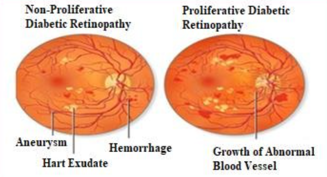
Fig. 1 Non-Proliferative (NPDR) and Proliferative Diabetic Retinopathy (PDR) [6]
3) Severe non-proliferative Retinopathy: In this case blood arteries are obstructing the flow of blood to some retinal regions. Growth factors generated by these affected regions instruct the retina to produce new blood vessels. Figure shows different stages of DR.
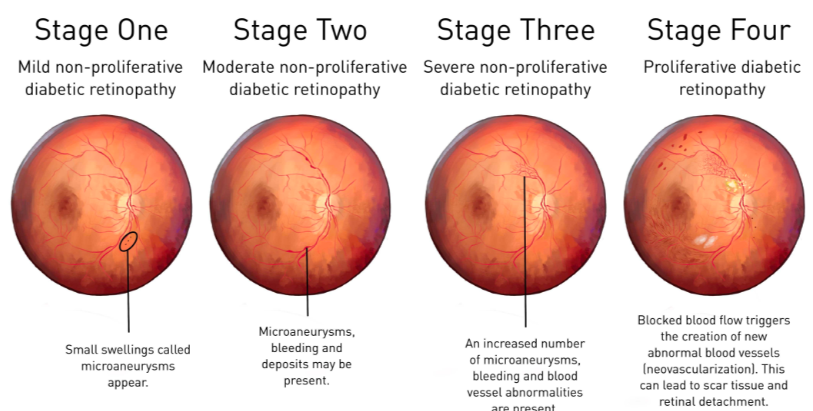
Fig. 2 Stages of Proliferative Diabetic Retinopathy (PDR) [8]
4) Proliferative Diabetic Retinopathy (PDR): The developing blood vessels are likely to bleed and leak at this advanced stage. Fresh blood vessels are triggered by the retina to grow along its inner surface and into vitreous gel, the liquid or blood that fills the eye. Due to their fragility, the fresh blood vessels are more likely to leak and drain. Retinal detachment may also result from the contraction of scar tissue. It is by no means a separator when the retina is pulled without end from hidden tissues, as in backdrop peeling. Because of retinal detachment, this may result in irreversible visual loss.
B. Diabetic Retinopathy Has Following Major Symptoms:
- Double or fluctuating vision.
- Development of a shadow in your field of view.
- Eye floaters and spots.
- Blurry and additionally misshaped vision.
- Vision loss, especially in the advanced stages.
This work offers an automated technique for "Detection of diabetic retinopathy (DR) using fundus images" in light of the urgency of the issue. Vascular segmentation, microaneurysms, exudates, and other pathological indications linked to diabetic retinopathy are among the pertinent elements that the system analyses and extracts from the Digital Retinal Fundus image [5]. Five stages of DR have been identified using the CNN classification: no DR, mild, moderate, severe, and proliferative DR.
II. LITERATURE REVIEW
Using fundus imaging, Avleen Malhi et al. [8] concentrate on the early diagnosis and grading of diabetic retinopathy (DR). It suggests an automated method for detecting exudates and microaneurysms, which are important markers of DR, using image processing, feature extraction, and machine learning models. The number of microaneurysms and the distance of exudates from the macula are used to grade. The study's accuracy for microaneurysm grading using decision trees was 99.9%, while its accuracy for exudate grading with SVM and KNN was 92.1%. Validation was conducted using public and real-time datasets, providing a straightforward but efficient DR diagnostic technique.
Butt et al.[9], presents a hybrid technique for detecting and classifying Diabetic Retinopathy (DR) using fundus images. It leverages transfer learning with pre-trained CNN models to extract features, creating a hybrid feature vector for classification. Performance was evaluated using various classifiers for binary and multiclass DR classification. The method achieved high accuracy, with 97.8% for binary classification and 89.29% for multiclass. This approach demonstrates significant improvements compared to recent DR detection methods, emphasizing its potential for early and accurate DR diagnosis.
Disadvantages: Combining features from multiple CNN models increases computational cost and may require significant processing power, making it less suitable for real-time applications or resource-constrained environments.
Omaisi Asia et al.[10] sheds a light on deep learning, specifically CNNs like ResNet-101, ResNet-50, and VggNet-16, to classify stages of Diabetic Retinopathy (DR) in fundus images. It addresses imbalanced and limited datasets by employing preprocessing, regularization, and augmentation to improve training quality. ResNet-101 achieved the highest accuracy of 98.88% with superior performance on multiple datasets compared to other methods. It demonstrates significant advances in DR detection using deep learning.
Disadvantages: ResNet-101 is computationally intensive, limiting scalability.
Diabetic Retinopathy (DR) is a complication of diabetes that can cause vision impairment and blindness if not detected early. While DR is irreversible, early detection and treatment can prevent vision loss. Manual diagnosis of DR through fundus images is time-consuming, costly, and prone to misdiagnosis, unlike computer-aided systems. Recently, deep learning, particularly Convolutional Neural Networks (CNNs), has improved performance in medical image analysis, including DR detection. Wejdan L. Alyoubi et al.[11] reviews and analyzes state-of-the-art deep learning methods for DR detection in color fundus images, explores available datasets, and discusses the challenges that require further research.
Moreover, Archana Senapati et al.[12] provides systematic review on artificial intelligence (AI) for diabetic retinopathy (DR) detection highlights AI's role in improving screening and diagnosis efficiency. AI models, particularly deep learning systems, demonstrate high accuracy in detecting DR from retinal fundus images, offering cost-effective solutions for large-scale screenings. These tools address challenges like accessibility in underserved areas and reduce reliance on human experts. Despite advancements, integration into clinical practice faces hurdles such as standardization, regulatory approval, and real-world validation.
Abdul Rahaman et al.[13] proposes an efficient and resource-friendly model for diabetic retinopathy (DR) detection and severity grading using the YOLO V7 framework alongside the APTOS and EyePACS datasets. The model integrates MobileNet V3 a compact and efficient CNN architecture optimized for low computational resource environments. It employs YOLO V7 to extract hierarchical features from retinal fundus images rapidly and effectively with accuracy 98%. A Quantum Marine Predator Algorithm (QMPA) refines feature selection, and the Adam optimizer tunes hyper parameters, improving classification performance while reducing over fitting. The study uses CLAHE (Contrast Limited Adaptive Histogram Equalization) and Wiener filters to enhance image quality and remove noise. This approach highlights the balance between achieving high accuracy and maintaining low computational demands, paving the way for widespread, cost-effective DR detection solutions in resource-constrained settings.
Disadvantages: The model's lightweight nature reduces training time, computational complexity, and energy consumption, making it suitable for mobile applications.
V. K. Bairagi et al.[14], develop a system that can accurately classify individuals suffering from diabetic retinopathy. Filtering algorithm is used to clean and preprocess the images collected by users, thereby ensuring the accuracy of the results and reducing the impact of noise on the diagnostic process. An efficient custom three layer CNN model with hyper-parameter tuning is used on kaggle ‘ilovescience’ dataset which gives promising accuracy of 94.45%.
Vatsala Anand et al.[15] has been proposed for quick and precise assessment for the diagnosis of diabetic retinopathy from fundus image. The proposed EfficientNetB0 model is compared with three transfer learning-based models, namely, ResNet152, VGG16, and DenseNet169. The experimental work is carried out using publicly available datasets from Kaggle consisting of 3,200 fundus images. Out of all the transfer learning models, the EfficientNetB0 model has outperformed with an accuracy of 91%, followed by DenseNet169 with an accuracy of 90%.
Sohini Roychowdhury, et al [16] discuss about the Classifiers such as Gaussian Mixture Model (GMM), k-nearest neighbor (KNN), support vector machine (SVM),and Ada Boost are analyzed for classifying retinopathy lesions from non - lesions. Gaussian Mixture Model (GMM) and KNN classifiers are found to be the best classifiers for bright and red lesion classification, respectively. A novel two-step hierarchical classification approach is proposed where the non-lesions or false positives are rejected in the first step. In the second step, the bright lesions are classified as hard exudates and cotton wool spots, and the red lesions are classified as hemorrhages and micro-aneurysms. The detection of neovascularization and vascular beading caused by proliferative DR, and druse caused by macular degeneration.
Disadvantages: The reduction in the number of features used for lesion classification by feature ranking using Adaboost where 30 top features are selected out of 78.
Mike Voets et al.[17] wanted to replicate the original 2016 JAMA study for detection of diabetic retinopathy using publicly available data sets. The original study had employed proprietary EyePACS images and additional datasets from hospitals in India, while the replication study used a publicly available Kaggle EyePACS dataset and Messidor-2 images for testing. The replicated model achieved an AUC of 0.951 on the Kaggle dataset and 0.853 on Messidor-2, falling short of the original AUC of 0.99 on both datasets. This performance discrepancy was attributed to differences in dataset characteristics and the grading methodology. The replication underscored the need for rigorous validation of deep learning models in medical imaging and better accessibility of data and methodologies for reproducibility. The researchers made their replication process open-source to promote further improvements in this domain.By addressing limitations in existing models while building on their strengths, the proposed system not only advances accuracy but also improves accessibility, cost-effectiveness, and scalability for real-world DR detection.
A. Comparison of Existing System
|
Author |
Journal |
Link |
Algorithm Used |
|
Avleen Malhi et al.[ 8] |
Springer |
https://link.springer.com/article/10.1007/s41315-022-00269-5 |
SVM and KNN |
|
Butt et al.[9], |
Diagnostics (Basel) |
https://pubmed.ncbi.nlm.nih.gov/35885512/
|
CNN |
|
Omaisi Asia et al.[10] |
Electronics
|
https://www.mdpi.com/2079-9292/11/17/2740
|
CNNs like ResNet-101, ResNet-50, and VggNet-16 |
|
Abdul Rahaman et al.[13] |
Diagnostics |
https://www.mdpi.com/2075-4418/13/19/3120
|
YOLO V7 |
|
V. K. Bairagi et al.[14], |
IJISAE |
CNN |
|
|
Vatsala Anand et al.[15] |
Machine Learning and Artificial Intelligence |
https://pubmed.ncbi.nlm.nih.gov/38694880/
|
ResNet152, VGG16, and DenseNet169 |
|
Sohini Roychowdhury, et al [16] |
IEEE(Journal of Biomedical and Health Informatics) |
https://ieeexplore.ieee.org/document/6680633
|
Gaussian Mixture Model (GMM) and KNN |
|
Akhilesh Kumar Gangwar et al.[18] |
https://www.researchgate.net/publication/344265006_diabetic_retinopathy_detection_using_transfer_learning_and_deep_learning |
CNN |
|
|
Sai Venkatesh Chilukoti et al.[19] |
ReseachGate |
https://www.researchgate.net/publication/358075386_ diabetic_retinopathy_detection_using_transfer_learning_from_pre-trained_convolutional_neural_network_models |
CNN |
|
Dr. Vijaylaxmi Mekali, et al.[20] |
JETIR |
https://www.jetir.org/papers/jetir2305477.pdf
|
CNN, (ResNet-50) |
III. PROPOSED WORK
- Data Collection: The proposed model is assessed using a large dataset of fundus image from Kaggle (https://www.kaggle.com/c/diabetic-retinopathy-detection/data\ ) to analyze the performances of CNN in order to detect type of diabetic retinopathy. Almost 9000 images for different classes were analyzed. Figure 3 shows the architecture of the proposed system.
- Pre-processing: Green plane extraction is a crucial pre-processing technique for retinal fundus pictures to enhance contrast and feature visibility. The green colour channel in RGB images provides the most precise representation of lesions and blood vessels, which are important indicators of DR. Isolating the green plane, makes blood vessels and microaneurysms, which are markers of the disease's progression, more visible. Convolutional neural networks (CNNs) are able to extract features more effectively as a result. This pre-processing step allows CNNs to focus on critical disease processes by eliminating noise and superfluous background information. Green plane extraction hence enhances CNN's ability to accurately detect early signs of DR.
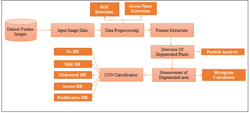
Fig 3: System Architecture
- Feature Extraction: Degenerated pixel detection and region of interest are the two most important factors to consider when determining the posibility of DR. Whereas, Degenerated pixel detection aids in identifying areas where blood vessel leakage have caused the retinal cells to stop functioning normally. Finding these pixels is essential for determining the extent of diabetic retinopathy and tracking its development. Area and volume are among the features of the degenerated area that are computed. The histogram, which provides the number of non-zero pixels and zero-pixels, is thus computed in order to determine the total degenerated area. The degenerated area is indicated by the number of non-zero pixel.
- Identification of type of eye Disease Using CNN: Typically, a CNN's hidden layers include convolutional, pooling completely connected, and normalisation layers. CNN will be used to train the image analytics engine to recognise important information in images. The CNN can identify the type of DR on a scale of 0 to 4 (No DR, Mild, Moderate, Severe, and Proliferative DR).
Convolutional Neural Networks (CNNs) have been shown to be particularly effective in diagnosing diabetic retinopathy (DR) due to its capacity to learn features from fundus images automatically. A CNN's hidden layers, which typically include convolutional layers, pooling layers, fully connected layers, and normalisation layers, will be utilised to extract significant data from images.
???????A. Technique Used
The amazing capacity of Convolutional Neural Networks (CNNs) to process and analyse images makes them perfect for the identification of diabetic retinopathy (DR). Their proficiency in feature extraction allows them to spot minute patterns in retinal fundus photos, including exudates, haemorrhages, and microaneurysms all of which are important markers of DR. Convolutional layers are used by CNNs to identify hierarchical characteristics, allowing for accurate illness severity categorisation. Figure 4 shows working of CNN.
- Convolutional Layers: The CNN's first layers, known as convolutional filters, look for low-level features like edges, colours, and textures in the images. Using non-linear activation functions, like ReLU, to introduce non-linearity allows the model to learn complex patterns.
- Pooling Layers: Max pooling layers reduce the dimensionality while retaining the most important information by down sampling the feature maps. This reduces the computational load and overfitting.
- Fully connected Layers: The high-level features are flattened before being routed through fully connected layers, which include a number of convolutional and pooling layers. The final categorisation about the presence and severity of diabetic retinopathy is established by merging the retrieved features in these layers.
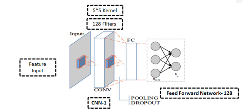
Fig. 4 Working CNN
4) Output Layer: The final layer typically uses a softmax activation function to produce probabilities for each class such as no DR, mild, moderate, severe DR.
IV. SYSTEM ANALYSIS
The 2000-image data set for classes 0, 2, and 3 as well as the 1200-image data set for class 1 (as shown in table 1) have been evaluated here. True positives (TP), false positives (FP), and false negatives (FN), values are used to calculate the accuracy and precision of the suggested system.
Table 1 : TP,TN,FP and FN
|
Class |
TP |
TN |
FP |
FN |
|
Class 0 |
1971 |
6331 |
26 |
28 |
|
Class 1 |
1129 |
7173 |
71 |
50 |
|
Class2 |
1891 |
6411 |
166 |
148 |
|
Class 3 |
1789 |
6516 |
126 |
202 |
|
Class 4 |
1525 |
6777 |
75 |
36 |
The values of TP, FP, and FN i.e truly positive, false positive and false negative can be used to calculate the accuracy, precision, sensitivity and specificity using formulas mentioned below:
- Precision=TP / (TP + FP)
- Sensitivity = TP / (TP + FN)
- Specificity = TN / (FP + TN)
- Accuracy = TP+ TN/ TP+ TN+ FP+ FN
Based on the above formulas, the system performance is calculated as shown in table 2 below. Thus, the model was proven to accurately identify cases of DR in 94.35% of cases. Each class represents a different severity level of diabetic retinopathy, and the accuracy of the model was also proven to encompass performance across all these classes.
Table 2: System Performance
|
Class |
Accuracy |
Precision |
Sensitivity |
|
|
Class 0 |
99.35 |
98.7 |
98.6 |
99.59 |
|
Class 1 |
98.56 |
94.08 |
95.76 |
99.02 |
|
Class 2 |
96.36 |
91.93 |
92.74 |
97.48 |
|
Class 3 |
96.2 |
93.41 |
89.84 |
98.1 |
|
Class 4 |
98.68 |
95.31 |
97.69 |
98.91 |
Figure 5 shows the system performance for each class.
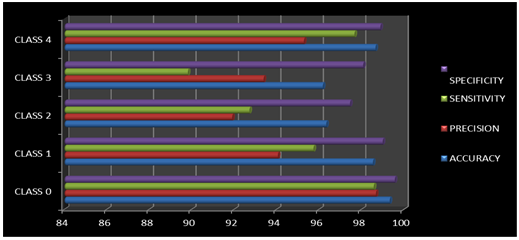 ???????
Fig.5 System Performance
Conclusion
Implementing a CNN for diabetic retinopathy (DR) detection achieved an impressive accuracy of 94.35%, showcasing its effectiveness in automating disease screening. This performance underscores the ability of CNNs to accurately identify and classify DR-related abnormalities in retinal fundus images, even under varying image qualities. The high accuracy aligns with the standards required for clinical relevance, demonstrating potential for deployment in real-world applications.This achievement highlights the robustness of CNNs in feature extraction and classification while reducing reliance on manual grading by specialists. Despite this success, future work should focus on improving generalization across diverse datasets, handling ungradable images, and addressing clinical integration challenges for wider adoption.
References
[1] Martina Kropp, Olga Golubnitschaja, Alena Mazurakova, Lenka Koklesova, Nafiseh Sarghein, Jiri Polivka Jr , Pavel Potuznik 7, Jiri Polivka 7, Ivana Stetkarova 8, Peter Kubatka 9, Gabriele Thumann 1,2 “Diabetic retinopathy as the leading cause of blindness and early predictor of cascading complications—risks and mitigation”, EPMA J. 2023 Feb 13;14(1):21–42. doi: 10.1007/s13167-023-00314-8 [2] J Huan Xiao1†Jinfan Tang&#x;Jinfan Tang2†Feng ZhangFeng Zhang3Luping LiuLuping Liu1Jing ZhouJing Zhou1Meiqi ChenMeiqi Chen1Mengyue LiMengyue Li1Xiaoxiao WuXiaoxiao Wu1Yingying NieYingying Nie1Junguo Duan Junguo Duan1, \"Global trends and performances in diabetic retinopathy studies: A bibliometric analysis\",https://www.frontiersin.org/journals/public-health/articles/10.3389/fpubh.2023.1128008/full [3] \"Facts & figures,\"https://idf.org/about-diabetes/diabetes-facts-figures/ [4] https://www.statista.com/topics/10473/diabetes-in-india/#:~:text=Diabetes%20belongs%20to%20one%20of,over%20124%20million%20by%202045. [5] Galal Mohamed Ismail University of Buraimi ,\"Diabetic RetinopathyJanuary 2017Sudanese Journal of Ophthalmology 9(2):35 DOI:10.4103/sjopthal.sjopthal_41_15\". [6] Hussein Khairallah, Lubna Alazzawi, Nabil Sarhan,\"Mobile Smart Screening and Remote Monitoring for Vision Loss Diseases\",International Journal of Engineering Research & Technology (IJERT) ISSN: 2278-0181 Vol. 8 Issue 10, October-2019 [7] By Sharon Peralta, \"Diabetic retinopathy\"https://www.allaboutvision.com/conditions/diabetic-retinopathy/ [8] Avleen Malhi, Reaya Grewal & Husanbir Singh Pannu ,\"Detection and diabetic retinopathy grading using digital retinal images\", International Journal of Intelligent Robotics and Applications Volume 7, pages 426–458, (2023). [9] Muhammad Mohsin Butt 1, D N F Awang Iskandar 1, Sherif E Abdelhamid 2, Ghazanfar Latif 3 4, Runna Alghazo , \"Diabetic Retinopathy Detection from Fundus Images of the Eye Using Hybrid Deep Learning Features\",2022 Jul 1;12(7):1607. doi: 10.3390/diagnostics12071607. [10] Omaisi Asia 1,Cheng-Zhang Zhu 1,2,*,Sara A. Althubiti 3ORCID,Dalal Al-Alimi 4ORCID,Ya-Long Xiao 1,2,Ping-Bo Ouyang 5 andMohammed A. A. Al-Qaness,\"Detection of Diabetic Retinopathy in Retinal Fundus Images Using CNN Classification Models\"Electronics 2022, 11(17), 2740; https://doi.org/10.3390/electronics11172740 [11] Wejdan L. Alyoubi , Wafaa M. Shalash, Maysoon F. Abulkhair,\"Diabetic retinopathy detection through deep learning techniques: A review\",Informatics in Medicine Unlocked Volume 20, 2020, 100377CMPSCI Tech. Rep. 99-02, 1999. [12] Archana Senapati, Hrudaya Kumar Tripathy a, Vandana Sharma b, Amir H. Gandomi \"Artificial intelligence for diabetic retinopathy detection: A systematic review\",Informatics in Medicine Unlocked Volume 45, 2024, 101445 [13] Abdul Rahaman Wahab Sait,\"A Lightweight Diabetic Retinopathy Detection Model Using a Deep-Learning Technique\",Diagnostics 2023, 13(19), 3120; https://doi.org/10.3390/diagnostics13193120. [14] V. K. Bairagi, Faaris Shaikh, Parmeshwar Randive, Sujeetsing More, Mrinai M. Dhanvijay, Priyanka Tupe-Waghmare \"Detecting Diabetic Retinopathy using Deep Learning\",https://ijisae.org/index.php/IJISAE/article/view/6227 Vol. 12 No. 4 (2024). [15] Vatsala Anand, Deepika Koundal,Deepika Koundal,Wael Y. AlghamdiWael Y. Alghamdi4Bayan M. AlsharbiBayan M. Alsharbi ,\"Smart grading of diabetic retinopathy: an intelligent recommendation-based fine-tuned EfficientNetB0 framework\",Front. Artif. Intell., 16 April 2024 Sec. Machine Learning and Artificial Intelligence. [16] Sohini Roychowdhury, Dara D. Koozekanani, and Keshab K. Parhi “DREAM: Diabetic Retinopathy Analysis using Machine Learning” IEEE Journal of Biomedical and Health Informatics 2013 IEEE [17] Mike Voets, Kajsa Mllersen, Lars Ailo Bongo, “Replication study: Development and validation of a deep learning algorithm for detection of diabetic retinopathy in retinal fundus photographs”, arXiv:1803.04337v3 [cs.CV] 30 Aug. [18] Akhilesh Kumar Gangwar,Ravi Vadlamani,\" Diabetic Retinopathy Detection Using Transfer Learning and Deep Learning\", September 2020Advances in Intelligent Systems and Computing DOI:10.1007/978-981-15-5788-0_64. [19] Sai Venkatesh Chilukoti, Dr. Anthony S Maida, and Dr. Xiali He,\"Diabetic Retinopathy Detection using Transfer Learning from Pre-trained Convolutional NeuralNetwork Models\",IEEE JOURNAL OF BIOMEDICAL AND HEALTH INFORMATICS, 2022. [20] Dr. Vijaylaxmi Mekali, 2Nithya S N, 2Nethra R, 2Priya E, 2Shanvi B P,\"DETECTION OF DIABETIC RETINOPATHY\",© 2023 JETIR May 2023, Volume 10, Issue 5
Copyright
Copyright © 2024 Ritesh Sansare, Tejaswi Ganesh Mangave. This is an open access article distributed under the Creative Commons Attribution License, which permits unrestricted use, distribution, and reproduction in any medium, provided the original work is properly cited.

Download Paper
Paper Id : IJRASET65554
Publish Date : 2024-11-26
ISSN : 2321-9653
Publisher Name : IJRASET
DOI Link : Click Here
 Submit Paper Online

