Ijraset Journal For Research in Applied Science and Engineering Technology
- Home / Ijraset
- On This Page
- Abstract
- Introduction
- Conclusion
- References
- Copyright
Detection of Parkinson’s Disease Using MRI and Spiral Images
Authors: Prof. V. V. Waykule, Siddhi Magdum, Abhishek Mule, Prachi Patil, Mayank Ughade
DOI Link: https://doi.org/10.22214/ijraset.2024.65238
Certificate: View Certificate
Abstract
Early diagnosis of Parkinson’s disease (PD), a neurodegenerative disease characterized by movement disorders, can be challenging. The project aims to develop a machine learning-based model for early diagnosis of Parkinson’s disease using MRI images and spiral models. Using advanced image processing techniques and machine learning algorithms, we analyze MRI scans to identify changes in the brain associated with Parkinson’s disease. In addition, the spiral test is a simple drawing technique used to identify poor motor skills, another early indicator of the disease. This work involves collecting and preprocessing MRI and spiral images and extracting features from both data types. To make the best decision, various machine learning models were used and compared, including convolutional neural networks (CNN) for MRI images and traditional methods for spiral graphs. Performance metrics such as accuracy, precision, recall, and F1 score were used to evaluate the model’s performance distinguishing PD patients from healthy individuals. This approach provides a non-invasive early diagnosis method for PD, which can help in timely treatment and control of the disease. The integration of MRI and motion analysis provides a more comprehensive diagnostic tool with useful clinical outcomes.
Introduction
I. INTRODUCTION
Many techniques are currently being employed in clinical environments as a tool for computer-aided diagnoses to evaluate patient data. PD or Parkinson's disease is an incurable progressive neurological condition that primarily affects the motor centres of the human brain and cannot yet be cured. Early diagnosis can offer patients temporary relief and may even have the potential to delay the progression of PD due to neurodegeneration. This area in the midbrain has relatively abundant dopamine neurons. The Dopamine-producing neurons here communicate with other neurons located near them within the brain by releasing dopamine. At the onset of the disease, the substantia nigra starts degenerating, and its symptoms include stiffness, bradykinesia, or slowing down, and resting tremors. Other symptoms result from the effects of fatigue and anxiety, depression, slowed cognitive activities, and voice difficulties. Parkinson's is supposed to be the second most common neurodegenerative disease among the elderly, after Alzheimer's disease. Both genetic and environmental factors cause the condition of PD, though the cause is still largely unclear. Owing to a lack of specialized medical laboratories, many diagnose this disease in the late stages of the illness. Generally, doctors rely on a patient's medical history and neurological assessments to diagnose. But this strategy often fails because most other neurodegenerative diseases present very similarly. Most diagnoses are made only when significant losses of dopamine have occurred; at least 25% of diagnoses may be false. Such false positive diagnosis may then allow the disease to progress in patients who actually do have PD or falsely label healthy individuals as having the disease, which complicates management.
Various clinical tests may be used to support the diagnosis of Parkinson's disease. Some are particularly relevant because they refer to biological functions in the brain. The common type of imaging technique used in the diagnosis of PD is Magnetic Resonance Imaging, which is non-invasive. This imaging technique has recently become better at detection through the newly improved MRI technology. That involves a decline in handwriting skill, particularly if the motor signs have started in the dominant hand. Handwriting is a complex task that requires coordination of cognitive, perceptual, and fine motor skills. In the recent years, new research is emerging to establish that changes in handwriting kinematics may serve as a biomarker for PD; consequently, handwriting analysis can be very effective, providing a window of opportunity for early diagnosis and tracking the course of the disease. MRI structural evaluation and movement function analysis through spiral test images is a new approach towards hybrid diagnostic models. It eliminates the flaws found in old single-source techniques used for the diagnosis of PD. Imaging and motor analysis separately present incomplete findings while, at the same time, it requires a lot of time for diagnosis. The hybrid model couples both approaches and utilizes the different strengths that the different methods will bring to give a better understanding of the disease. The active interaction between the structural information gained from MRI and the functional data gathered using the spiral test lends an enriched view of the disease, which further helps in building a degree of accuracy in diagnosis.
The clinical signs can distinguish PD patients from healthy people by many different machine learning methods, such as Support Vector Machines, Random Forest, and Convolutional Neural Networks. CNNs are very effective in many tasks because they can automatically learn important features. Many tests have been carried out on CNN-based models that sorted biomedical images with a very high success rate in almost all uses. We used CNNs as classifiers, mixing MRI scans with spiral test images, to identify PD. Adding the spiral images was very helpful for making our model work better because CNNs are known to work well with MR images. This mixed method has also performed well at the top level and shows promise in CNN for correctly diagnosing PD from various types of images.
II. MOTIVATION AND OBJECTIVES
The rising prevalence of Parkinson's disease throws the medical fraternity into an urgent need to enhance early diagnosis and manage the condition better. While conventional methods of diagnosis are indeed well-valued, they fail miserably in accurately diagnosing this disease, especially during its early stages. The chaotic symptoms of PD overlapping with other neurodegenerative diseases depict the urgency of having robust diagnostic tools. Recent advancements in imaging technologies particularly the use of MRI along with other novel motor assessments using spiral test images pose a good opportunity to improve the accuracy of diagnosis. It was from these motivations that our research aimed at developing a hybrid diagnostic model, exploiting the advantages brought forward by MRI and spiral test images.
A. Motivation
Parkinson's disease is one of the most prevalent neurodegenerative diseases; millions of patients exist globally. Early diagnosis, however, represents the hallmark of managing the disease and slowing its progression; however, the existing methods of diagnosis are clinically subjective and postpone diagnosis and appropriate treatment, oftentimes not properly oriented. Conventional approaches focus entirely on motor symptoms of the disease, with no manifestations before the advanced state of the disease.
This project is motivated by the need for a more objective, accurate, and early diagnostic tool that integrates neurological and motor function analyses. MRI images provide insights into structural brain changes associated with PD, while spiral test drawings reveal motor impairments that can serve as early indicators. The project aims to develop a comprehensive detection system that improves diagnostic accuracy, reduces reliance on subjective evaluations, and enables timely intervention by utilizing machine learning algorithms to process and combine these data sources.
Additionally, the motivation stems from the growing potential of machine learning in medical diagnostics, where combining multiple data modalities can yield more robust results. This project seeks to harness that potential, offering a scalable and adaptive solution to help clinicians diagnose PD more effectively and at earlier stages.
???????B. Objectives
The Parkinson’s Disease Detection System has the following key objectives:
- The model should be capable of distinguishing between individuals with Parkinson's disease and healthy individuals based on features extracted from the spiral drawings & MRI images.
- Integrating and optimizing machine learning models that analyze spiral drawing and MRI images will enhance the accuracy of Parkinson’s disease detection.
- Combine features from spiral drawings and MRI images to create a more robust diagnostic tool that can generalize well across different patient demographics and variations in data quality.
- Refine feature extraction methods to capture the most relevant patterns in spiral drawings and MRI images that are indicative of Parkinson's Disease, leading to improved model performance.
- Validate the methodology by comparing its diagnostic accuracy with traditional diagnostic methods.
II. LITERATURE SURVEY
Zhu Li et.al [1] have suggested that captures relevant PD-related handwriting features. Additionally, the paper introduces an innovative method using CC-Net to classify handwriting samples without templates, aiming to improve PD diagnosis accuracy while enhancing patient accessibility. Additional studies have indicated that PD patients exhibit significant tremors or bradykinesia when composing basic sentences. Consequently, researchers can differentiate between healthy individuals and PD patients based on features extracted from handwriting samples.
Md.Ariful Islam et.al [2] The paper reviews the application of machine learning (ML) and deep learning (DL) techniques for Parkinson's disease (PD) detection, focusing on handwriting and voice datasets. It highlights the importance of early diagnosis for effective treatment and the challenges of distinguishing PD from similar conditions. Various classifiers such as Random Forest (RF), Support Vector Machine (SVM), and Convolutional Neural Networks (CNN) are evaluated for their effectiveness, with some models achieving over 90% accuracy. The study emphasizes the need for larger datasets and improved biomarkers to enhance diagnostic accuracy, providing a roadmap for future research in developing more reliable ML/DL tools for PD diagnosis
Sabyasachi Chakraborty et.al [3] utilize Spiral sketch-based CNN and Wave sketch-based CNN models for the analysis and detection of Parkinson's Disease from drawing patterns. Using machine learning algorithms, the data was collected using telemetry touch screen devices in the home environments to distinguish off episodes and peak dose dyskinesia. Several features were extracted from the data and used as input to the machine learning classifiers. Classifiers like Support Vector Machine, Logistic Regression, Random Forest, and Multi-Layer Perceptron (MLP) for feature extraction and classification. The model achieved an accuracy of 93.30% in the detection of Parkinson’s Disease from spiral drawing and wave drawing.
Chakraborty et.al [5] explore the utilization of a 3D convolutional neural network (CNN) for Parkinson's Disease (PD) diagnosis using T1-weighted MRI scans from the PPMI database. Their 35-layer network, incorporating 12 3D convolutional layers, achieved impressive results: 95.29% accuracy, 0.943 recall, 0.927 precision, and an AUC score of 0.98. To enhance model interpretability, class activation maps (CAMs) confirmed the network's focus on the substantia nigra, a region significantly impacted by PD. The model's reliability was established through a 5-split cross-validation process. Although the findings are promising, the researchers emphasize the necessity of expanding the dataset and conducting further validation to ensure the model's efficacy across diverse patient groups.
Sayyed Shahid Hussain et.al [13] have suggested the application of Convolutional Neural Networks (CNNs) in identifying Parkinson's Disease (PD) from MRI scans. PD diagnosis presents difficulties due to its symptoms overlapping with other neurological conditions, and conventional diagnostic approaches, which rely on clinical history and neurological evaluations, often lead to incorrect diagnoses. While imaging methods such as PET and SPECT are beneficial, their invasive nature and high costs make MRI a preferable, non-invasive alternative. Unlike traditional machine learning techniques, such as SVM and decision trees, which require manually extracted features and are prone to errors, CNNs automatically learn features from MRI data, enhancing their effectiveness in complex medical image analysis. Utilizing the Parkinson Progression Markers Initiative (PPMI) dataset, the proposed CNN model demonstrated an accuracy range of 95% to 99%, surpassing conventional methods like SVM and logistic regression. This outcome underscores CNN's ability to automate feature extraction and enhance the accuracy of PD detection.
Babita Majhi et.al [10] have suggested the difficulties in diagnosing Parkinson's disease (PD), particularly in differentiating it from similar conditions and identifying it early. It emphasizes the growing importance of machine learning (ML) in enhancing diagnostic precision through the analysis of imaging data from MRI, SPECT, and PET scans, as well as clinical information. This multifaceted approach addresses the shortcomings of conventional methods that rely solely on clinical evaluations. The authors describe how algorithms such as Support Vector Machines (SVMs) and Convolutional Neural Networks (CNNs) are utilized to examine imaging data, with CNNs identifying PD-related changes in brain structure and SVMs demonstrating enhanced classification accuracy in dopaminergic imaging. The paper also explores advanced ML techniques, including deep learning and multimodal analysis, noting that combining imaging and clinical data strengthens PD detection models. Various imaging modalities, such as resting-state fMRI, diffusion tensor imaging (DTI), and dopamine transporter scans, are discussed for their potential in early PD detection. The article concludes by acknowledging current ML limitations, including the need for additional validation and clinical integration, while proposing future research directions to enhance model generalizability and diagnostic accuracy.
Lucas Salvador et.al [8] have suggested the application of handwriting analysis, particularly spiral drawings, as a diagnostic tool for identifying Parkinson's disease (PD) by examining motor symptoms like tremors, which are apparent in PD patients' handwriting. It introduces a hybrid model that combines SqueezeNet, a deep learning-based CNN for feature extraction, with an SVM for classification. SqueezeNet is chosen for its efficiency and low computational requirements in processing spiral drawings, while the SVM effectively classifies PD and healthy individuals, achieving 91.26% accuracy and surpassing models like VGG11 and ResNet. The authors also consider other machine learning methods, such as Naive Bayes and Random Forest, and address challenges including small datasets and overfitting. The proposed hybrid approach mitigates these issues by utilizing both paper and digital spiral data.
M Shaban et.al [25] have suggest the dataset comprising 204 handwriting samples, evenly split between spiral and wave patterns, collected from 55 control subjects and Parkinson's disease (PD) patients at various stages, matched for age and gender.
The proposed convolutional neural network (CNN) design includes a preprocessing stage that standardizes image dimensions and employs data augmentation through 90-degree rotations to enhance classifier performance. The network architecture consists of five convolutional layers followed by fully connected layers, with the final layer adapted for binary classification between PD and control groups. The research implemented both 4-fold and 10-fold cross-validation methods, yielding validation accuracies of approximately 87% and 88%, respectively, across datasets. The CNN model demonstrated high sensitivity (87% for spiral and 89% for wave datasets) and area under the curve (AUC) values nearing 93% and 94%, indicating effective discriminatory capabilities. While the results are promising, the reliance on handwriting examinations may limit early-stage PD diagnosis, prompting future investigations into electroencephalogram (EEG) analysis for identifying early biomarkers.
Saman Sarraf et.al [14] have suggested that advanced machine-learning approach incorporating a zero-masking method to preserve all pertinent information from imaging datasets. Stacked auto-encoder (SAE) networks were employed to extract high-level features, while support vector machines (SVM) were used for classifying multimodal and multiclass MR/PET data. The research also examined subject-level classification, which yielded high accuracy rates. The introduction of a decision-making algorithm substantially enhanced the precision of subject-level identification, reaching 100% accuracy for MRI pipelines after adjustment. This study integrated cutting-edge preprocessing techniques, deep learning frameworks, and thorough validation procedures to effectively categorize Alzheimer's disease using MRI and fMRI data. The outcomes demonstrate a notable improvement in classification accuracy, underscoring the efficacy of the proposed methodologies. Although the DeepAD pipeline showed promising results in Alzheimer's disease classification, these constraints highlight the necessity for additional research to tackle these issues and enhance the models' robustness and applicability.
Elisabetta Sarasso el.at [15] evaluated 39 studies investigating the progressive changes in Parkinson's disease (PD) patients using magnetic resonance imaging (MRI). The study explored structural changes in both grey and white matter, highlighting the importance of disease progression, severity, and cognitive factors when interpreting MRI findings and their clinical relevance for individuals with PD. The researchers employed various MRI analysis methods, including voxel-based morphometry (VBM), diffusion tensor imaging (DTI), and FreeSurfer for structural assessments. Initially, 504 articles were identified through a literature search, which was reduced to 502 after removing duplicates. The final qualitative analysis included 39 articles, although the exact number of images analyzed across these studies was not specified in the provided information.
III. METHODOLOGY
A. Dataset
The system receives two types of input data: spiral drawing images from patients and MRI scans. Spiral drawings will be collected on paper, and scanned to create digital images while the MRI scans are sourced from medical imaging systems or public datasets like the PPMI.[25][1][8][10]
1) Spiral Images
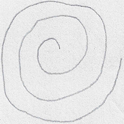
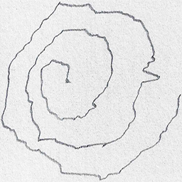
Healthy Parkinson
Fig. 1. Spiral Images
2) MRI Scans
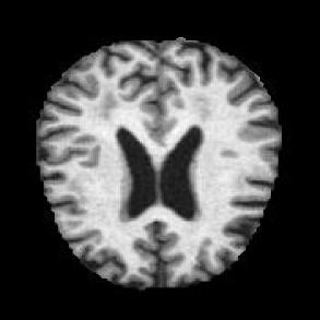
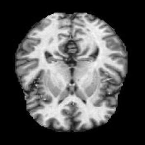
Healthy Parkinson
Fig. 2 MRI Scans
B. Preprocessing
For spiral drawings, noise reduction, resizing, and grayscale conversion are applied to optimize feature extraction. MRI scans undergo preprocessing steps, including skull stripping, intensity normalization, and slicing into 2D sections or processing as 3D volumes.[5]
C. Feature Extraction
Spiral images are fed into a convolutional neural network (CNN) architecture, such as SqueezeNet, to extract relevant motor features related to PD tremors and dyskinesia. Meanwhile, MRI scans are passed through a 3D CNN to detect structural brain changes, especially in regions like the substantia nigra.[8]
D. Fusion
Feature Level Fusion is utilized to combine two extracted datasets. This approach incorporates features from different data sources, including spiral images and MRI scans, to create a more comprehensive data representation. This process can enhance the performance of machine learning algorithms in detecting Parkinson's disease (PD).[2]
E. Model Training and Testing
In the training and evaluation of the system, images were processed in batches. Both the Spiral Images and MRI Scans utilized batches of 50 images each. The entire procedure incorporated 10 batches: 7 were allocated for training, 2 for validation, and 1 for testing. This methodology was applied uniformly across both model types.
F. Classification
Convolutional neural networks excel at detecting Parkinson's disease due to their exceptional feature extraction capabilities and ability to identify patterns in image data. These patterns include noticeable alterations in brain structure visible in MRI scans or atypical movement patterns in spiral drawings indicative of Parkinson's symptoms. The CNN's capacity to handle complex, high-dimensional data makes them particularly suitable for intricate medical imaging tasks. Furthermore, CNNs can integrate multiple data sources, such as MRI and spiral images, into a single model, enabling comprehensive analysis. Their success in various medical diagnostic applications promotes end-to-end learning, minimizing manual intervention and allowing for real-time predictions in clinical settings. CNNs also provide flexibility and efficiency, leading to improved early diagnosis with accurate and useful identification of Parkinson's disease.[1],[3],[10]
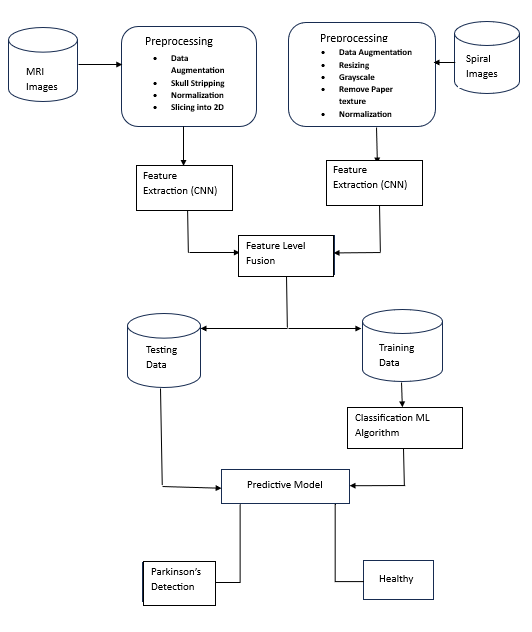
Fig. 3 System Architecture
V. RESULT
A. Spiral Images
|
Model |
Accuracy |
Precision |
F1 |
Recall |
|
CNN |
95% |
91.25% |
94.32% |
94% |
|
AlexNet |
88.9% |
88.40% |
86% |
89.30% |
|
SVM |
70.16% |
71.20% |
75% |
72% |
|
Random Forest |
87.38% |
86% |
87% |
88% |
Table No. 1
B. MRI Scan
|
Model |
Accuracy |
Precision |
F1 |
Recall |
|
CNN |
95.27% |
92.70% |
91.20% |
94.30% |
|
VGG16 |
82.90% |
79.30% |
79.80% |
88.60% |
|
Inception V3 |
82.70% |
81.70% |
80.20% |
84.40% |
|
ResNet |
88.20% |
88.10% |
79.80% |
76.90% |
Table No.2
According to Table No. 1 and Table No.2, the CNN model demonstrates superior performance in predicting Parkinson's Disease compared to other Machine Learning models.
Conclusion
The project on Machine Learning-based Detection of Parkinson\'s Disease significantly advances medical diagnostic techniques. By combining sophisticated machine learning algorithms with data from MRI scans and spiral test drawings, the system enables early and accurate identification of Parkinson\'s disease. This comprehensive approach enhances the diagnostic process, leading to improved patient outcomes and more informed clinical decision-making. While the system boasts numerous advantages, including high accuracy, user-friendly design, and scalability, it also faces challenges such as data dependence, technical hurdles, and the inherent complexity of diagnosing neurodegenerative disorders. Addressing these issues is crucial for ensuring the system\'s optimal performance, effectiveness, and accessibility to a wide range of users. This innovative approach has the potential to revolutionize Parkinson\'s Disease diagnosis, offering valuable insights for both medical professionals and researchers. By facilitating early detection, the system contributes to better patient care and represents a significant advancement in medical technology. It holds promise for enhancing the quality of life for those affected by the disease. As healthcare trends evolve, this system is likely to adapt, making continued strides in addressing neurodegenerative disorders through ongoing updates, user engagement, and adherence to regulatory standards.
References
[1] Zhu Li, Jiayu Yang, Yanwen Wang, Miao Cai, Xiaoli Liu, Kang Lu, “Early diagnosis of Parkinson\'s disease using Continuous Convolution Network: Handwriting recognition based on off-line hand drawing without template”, Journal of Biomedical Informatics 2022 [2] Md.Ariful Islam, Md.Ziaul Hasan Majumder, Md.Alomgeer Hussein , Khondoker Murad Hossain, Md.Sohel Miah, A review of machine learning and deep learning algorithms for “Parkinson\'s disease detection using handwriting and voice datasets”, Heliyon 2024 [3] Sabyasachi Chakraborty, Satyabrata Aich, Jong-Seong-Sim, Eunyoung Han, Jinse Park, Hee-Cheol Kim “Parkinson’s Disease Detection from Spiral and Wave Drawings using Convolutional Neural Networks: A Multistage Classifier Approach” International Conference on Advanced Communications Technology (ICACT) 2020. [4] Nanziba Basnin, Nazmun Nahar, Fahmida Ahmed Anika, Mohammad Shahadat Hossain & Karl Anderson “Deep Learning Approach to Classify Parkinson’s Disease from MRI Samples” International conference on brain informatics, 2021. [5] Sabyasachi Chakraborty, Satyabrata Aich, Hee-Cheol Kim “Detection of Parkinson’s Disease from 3T T1 Weighted MRI Scans Using 3D Convolutional Neural Network” Diagnostics, 2020 [6] Milton Camacho, Matthias Wilms, Pauline Mouches, Hannes Almgren, Raissa Souza, Richard Camicioli, Zahinoor Ismail, Oury Monchi, Nils D. Forkert “Explainable classification of Parkinson\'s disease using deep learning trained on a large multicenter database of T1-weighted MRI datasets” Elsevier Inc. 2023 [7] Yingcong Huang, Kunal Chaturvedi, Al-Akhir Nayan, Mohammad Hesam Hesamian, Ali Braytee, Mukesh Prasad “Early Parkinson’s Disease Diagnosis through Hand-Drawn Spiral and Wave Analysis Using Deep Learning Techniques” Information 2024 [8] Lucas Salvador, Bernardo, Robertas, Victor Hugo “System for Parkinson’s Disease Detection Based on Handwritten and Spiral Patterns” Sciendo 2021 [9] LS Bernardo, R Damasevicius, VHC De Albuquerque, R Maskeliunas “A hybrid two-stage SqueezeNet and support vector machine system for Parkinson\'s disease detection based on handwritten spiral patterns” International Journal of Applied Mathematics and Computer Science, 2021 [10] Babita Majhi, Aarti Kashyap, Siddhartha Suprasad Mohanty, Sujata Dash, Saurav Mallik, Aimin Li, and Zhongming Zhao “An improved method for diagnosis of Parkinson’s disease using deep learning models enhanced with metaheuristic algorithm” Springer BMC medical imaging, 2024 [11] G.Priyadharshini, T.Gowtham, M.Harshavardhan Bhoopathi, M.Reshma, V.Tamilarasi, P.Nandhini “ Detection of Parkinson\'s Disease Using Machine Learning” ISSN 2581-8678, Volume IV, Issue I, Jan 2022, ETJRI. [12] Ferdib-AlIslam, Laboni Akter “Early Identification of Parkinson\'s Disease from Hand-drawn Images using Histogram of Oriented Gradients and Machine Learning Techniques” 2020 Emerging Technology in Computing, Communication and Electronics (ETCCE), 16 February 2021, IEEE. [13] Sayyed Shahid Hussain, Xu Degang, Pir Masoom Shah, Saif Ul Islam, Izaz Ahmad Khan, Fuad A. Awwad, and EmadA.A.Ismail “ Classification of Parkinson’s Disease in Patch-Based MRI of Substantia Nigra” Diagnostics 2023. [14] Saman Sarraf, Danielle D. DeSouza, John Anderson, Ghassem Tofighi “ DeepAD: Alzheimer’s Disease Classification via Deep Convolutional Neural Networks using MRI and fMRI ” BioRxiv, 2016 . [15] Elisabetta Sarasso1, Federica Agosta, Noemi Piramide, Massimo Filippi “ Progression of grey and white matter brain damage in Parkinson’s disease: a critical review of structural MRI literature”Journal of neurology, 2021 Springer. [16] Pugalenthi, R., Rajakumar, M., Ramya, J., Rajinikanth, V “Evaluation and classification of the brain tumour MRI using machine learning technique” J. Control Eng. Appl. Inform. 21(4), 12–21 (2019) [17] Bhat S, Acharya UR, Hagiwara Y, Dadmehr N, Adeli H. “Parkinson’s disease: cause factors, measurable indicators, and early diagnosis” Comput Biol Med.2018;102:234–41. [18] Stefano C, Fontanella F, Impedovo D, et al., “Handwriting analysis to support neurodegenerative diseases diagnosis: A review” Pattern Recognition Lett, 2018, 121(APR.) [19] Y. Wan, Y. Zhu, Y.i. Luo, X. Han, Y. Li, J. Gan, N.a. Wu, A. Xie, Z. Liu, “Determinants of diagnostic latency in Chinese people with Parkinson’s disease”, BMC Neurology 19 (1) (2019) [20] C. Fearon, A. Fasano, “Parkinson’s Disease and the COVID-19 Pandemic”, J Parkinsons Dis 11 (2) (2021) 431–444. [21] Rachel Saunders-Pullman, Carol Derby, Kaili Stanley, Alicia Floyd, Susan Bressman, Richard B. Lipton, Amanda Deligtisch, Lawrence Severt, Qiping Yu, Mónica Kurtis, Seth L. Pullman, “Validity of spiral analysis in early Parkinson\'s disease”, Movement Disorders: Official J Movement Disorder Soc 23 (4) (2008) 531–537. [22] Xiao, Y., Fonov, V., Chakravarty, M.M., Beriault, S., al Subaie, F., Sadikot, A., Pike, G.B., Bertrand, G., Collins,” A dataset of multi-contrast population-averaged brain MRI atlases of a Parkinson?s disease cohort”. Data in brief, 2017 Elsevier. [23] Sarasso, E., Agosta, F., Piramide, N., Filippi, “Progression of grey and white matter brain damage in Parkinson’s disease: a critical review of structural MRI literature”, Journal of Neurology, 2021 Springer. [24] Sarraf, S.; Tofighi, G. ”For the Alzheimer’s Disease Neuroimaging Initiative. DeepAD: Alzheimer’s Disease Classification via Deep Convolutional Neural Networks Using MRI and fMRI”, BioRxiv, 2016 biorxiv.org. [25] M Shaban “Deep convolutional neural network for Parkinson\'s disease based handwriting screening” 2020 IEEE 17th International Symposium on Biomedical Imaging ieeexplore.
Copyright
Copyright © 2024 Prof. V. V. Waykule, Siddhi Magdum, Abhishek Mule, Prachi Patil, Mayank Ughade. This is an open access article distributed under the Creative Commons Attribution License, which permits unrestricted use, distribution, and reproduction in any medium, provided the original work is properly cited.

Download Paper
Paper Id : IJRASET65238
Publish Date : 2024-11-13
ISSN : 2321-9653
Publisher Name : IJRASET
DOI Link : Click Here
 Submit Paper Online

