Ijraset Journal For Research in Applied Science and Engineering Technology
- Home / Ijraset
- On This Page
- Abstract
- Introduction
- Conclusion
- References
- Copyright
Enhancement of X-Ray Images by Gradient Domain Guided Filtering and Non Sub Sampled Shearlet Transform
Authors: G. Rajesh Babu, J. Jaya Durga, Sk. Abdul Khadar, Ch. Hemani, N.V.V. Seshu Babu
DOI Link: https://doi.org/10.22214/ijraset.2024.59765
Certificate: View Certificate
Abstract
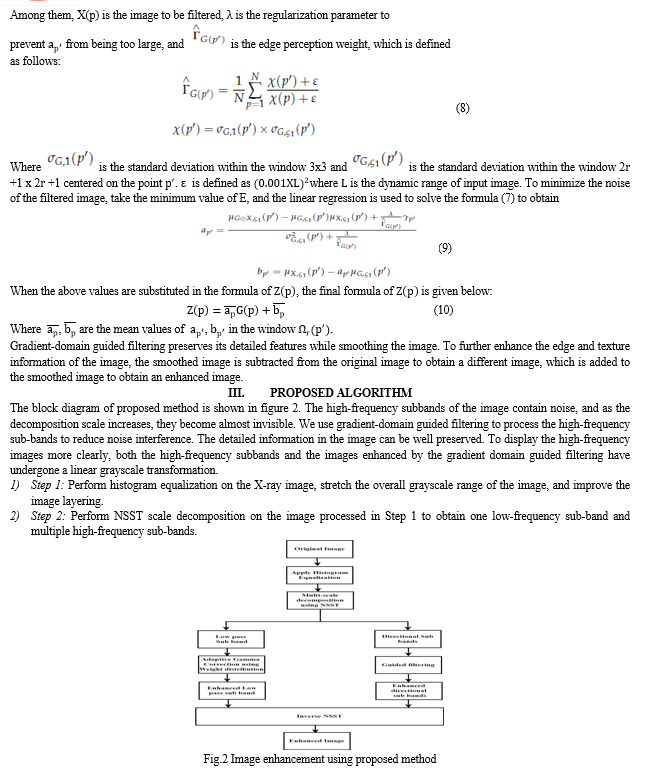
In order to address the issues of low resolution, noise amplification, missing details, and weak edge gradient retention in the X-ray image improvement process, we propose in this study an image enhancement technique that combines non-Subsampled Shearlet transform with gradient-domain guided filtering. We start by breaking down the non Subsampled Shearlet transform and histogram equalization into the original image. We receive multiple high-frequency sub-bands as well as a low-frequency sub-band. To enhance the overall contrast of the image and bring out the contour information in the low-frequency sub-band, adaptive gamma correction with weighting distribution is applied. To reduce image noise and emphasize edge information and detail, gradient-domain guided filtering is applied to the high-frequency sub-bands. In order to generate the final enhanced image, we use the inverse non-Subsampled Shearlet transform to reconstruct all of the sub-bands that were successfully processed. The experimental findings demonstrate the effectiveness of the suggested algorithm in improving X-ray images, as well as the clear advantages its objective index provides over certain conventional algorithms.
Introduction
I. INRODUCTION
With advancements in photoelectric detecting and image analysis technologies, X-ray images are now often used in medical diagnosis, security inspection, aerospace, defect detection, machinery manufacture, and other industries. Low dynamic range, low definition, low contrast, and excessive noise are present in the X-ray instrument's image, the hardware equipment's image, and the detecting principle of the radiographic inspection system. The acquired image is used directly for defect detection and image analysis. The detection results become noticeably inaccurate as a result. Therefore, it is advantageous to analyze X-ray images using image enhancement techniques [1,2], as this improves image quality and visual effects and facilitates subsequent detection. The most popular image enhancement algorithms are spatial domain pixel enhancement and transform domain multi-scale coefficient improvement enhancement techniques. Through pixel-by-pixel processing techniques including histogram equalization [3,4,5], image sharpening and grayscale stretching [6,7,8], and retinex theory [9], the spatial domain can be improved. A gray-level information histogram for X-ray image contrast enhancement was proposed by Zeng et al. [10], and it significantly enhances the performance of several histogram-based enhancement algorithms. But, there will be an over-enhancement issue that results in distorted images when an image is enhanced. Nonlinear unsharp masking was first presented for mammography enhancement by Panetta et al. [11]. When it comes to bringing out the small features in the original photographs, this method works well. But at the same time, it overshoots the crisp details and amplifies noise. A retinex-based framework for improving medical X-ray images was presented by Tao et al. [12]. The dark area pixels in the original image distort the contour borders of the image, showing shadows that never were. Despite this, the framework can improve details, reduce noise, and increase contrast.
The original image is transformed into the frequency domain for multi-scale decomposition first, and then the enhanced image is inversely transformed after amplifying or filtering the decomposed sub-image. Frequently employed transformations include the wavelet transform [13], ridgelet transform [14], curvelet transform [15], wedgelet transform [16], contourlet transform [17], nonsubsampled contourlet transform (NSCT) [18,19], Shearlet transform [20,21], nonsubsampled Shearlet transform (NSST) [22], and others. An approach to improve the details at various scales in screening mammography was presented by Tang et al. [23] and was based on a multiscale measure in the wavelet domain.
Wavelet transform, however, is not very good at capturing anisotropic singular features in images since it can only record data in three directions: horizontal, vertical, and diagonal. An intensity adaptive nonlinear multiscale detail and contrast enhancement method was proposed by Ostojié et al. [24] for digital radiography. The technique adjusts to the local exposure level, which lessens the saliency of the artifacts; nonetheless, the detail improvement adaption to image pixel intensity has to be reinforced. A medical image enhancement technique based on enhanced gamma correction in the Shearlet domain was presented by Zhou et al. [25]. This technique greatly improves overall contrast and highlights the image's textural features. However, because this approach lacks translation invariance, the picture will result in a pseudo-Gibbs phenomena.
Images following NSST may achieve optimal sparse representation and nonlinear error approximation due to the unconstrained shearing directions. Numerous successes have been made while using the NSST for image enhancement. In order to improve blur, Zhang et al. [26] used NSST and tetrolet transform on remote sensing photos, successfully preserving the image's edges and details while greatly enhancing the information entropy and mean. Li et al. [27] investigated the NSST domain. The pseudo-Gibbs phenomenon is successfully eliminated from the image during the enhancement process by the contrast enhancement method, which leverages the remote sensing image enhancement coefficient as an adjustable pattern recognition job. A phase stretching transformation and NSST-based visual sensor image enhancement technique was presented by Tong et al. [28]. In this approach, the author processes the various scale components following NSST decomposition using nonlinear models with various thresholds. The algorithm has the ability to reduce noise and successfully boost the image's contrast. The parameter selection has some degree of randomness, and none of the aforementioned research have examined the parameters and effects of the NSST's shearing orientations and decomposition levels.
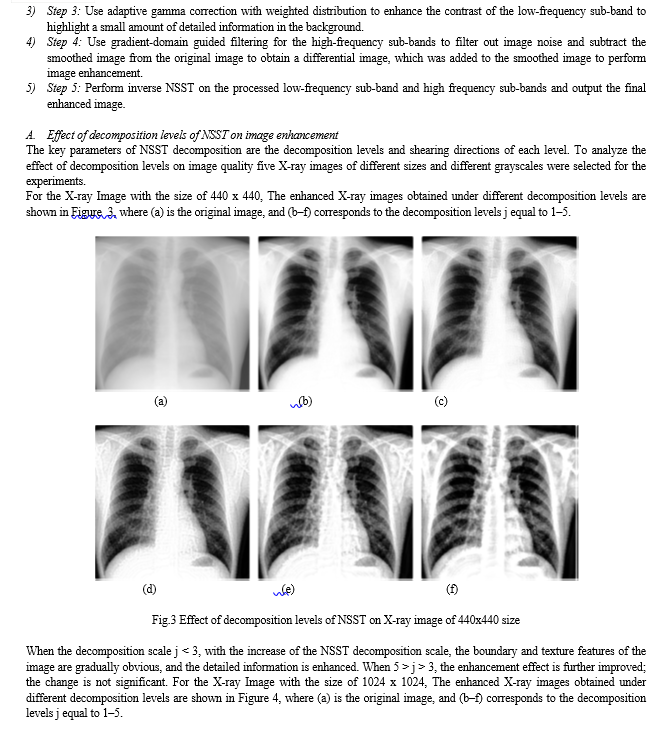
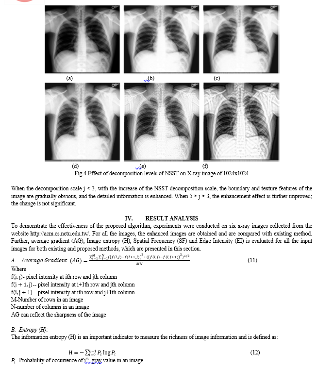
A linear edge-preserving guided image filtering technique was proposed by He et al. [29]. Detailed information won't be blurred in the filtered image. In order to solve the issue of abiding by halo artifacts, Li et al. [30] proposed a weighted guided image filter by adding an edge-aware weighting into an already-existing guided image filter. In order to lessen the impact of image edge smoothing, Kou et al. [31] suggested gradient-domain guided filtering and added first-order edge-aware restrictions to image processing, which can better maintain image edges. However, edge retention may be decreased and edges may be too smoothed when intensity domain limitations are applied to edges and details. We suggest an image enhancing technique to deal with the aforementioned problems. It is applied to X-ray pictures and is based on gradient-domain guided filtering and NSST. The approach combines gradient-domain guided filtering for enhancing picture detail with the benefits of NSST for sparse image representation. The low-frequency sub-band highlights minute details in the background by boosting contrast using adaptive gamma correction with a weighted distribution. By evaluating the image quality of the four-levels direction decomposition under various scale decomposition levels and the image quality of different direction decomposition sequences under the four-levels scale decomposition, the high-frequency sub-bands use gradient-domain guided filtering to remove image noise and extract edge and texture information. In order to determine the ideal NSST decomposition parameters, we compare the execution times of several decompositions. The suggested approach may produce high-quality images for further research and analysis by enhancing the contrast, details, and texture information of medical and industrial X-ray images, as demonstrated by the enhancement experiments conducted on these images.
II. RELATED WORK
A. Non Subsampled Shearlet Transform
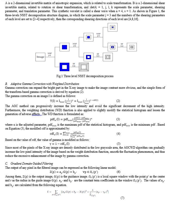
Non Subsampled Shearlet transform (NSST) is a kind of non Subsampled multiscale transform, which was introduced based on the theory of Shearlet transform [11]. The image is decomposed by NSST into multiple scales with multiple directions by multiscale and multidirectional decompositions. Firstly, the nonsubsampled pyramid (NSP) is adopted as the multiscale decomposition filter to decompose the image into one low-frequency sub-band and one high-frequency sub-band. Then, the high-frequency sub-band is decomposed by the shearing filter (SF) to achieve the multidirectional sub-bands. Due to the NSST decomposition process having no subsampling for the NSP and the SF, the NSST is shift-invariant. Its construction is simple and anisotropic, and in dimension n = 2, the affine systems with composite dilations are collections of the form:

Conclusion
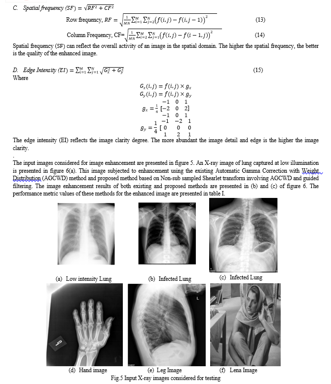
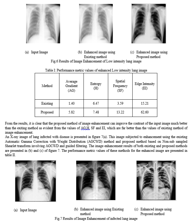
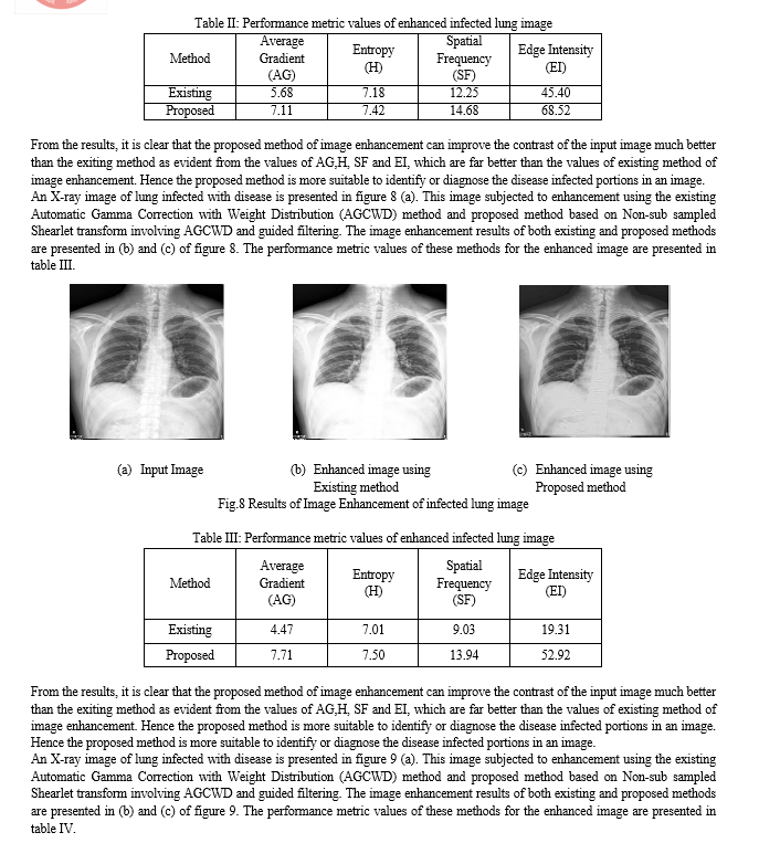
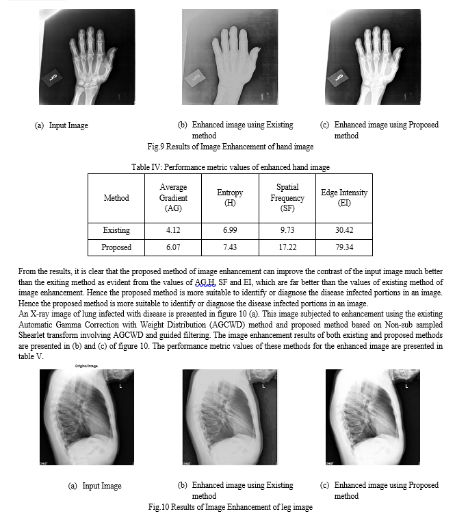
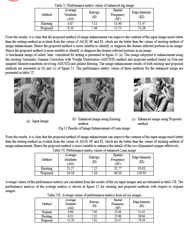
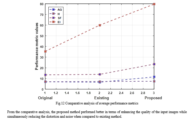
The low contrast of X-ray images makes it difficult to identify tiny and abnormal details. In order to solve this problem, a novel X-ray image enhancement method based on Non-Sub sampled Shearlet transform is proposed in this paper. The low frequency sub bands of decomposed input image after NSST is enhanced using Automatic Gamma Correction with Weight Distribution (AGCWD) technique while the directional sub bands are enhanced using gradient domain guided filtering technique. Later these bands are combined to reconstruct the enhanced image in spatial domain. The performance of the proposed image is tested on six different images in terms of average gradient (AG), Image entropy (H), Spatial Frequency (SF) and Edge Intensity (EI) and compared with existing method of image enhancement. Result analysis showed that proposed method performed better in terms of enhancing the quality of the input images while simultaneously reducing the distortion and noise when compared to existing method.
References
[1] Zhao, Tao, and Si-Xiang Zhang. 2022. \"X-ray Image Enhancement Based on Nonsubsampled Shearlet Transform and Gradient Domain Guided Filtering\" Sensors 22, no. 11: 4074. https://doi.org/10.3390/s22114074. [2] Yang, Y., Su, Z. and Sun, L., “Medical image enhancement algorithm based on wavelet transform”, IET Electronics Letters, Sun L. Medical image enhancement algorithm based on wavelet transform, Vol. 46, No. 2, pp. 120-121, 2010. [3] Li Yang, Yanmei Liang and Hailun Fan, “Study on the methods of image enhancement for liver CT images”, Optik - International Journal for Light and Electron Optics, Vol. 121, No 19, pp.1752-1755, 2010. [4] Peng Feng, Yingjun Pan, Biao Wei and Wei Jin Deling Mi, “Enhancing Retinal Image by the Contourlet Transform”, Pattern Recognition Letters, Vol. 28, No. 4, pp. 516-522, 2007. [5] Sundaram M., Ramar K., Arumugam N. and Prabin G. “Histogram Modified Local Contrast Enhancement for mammogram images”, Applied Soft Computing, Vol. 11, No. 8, pp. 5809-5816, 2011. [6] Tay, P.C., Garson, C.D., Acton, S.T. and Hossack, J.A. “Ultrasound Despeckling for Contrast Enhancement”, IEEE Transactions on Image Processing, Vol. 19, No. 7, pp. 1847-1860, 2010. [7] Bhutada, G.G., Anand, R.S. and Saxena, S.C., “Edge preserved image enhancement using adaptive fusion of Images Denoised by Wavelet and Curvelet Transform”, Digital Signal Processing, Vol. 21, No.1, pp.118-130, 2011. [8] Karen F. Panetta, Yicong Zhou, Sos Agaian and Hongwei Jia, “Nonlinear Unsharp Masking for Mammogram Enhancement”, IEEE Transactions on Information Technology in Biomedicine, Vol. 15, No.6, pp.918-928, 2011. [9] Zhiguo Gui and Yi Liu, “An Image Sharpening Algorithm based on Fuzzy Logic”, International Journal for Light and Electron Optics, Vol. 122, pp. 697-702, 2011. [10] Eltoukhy, M. M., Faye, I. and Samir, B.B., “Breast cancer diagnosis in digital mammogram using multiscale curvelet transform”, 171 Computerized Medical Imaging and Graphics. Vol. 34, No.4, pp. 269-276, 2010. [11] Phan, T. H. Truc, Md. A. U. Khan, Young-Koo Lee, Sungyoung Lee and Tae-Seong Kim, “Vessel Enhancement Filter Using Directional Filter Bank”, Computer Vision and Image Understanding, Vol.113, pp. 101-112, 2009. [12] Akram, M. U., Atzaz, A., Aneeque, S. F. and Khan, S. A., “Blood Vessel Enhancement and Segmentation using Wavelet Transform”, IEEE International Conference on Digital Image Processing, pp. 34-38, 2009. [13] Louis D.N., Ohgaki H., Wiestler O.D, Cavenee W.K. (Eds.), “WHO Classification of Tumors of the Central Nervous System”, International Agency for Research on Cancer (IARC), Lyon, France, 2007. [14] Jan C. Buckner, “Central Nervous System Tumors”, Mayo Clinic Proceedings, Vol. 82, No. 10, pp. 1271-1286. 2007. [15] Vidyarthi, Mittal, “Probabilistic mutual information based extraction of malignant brain tumors in MR images”, 9th IEEE International Conference on Industrial and Information Systems (ICIIS), pp. 1-6, 2014. [16] Shabana Urooj , Amir, “An Automated Approach of CT Scan Image Processing for Brain Tumor Identification and Evaluation”, Journal of Advances in Biomedical Engineering and Technology, vol.2, pp. 11-16, 2015. [17] R.C. Gonzalez and R.E. Woods, Digital Image Processing: 3rd ed., 2008, Pearson Education Inc. [18] A. Beghdadi and A. L. Négrate, “Contrast enhancement technique based on local detection of edges,” Computer Vision, Graphics, and Image Processing, Vol. 46, No. 2, pp. 162–174, 1989. [19] Robert H. Sherrier and G. A. Johnson, “Regionally Adaptive Histogram Equalization of the Chest”, IEEE Transactions on Medical Imaging, Vol. 6, No. 1, pp. 1 -7, 1987. [20] Andrea Polesel, Giovanni Ramponi, and V. John Mathews, “Image Enhancement via Adaptive Unsharp Masking”, IEEE Transactions on Image Processing, Vol. 9, No. 3, pp. 505 – 510, 2000. [21] Jean-Luc Starck, Fionn Murtagh, Emmanuel J. Candès, and David L. Donoho, “Gray and Color Image Contrast Enhancement by the Curvelet Transform”, IEEE Transactions on Image Processing, Vol. 12, No. 6, pp. 706 – 717, 2003. [22] Huang Kaiqi, Wu Zhenyang and Wang Qiao, “Image enhancement based on the statistics of visual representation”, Image and Vision Computing, Vol. 23, No.1, pp. 51–57, 2005. [23] Nagesha and G. Hemantha Kumar, “A level crossing enhancement scheme for chest radiograph images”, Computers in Biology and Medicine, Vol. 37, No. 10, pp. 1455 – 1460, 2007. [24] Aditi Majumder and Sandy Irani, “Perception-Based Contrast Enhancement of Images”, ACM Transactions on Applied Perception, Vol. 4, No. 3, Article 17, 2007. [25] Sos S. Agaian, Blair Silver and Karen A. Panetta, “Transform Coefficient Histogram-Based Image Enhancement Algorithms Using 164 Contrast Entropy”, IEEE Transactions on Image Processing, Vol. 16, No. 3, pp. 741 – 758, 2007. [26] Buyue Zhang and Jan P. Allebach, “Adaptive Bilateral Filter for Sharpness Enhancement and Noise Removal”, IEEE Transactions on Image Processing, Vol. 17, No. 5, pp. 664 – 678, 2008. [27] T.L. Economopoulos, P.A. Asvestas and G.K. Matsopoulos, “Contrast enhancement of images using Partitioned Iterated Function Systems”, Image and Vision Computing, Vol. 28, No. 1, pp. 45–54, 2010. [28] D.H. Kim and E.Y. Cha, “Intensity surface stretching technique for contrast enhancement of digital photography”, Multidimensional Systems and Signal Processing, Vol. 20, No. 1, pp. 81 – 95, 2009. [29] H. Huang and X. Xiao, “Example-based contrast enhancement by gradient mapping”, Visual Computer, Vol. 26, No. 6-8, pp. 731–738, 2010. [30] Chi-Yi Tsai and Chien-Hsing Chou, “A novel simultaneous dynamic range compression and local contrast enhancement algorithm for digital video cameras”, EURASIP Journal on Image and Video Processing, Vol. 2011, No. 6, pp. 1 – 19, 2011. [31] John B. Zimmerman, Stephen M. Pizer, Edward V. Staab, J. Randolph Perry, William Mccartney and Bradley C. Brenton, “An Evaluation of the Effectiveness of Adaptive Histogram Equalization for Contrast Enhancement”, IEEE Transactions on Medical Imaging, Vol. 7, No. 4, pp. 304 – 312, 1988. [32] Y. T. Kim, “Contrast Enhancement Using Brightness Preserving BiHistogram Equalization”, IEEE Transactions on Consumer Electronics, Vol. 43, No. 1, pp. 1 – 8, 1997. [33] Yu Wang, Qian Chen and Baomin Zhang, “Image Enhancement Based on Equal Area Dualistic Sub-Image Histogram Equalization Method”, IEEE Transactions on Consumer Electronics, Vol. 45, No. 1, pp. 68 –75, 1999. [34] JC. Fu, H.C. Lien and S.T.C. Wong, “Wavelet-based histogram equalization enhancement of gastric sonogram images”, Computerized Medical Imaging and Graphics, Vol. 24, No. 2, pp. 59 – 68, 2000. [35] Sarif Kumar Naik and C. A. Murthy, “Hue-Preserving Color Image Enhancement Without Gamut Problem”, IEEE Transactions On Image Processing, Vol. 12, No. 12, pp. 1591 – 1598, 2003. [36] Chang-Jiang Zhang and Min Hu, “Contrast enhancement for image by WNN and GA combining PSNR with information entropy”, Fuzzy Information and Engineering - Advances in Soft Computing, Vol. 40,pp. 3 - 15, 2007. [37] Rui,W.; Guoyu,W. Medical X-Ray Image Enhancement Method Based on TV-Homomorphic Filter. In Proceedings of the 2017, 2nd International Conference on Image, Vision and Computing (ICIVC), Chengdu, China, 2–4 June 2017; pp. 315–318. [38] Wang, X.; Chen, L. Contrast enhancement using feature-preserving bi-histogram equalization. Signal Image Video Process. 2018, 12, 685–692. [39] Wu, W.; Yang, X.; Li, H.; Liu, K.; Jian, L.; Zhou, Z. A novel scheme for infrared image enhancement by using weighted least squares filter and fuzzy plateau histogram equalization. Multimed. Tools Appl. 2017, 76, 24789–24817. [40] Xiao, B.; Tang, H.; Jiang, Y.; Li, W.; Wang, G. Brightness and contrast controllable image enhancement based on histogram specification. Neurocomputing 2018, 275, 2798–2809. [41] Lombardo, P.; Oliver, C.J.; Pellizzeri, T.M.; Meloni, M. A newmaximum-likelihood joint segmentation technique for multitemporal SAR and multiband optical images. IEEE Trans. Geosci. Remote Sens. 2003, 41, 2500–2518. [42] Magudeeswaran, V.; Ravichandran, C.; Thirumurugan, P. Brightness preserving bi-level fuzzy histogram equalization for MRI brain image contrast enhancement. Int. J. Imaging Syst. Technol. 2017, 27, 153–161. [43] Mohan, S.; Ravishankar, M. Modified contrast limited adaptive histogram equalization based on local contrast enhancement for mammogram images. In Proceedings of the International Conference on Advances in Information Technology and Mobile Communication, Bangalore, India, 27–28 April 2012; pp. 397–403. [44] Al-Ameen, Z.; Sulong, G. Ameliorating the Dynamic Range of Magnetic Resonance Images Using a Tuned Single-Scale Retinex Algorithm. Int. J. Signal Processing Image Process. Pattern Recognit. 2016, 9, 285–292. [CrossRef] [45] Zeng, M.; Li, Y.; Meng, Q.; Yang, T.; Liu, J. Improving histogram-based image contrast enhancement using gray-level information histogram with application to X-ray images. Optik 2012, 123, 511–520. [CrossRef] [46] Panetta, K.; Zhou, Y.; Agaian, S.; Jia, H. Nonlinear unsharp masking for mammogram enhancement. IEEE Trans. Inf. Technol. Biomed. 2011, 15, 918–928. [CrossRef] [47] Tao, F.; Yang, X.; Wu, W.; Liu, K.; Zhou, Z.; Liu, Y. Retinex-based image enhancement framework by using region covariance filter. Soft Comput. 2018, 22, 1399–1420.
Copyright
Copyright © 2024 G. Rajesh Babu, J. Jaya Durga, Sk. Abdul Khadar, Ch. Hemani, N.V.V. Seshu Babu. This is an open access article distributed under the Creative Commons Attribution License, which permits unrestricted use, distribution, and reproduction in any medium, provided the original work is properly cited.

Download Paper
Paper Id : IJRASET59765
Publish Date : 2024-04-03
ISSN : 2321-9653
Publisher Name : IJRASET
DOI Link : Click Here
 Submit Paper Online

