Ijraset Journal For Research in Applied Science and Engineering Technology
- Home / Ijraset
- On This Page
- Abstract
- Introduction
- Conclusion
- References
- Copyright
Enhancing Brain Tumor Diagnosis: A Synergistic Approach with Support Vector Machines and Decision Trees for Improved Detection and Segmentation
Authors: Srikumaran R, Sanjay Kumaran V, Vijay M, Balamurugan A
DOI Link: https://doi.org/10.22214/ijraset.2024.58595
Certificate: View Certificate
Abstract
Brain tumor identification and segmentation are critical components of modern medical imaging, facilitating timely diagnoses and effective therapy planning. This paper introduces an innovative methodology that harmoniously integrates Support Vector Machines (SVM) and Decision Tree methods. SVM adeptly classifies brain images into tumor and non-neoplastic regions, harnessing its proficiency with complex datasets. Decision Tree methods complement this process, providing transparent segmentation that accurately delineates intricate tumor boundaries. The synergy between SVM and Decision Tree methods establishes a robust framework for brain tumor identification. This novel approach holds promise for enhanced diagnosis speed, particularly valuable in time-sensitive medical scenarios. Emphasizing potential advancements in patient care outcomes, the methodology enables tailored treatment plans with greater precision. Positioned at the core of brain image analysis, this novel methodology signifies a substantial step forward in healthcare practices, fostering personalized approaches to brain tumor diagnosis and treatment.
Introduction
I. INTRODUCTION
The landscape of clinical imaging and medical care has undergone a remarkable revolution with the advent of sophisticated methodologies, notably artificial intelligence (AI) and machine learning. In this era of medical innovation, the identification and classification of brain cancers emerge as pivotal applications, holding profound implications for patient care and outcomes. Brain cancers, often posing imminent threats and demanding prompt attention, have historically presented formidable diagnostic challenges. However, the integration of intelligent techniques into clinical practice heralds a new era marked by heightened precision and efficiency.
A brain tumor is an abnormal tissue growth in the brain that poses a significant risk to one's health due to its ability to grow exponentially. In order to detect brain tumors, Deep Learning and Machine Learning algorithms can be applied to MRI scans. Using artificial neural networks (ANN) and sophisticated image processing methods, this study presents a novel approach to brain tumor detection and classification. This overview delves into the realm of brain tumor detection and classification, unraveling the methodologies, challenges, and remarkable advancements achieved through the application of intelligent algorithms. The imperative role of medical imaging, accentuated as one of the most significant breakthroughs in clinical cancer diagnoses, serves as the cornerstone for developing innovative diagnostic strategies.
The urgency of addressing global health challenges, particularly the escalating incidences of cardiovascular diseases and cancer, underscores the need for transformative approaches [2]. Brain tumors, constituting a substantial portion of cancer-related deaths, amplify the call for innovative diagnostic methodologies that can navigate the intricate landscape of these life-threatening conditions [4]. Notably, a comprehensive study in 2019 reported that 17,000 individuals succumb to brain cancer annually in the United States alone [2].
Survival rates following brain tumor diagnoses underscore the critical importance of early detection. The five-year survival rates, standing at 34% for men and 36% for women, emphasize the direct correlation between early diagnosis, timely intervention, improved rehabilitation outcomes, and extended survival periods [2].
Recent studies [5-14] explore the transformative potential of intelligent algorithms in the identification and segmentation of brain tumors. Proposed synergistic approach, intertwining Support Vector Machines and Decision Tree algorithms, seeks to contribute to ongoing advancements in this critical domain. By amalgamating medical imaging breakthroughs, sophisticated algorithms, and a commitment to addressing diagnostic challenges, this research aspires to be a catalyst for precision, efficiency, and improved patient outcomes in the realm of brain tumor diagnostics.
A. Image Classification
In an undeniably computerized world, where the sheer volume of visual information outperforms the capacity to handle it physically, picture characterization arises as a vital innovation [4]. From perceiving recognizable countenances in photographs to diagnosing ailments from sweeps and, surprisingly, directing self-driving vehicles, picture characterization assumes an extraordinary part across a range of enterprises and applications. At its center, picture order is the craftsmanship and study of training PCs to "see" and grasp visual substance. From the perspective of man-made brainpower and AI, it empowers machines to unravel unpredictable examples, shapes, and highlights inside pictures, getting a handle on the visual world in manners that were once solely human spaces.
B. Image Segmentation
In the domain of computerized symbolism, there exists a basic undertaking that is likened to a craftsman's brushstroke on a material, carefully characterizing the limits and locales inside a picture. This errand, known as "picture division," is a strong computational procedure that presents to machines the capacity to see and grasp the visual world with surprising accuracy [7]. Picture division is the most common way of dividing a picture into unmistakable and significant districts or articles, disconnecting them from the encompassing foundation. Maybe defining boundaries around unambiguous components inside an image, changing a perplexing embroidery of pixels into an organized guide of individual elements. While this could appear to be a straightforward creative undertaking, it holds huge importance in different fields, going from clinical imaging and independent mechanical technology to satellite picture examination and PC vision.
C. Deep Learning
In the consistently propelling scene of man-made reasoning, a significant change is happening - one that reflects the human mind's unpredictable functions and can possibly reform the capacities of machines. This change is known as "profound learning," and it remains as the apex of AI, empowering PCs to accomplish accomplishments once viewed as the domain of sci-fi [12]. Profound learning, at its center, addresses a jump forward in the journey to make machines figure out, reason, and cooperate with the world in more nuanced and human-like ways. It is a subset of AI, recognized by its capacity to gain and concentrate unpredictable examples and portrayals from immense measures of information consequently. At the core of profound learning are counterfeit brain organizations, propelled by the unpredictable trap of neurons in the human mind. In the exploration of the profound learning universe, one will unravel its essence and delve into the extraordinary power it holds.
This paper concentrated on developing the deep learning approach for brain tumor detection in MR images. Section 2 deals with the literature review regarding the deep learning mechanisms in brain tumor detection. Section 3 deals with the proposed method which contains the step by step procedures of the method. Section 4 is Result analysis which will have the accuracy and speed of the algorithms. Section 5 is the conclusion of this paper. Section 6 will have the Future Scopes of the Proposed method.
II. LITERATURE REVIEW
This section contains the current landscape of brain tumor diagnosis methodologies, exploring a spectrum of approaches from traditional techniques to modern deep learning models. Examining the strengths and limitations of existing studies lays the groundwork for the novel integration of Support Vector Machines (SVM) and Decision Tree algorithms in advancing brain tumor identification and segmentation.
A. Brain Tumor Magnetic Resonance Image Classification: A Deep Learning Approach
Machiraju Jaya Lakshmi et.al [1]. Has proposed in this paper, From the previous ten years, numerous analysts are centered around the cerebrum cancer recognition component utilizing attractive reverberation pictures. The conventional methodologies follow the component extraction process from base layer in the organization. This situation isn't reasonable to the clinical pictures. To resolve this issue, the proposed model utilized Commencement v3 convolution brain network model which is a profound learning method. This model concentrates the staggered elements and orders them to track down the early location of cerebrum growth. The proposed model purposes the profound learning approach and hyper boundaries. These boundaries are improved utilizing the Adam Streamlining agent and misfortune capability. The misfortune capability assists the machines with demonstrating the calculation with input information. The delicate max classifier is utilized in the proposed model to order the pictures in to various classes. It is seen that the precision of the Beginning v3 calculation is recorded as 99.34% in preparing information and 89% exactness at approval information. The improvements in the clinical field support the clinical specialists to really work with the patients. With the use of computerized reasoning (computer-based intelligence) in the medical care assists the clinical area with serving more to the patients.
B. Brain Tumor Classification Based on Attention Guided Deep Learning Model
Wen Jun et.al.[2] has proposed in this paper Malignant growth is the subsequent driving reason for death around the world. Mind growths exclude for one of each and every four disease passings. Giving an exact and ideal finding can bring about opportune medicines. As of late, the fast advancement of picture order has worked with PC helped determination. The convolutional brain organization (CNN) is one of the most broadly utilized brain network models for characterizing pictures. In any case, its adequacy is restricted in light of the fact that it can't precisely recognize the point of convergence of the sore. This paper proposes an original mind cancer characterization model that coordinates a consideration instrument and a multipath organization to settle the above issues. A consideration instrument is utilized to choose the basic data having a place with the objective locale while overlooking superfluous subtleties. A multipath network relegates the information to different channels, prior to changing over each channel and combining the consequences, everything being equal.
C. A Deep Learning Framework for Automatic Brain Tumor Classification Using Transfer Learning
Archie Rehman et.al.[3] Has proposed in this framework, Cerebrum cancers are the most horrendous sickness, prompting an exceptionally short future in their most elevated grade. The misdiagnosis of cerebrum growths will bring about off-base clinical intervention and diminish chance of endurance of patients. The precise finding of mind growth is a central issue to make a legitimate treatment intending to fix and work on the presence of patients with cerebrum cancers sickness. The PC supported cancer recognition frameworks and convolutional brain networks gave examples of overcoming adversity and have taken significant steps in the field of AI. The profound convolutional layers extricate significant and powerful elements naturally from the information space when contrasted with customary ancestor brain network layers. In the proposed method, three examinations are conducted using three designs of convolutional brain networks (Alex Net, Google Net, and VGG Net) to classify brain tumors such as meningioma, glioma, and pituitary.
D. Deep Learning for Medical Anomaly Detection – A Survey
Tharindu Fernando et.al.[4] Has proposed in this framework AI based clinical peculiarity identification is a significant issue that has been broadly contemplated. Various methodologies have been proposed across different clinical application areas, and similarities are observed across these specific applications. Despite this resemblance, there is a lack of organized association among these diverse examination applications, hindering the comprehensive study of their benefits and constraints. The primary objective of this overview is to offer a thorough theoretical analysis of popular deep learning techniques in clinical anomaly identification. Specifically, a systematic and comprehensive review of state-of-the-art procedures is presented, analyzing not only their architectural differences but also their training algorithms. Additionally, a comprehensive overview of deep model interpretability techniques is provided, offering insights into methods used to interpret model decisions.
E. Brain Tumor Segmentation Using Deep Learning on MRI Images
Almetwally M. Mostafa to et.al.[5] Has proposed in this framework, Mind cancer (BT) conclusion is an extensive cycle, and extraordinary ability and mastery are expected from radiologists. As the quantity of patients has extended, so has how much information to be handled, making past methods both expensive and ineffectual. Numerous scholastics have inspected a scope of solid and fast strategies for recognizing and classifying BTs. As of late, profound learning (DL) strategies have acquired ubiquity for making PC calculations that can rapidly and dependably analyze or section BTs. To recognize BTs in clinical pictures, DL allows a pre-prepared convolutional brain organization (CNN) model. The recommended attractive reverberation imaging (X-ray) pictures of BTs are remembered for the BT division dataset, which was made as a benchmark for creating and assessing calculations for BT division and conclusion.
F. Observation from The Literature Review
The literature underscores significant advancements in deep learning techniques for brain tumor detection, showcasing the efficacy of models like Commencement v3 with impressive accuracy rates [1]. Attention-guided classification, integrating attention mechanisms and multipath networks, offers a precision-focused approach [2]. Transfer learning, employing convolutional neural networks (AlexNet, GoogleNet, and VGGNet), enhances specificity in brain tumor classification [3]. A thorough survey on deep learning for medical anomaly detection reveals insights into various techniques, their architectural nuances, and training algorithms [4]. Additionally, the application of convolutional neural networks proves instrumental in efficient brain tumor segmentation on MRI images, promising improved diagnostic practices [5].
III. PROPOSED METHOD
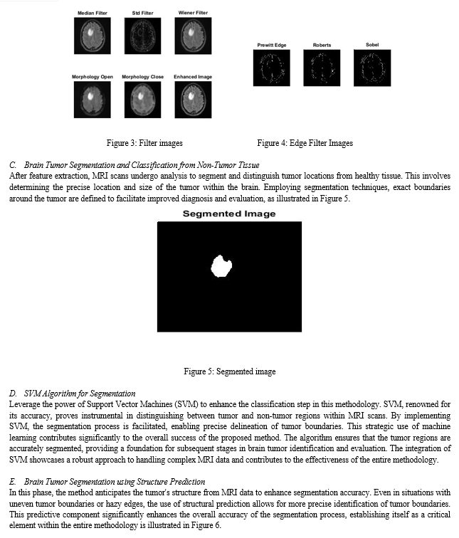
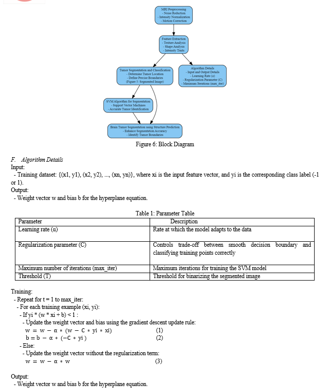
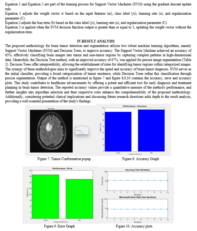
Better patient outcomes may result from the proposed method's potential to dramatically increase the precision and effectiveness of brain tumor detection and segmentation. This is due to the fact that SVM is a potent machine learning method that works well for picture segmentation and classification applications. Additionally, medical image processing and segmentation may be done quickly and effectively, which is necessary for precise tumor diagnosis and segmentation. The suggested method preprocesses the brain MRI image in order to get rid of noise and artifacts. The preprocessed image is then used to extract a set of useful and representative features. These characteristics may be intensity, texture, shape, or a mix of these. To determine whether each pixel in the image corresponds to the tumor region or not, the method then trains an SVM classifier. The tumor region in the input image is segmented using the SVM classifier once it has been trained.
A. MRI Preprocessing
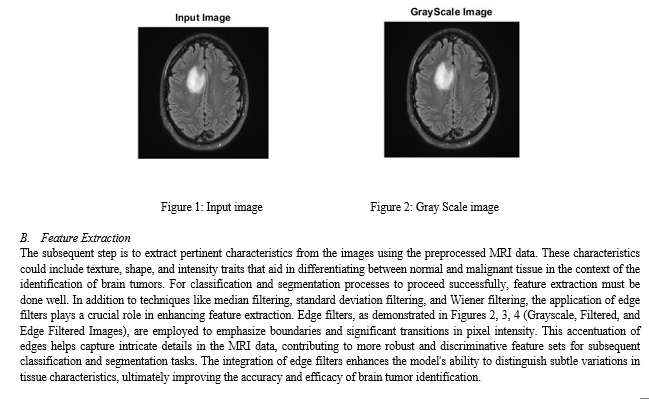
The first procedure in the pipeline is MRI preprocessing. It uses a number of strategies to improve the MRI pictures' accuracy and dependability. To guarantee that the images are in a consistent and acceptable form for future analysis, these approaches may include noise reduction, intensity normalization, and motion correction. The initial step involves MRI preprocessing to enhance accuracy and reliability. MRI image is shown in Figure 1. Ensure images are in a consistent and acceptable form for subsequent analysis.


V. FUTURE WORK
Increase the method's precision and effectiveness. More sophisticated machine learning algorithms, such as deep learning algorithms, can be used to do this. Enhance the method's resistance to noise and visual artifacts. This can be accomplished by creating fresh picture preprocessing methods. Expand the method to segment gliomas and meningiomas in addition to other kinds of brain tumors. Create a user-friendly interface for the method so that medical professionals may utilize it with ease.
VI. AUTHORS' BIOGRAPHY
Sanjay Kumaran, Srikumaran R, and Vijay M are Computer Science and Engineering students at KPR Institute of Engineering and Technology, contributing their expertise to this paper. Guided by Professor Balamurugan A, the team benefits from his extensive experience in the field.
Conclusion
The suggested method for brain tumor detection and segmentation, which employs SVM and Decision Tree algorithms, has the potential to significantly advance the field of brain tumor diagnosis and therapy. Using SVM and Decision Tree methods, the method may achieve good accuracy and efficiency in tumor identification and segmentation. The suggested approach has the potential to improve patient care in a variety of ways, including early detection, treatment planning, and tracking disease progression. Overall, the suggested method represents a promising novel approach to brain tumor identification and segmentation. Continuing research and development in this field allows us to aim for the creation of more accurate, efficient, and resilient brain tumor detection and segmentation method. This, in turn, leads to improved patient care and outcomes for individuals with brain tumors.
References
[1] M. J. Lakshmi and S. N. Rao, \'\'Brain tumor magnetic resonance image classification: A deep learning technique Soft Computing, vol. 26, no. 13, pp. 6245–6253, July 2022. [2] W. Jun and Z. Liana, \'\'Brain tumor classification based on attention driven deep learning model, International Journal of Computing and Intelligence Systems, vol. 15, no. 1, p. 35, December 2022. [3] Rahman, S. Naz, M. I. Raza, F. Aram, and M. Imran, \'\'A deep learning-based system for automatic brain tumor categorization utilizing transfer learning, Circuits, Systems, and Signal Process., vol. 39, no. 2, pp. 757-775, February 2020. [4] Fernando, H. Gamboled, S. Denman, S. Sridhar, and C. Folks, \"Deep learning for medical anomaly detection—A survey,” ACM Computing Surveys, vol. 54, no. 7, pp. 1–37, September 2022. [5] Mostafa, Almetwally M., Mohammed Zakariah, and Eman Abdullah Aldakheel. \"Brain Tumor Segmentation Using Deep Learning on MRI Images.\" Diagnostics 13, no. 9 (2023): 1562. [6] S. Undersold and A. Lundervold, \"An overview of deep learning in medical imaging concentrating on MRI,\'\' Zeitschrift für Medizinische Physik, vol. 29, no. 2, pp. 102-127, 2019. [7] R. Ranjbarzadeh, A. B. Kasugai, S. J. Ghoushchi, S. Ansari, M. Naseri, and M. Bendechache. \"Brain tumor segmentation based on deep learning and an attention mechanism using MRI multi-modalities brain images.\" Sci. Rep., vol. 11, no. 1, pp. 10930, May 2021. [8] Ruba, R. Tamilselvi, and M. P. Beham, \'\'Brain tumor segmentation in multimodal MRI images utilizing novel LSIS operator and deep learning, J. Ambient Intelligence and Humanized Computing, pp. 1-15, March 2022. [9] P. K. Ramtekkar, A. Pandey, and M. K. Pawar, \"Innovative brain tumor detection using optimized deep learning techniques,\" International Journal of System Assurance and Engineering Management, vol. 14, no. 1, pp. 459-473, Feb. 2022. [10] Y. Suter, A. Jungo, M. Rebsamen, U. Knecht, E. Herrmann, R. Wiest, and M. Reyes, \"Deep learning versus classical regression for brain tumor patient survival prediction,\" in Proc. Int. MICCAI Brainlesion Workshop, 2019, pp. 429–440. [11] G. Chetty, M. Yamin, and M. White, \'\'A low resource 3D U-Net based deep learning model for medical image interpretation,\'\' International Journal of Information Technology, vol. 14, no. 1, pp. 95-103, February 2022, [12] Z. Shaukat, Q. U. A. Farooq, S. Tu, C. Xiao, and S. Ali, \"A state-of-the-art technique to perform cloud-based semantic segmentation using deep learning 3D U-Net architecture,\" in BMC Bioinf., vol. 23, no. 1, pp. 251-272, December 2022. [13] J. N. Stember and H. Shalu, \'\'Deep reinforcement learning with automated label extraction from clinical reports accurately classifies 3D MRI brain volumes J. Digital Imaging, vol. 35, no. 5, pp. 1143-1152, October. [14] P. Agrawal, N. Katal, and N. Hooda, \"Segmentation and classification of brain tumor using 3D-UNet deep neural networks,\" International Journal of Cognition and Computer Engineering, vol. 3, pp. 199-210, June 2022. [15] A. S. Akbar, C. Fatichah, and N. Suciati. \"Single level UNet3D with multipath residual attention block for brain tumor segmentation.\" J. King Saud Univ. Computer and Information Science, vol. 34, no. 6, pp. 3247-3258, June 2022.
Copyright
Copyright © 2024 Srikumaran R, Sanjay Kumaran V, Vijay M, Balamurugan A. This is an open access article distributed under the Creative Commons Attribution License, which permits unrestricted use, distribution, and reproduction in any medium, provided the original work is properly cited.

Download Paper
Paper Id : IJRASET58595
Publish Date : 2024-02-24
ISSN : 2321-9653
Publisher Name : IJRASET
DOI Link : Click Here
 Submit Paper Online

