Ijraset Journal For Research in Applied Science and Engineering Technology
- Home / Ijraset
- On This Page
- Abstract
- Introduction
- References
- Copyright
Enhancing Breast Cancer Detection: Leveraging Convolutional Neural Networks
Authors: Mohith K P, Rahil Hussain, Anzar Iqbal, Vijay Kumar Gottipati, Gella Nagendram, V Mano Priya, Sushma Shetty
DOI Link: https://doi.org/10.22214/ijraset.2024.63683
Certificate: View Certificate
Abstract
Breast cancer continues to be a critical health concern, necessitating early detection and accurate classification for effective treatment. This study presents a comparative analysis between a custom-designed Convolutional Neural Network (CNN) and the pre-trained DenseNet121 model for breast cancer detection and classification. We compiled a comprehensive dataset of breast cancer images and applied appropriate preprocessing techniques to optimize the input for the models. The dataset was divided into training, validation, and testing sets to evaluate the models\' performance. The CNN model comprises multiple convolutional and pooling layers followed by fully connected layers for classification, while the DenseNet121 model is fine-tuned specifically for breast cancer detection. The models were evaluated based on metrics such as accuracy, precision, recall, F1 score, and AUC-ROC. The DenseNet121 model outperformed the custom CNN model, achieving higher accuracy and reliability. For wider accessibility, we integrated the superior DenseNet121 model into a user-friendly web-based interface using Python Flask, enabling real-time breast cancer predictions. Ethical considerations were paramount, ensuring data privacy, security, and transparency in all model predictions. This study highlights the effectiveness of the DenseNet121 model and contributes to improved breast cancer diagnosis and patient care.
Introduction
I. INTRODUCTION
Breast cancer continues to be one of the most significant health challenges for women globally, also affecting a considerable number of men. According to recent data from the American Cancer Society, breast cancer remains a major cause of mortality, with thousands of deaths reported each year. This disease can develop into either benign or malignant tumors, with malignant tumors leading to aggressive and uncontrolled cancerous growths. The prognosis for breast cancer patients significantly improves with early detection, underscoring the necessity for effective diagnostic methods. However, despite the vital role of traditional diagnostic tools like mammography, early-stage detection often faces obstacles due to insufficient awareness and limited screening availability.
The gold standard for confirming breast cancer involves histopathological examination of tissue samples obtained through biopsies. While this method is highly reliable, it is invasive and often requires significant time for diagnosis. In recent years, advancements in medical image analysis, particularly through the use of Convolutional Neural Networks (CNNs), have opened new avenues for enhancing diagnostic accuracy and efficiency. CNNs are designed to automatically and adaptively learn spatial hierarchies of features, making them particularly suitable for analyzing complex medical images.
This research focuses on employing CNNs and the DenseNet121 architecture, a densely connected convolutional network, for the detection and classification of breast cancer. We have meticulously curated a comprehensive dataset of breast cancer images and applied various preprocessing techniques to optimize these images for model training. The dataset was split into training, validation, and testing subsets to ensure a thorough evaluation of the model's performance. A Novel Skin and Mole Pattern Identification Using a Deep Residual Pooling Network (DRPN) helped our research work to understand more about the deep learning-based approach [15].
Our approach includes a comparative analysis of a custom-designed CNN and the pre-trained DenseNet121 model. We evaluated the models using several performance metrics, such as accuracy, precision, recall, F1 score, and the area under the receiver operating characteristic curve (AUC-ROC). Our objective is to develop a reliable and accessible diagnostic tool that enhances breast cancer detection and contributes to better patient care outcomes.
To increase accessibility and usability, we have integrated the superior DenseNet121 model into a user-friendly web-based application using Python Flask.
This interface allows for real-time predictions of breast cancer, making advanced diagnostic capabilities available to a broader audience. Ethical considerations, including data privacy, security, and transparency, have been carefully addressed to ensure the integrity of the diagnostic process.
In summary, this study aims to leverage state-of-the-art deep learning techniques to improve breast cancer diagnosis. By providing a detailed comparative analysis of CNN and DenseNet121 models, we seek to highlight the potential of these technologies in transforming medical diagnostics and ultimately enhancing patient care.
II. RELATED WORK
A. Breast Cancer Detection Using Artificial Intelligence Techniques
Integrating artificial intelligence (AI) in breast cancer detection promises significant advancements. AI techniques, including deep learning, have demonstrated the potential to improve diagnostic accuracy [1]. However, challenges remain, particularly in achieving high precision in image classification. This study highlights AI's transformative potential while acknowledging the need for further refinement to address existing limitations.
B. Classification of Breast Cancer Histology Images Using Convolutional Neural Networks
The use of CNNs for classifying breast cancer histology images presents a cost-effective and efficient diagnostic solution [2,3]. This study proposes a CNN-based approach for classifying hematoxylin and eosin-stained images into normal tissue, benign lesions, in situ carcinoma, and invasive carcinoma. The results demonstrate the method's effectiveness, with high sensitivity and accuracy, showcasing the potential of deep learning in medical diagnostics [4].
C. Breast Cancer Detection Using K-nearest Neighbour Machine Learning Algorithm
Detecting breast cancer at early stages is vital for saving lives. This study proposes a method combining image processing techniques with K-nearest Neighbour (KNN) and Logistic Regression models for high-accuracy detection [6]. By extracting features from mammography images and applying these models, the research aims to enhance diagnostic accuracy and reduce the rate of missed detections by radiologists.
D. Abnormal Breast Identification Using Nine-Layer Convolutional Neural Network with Parametric Rectified Linear Unit and Rank-Based Stochastic Pooling
This study enhances breast cancer detection using a nine-layer CNN with various activation functions and pooling techniques [7]. By applying data augmentation and cost-sensitive learning, the model achieves high sensitivity and specificity, outperforming traditional AI methods. The combination of parametric ReLU and rank-based stochastic pooling yielded the best results, highlighting deep learning's superiority in medical image analysis.
E. Breast Cancer Mass Detection in Mammograms Using K-means and Fuzzy C-means Clustering
This research employs fuzzy K-means and Fuzzy C-means algorithms to improve breast cancer mass detection in mammograms [8]. By addressing the limitations of human analysis and enhancing image processing techniques, the study aims to reduce diagnostic errors and improve patient outcomes. The approach underscores the importance of advanced image processing methodologies in accurate tumor detection.
F. Earlynet: Early Breast Cancer detection with VGG11 and EfficientNet
This research proposed a novel early neural network based on transfer learning named ‘EARLYNET’ to automate breast cancer prediction [9]. In this research, the new hybrid deep learning model was devised and built for distinguishing benign breast tumors from malignant ones
G. SANAS-net: Spatial Attention Neural Architecture search for Breast Cancer Detection
The primary contribution of this paper is the proposal of a spatial attention-based neural architecture search network (SANAS-Net) technique that incorporates a spatial attention mechanism, enabling the model to learn and prioritize key regions within mammograms (MMs) [13]
III. METHODOLOGY
A. Data Collection
We gathered mammogram images from multiple reputable sources, ensuring a comprehensive dataset that includes both normal tissue samples and carcinoma categories. These sources included hospitals, research institutions, and public databases, providing a diverse array of images. Each image was carefully annotated by expert radiologists to ensure accuracy and reliability. The dataset was then pre-processed to standardize image formats and enhance quality. This comprehensive collection allowed for robust training and validation of our diagnostic model.
B. Data Preprocessing
Several image processing techniques were employed to prepare the images for analysis. This included enhancement to improve image quality by adjusting contrast and reducing noise. Segmentation techniques were used to isolate regions of interest, such as the breast tissue, ensuring that irrelevant areas were excluded from the analysis. Color space conversion was performed to standardize the image format, facilitating consistent analysis across the dataset. These preprocessing steps were crucial in ensuring that the images were in optimal condition for subsequent analysis by our diagnostic model.
C. Feature Extraction
The collected images were converted to grayscale to simplify the data and reduce computational complexity. Each image was resized to a uniform dimension of 256x256 pixels, ensuring consistency across the dataset. Pixel values were normalized to the range [0, 1] to standardize the data and facilitate efficient model training. These preprocessing steps helped in extracting relevant features from the images, enabling the model to focus on significant patterns associated with breast cancer. By reducing variability and enhancing key characteristics, we aimed to improve the model's accuracy and performance.
D. Data Splitting
The dataset was carefully divided into training and testing sets to ensure a balanced and unbiased evaluation of the models. This segregation included both input images and their corresponding labels, with a representative distribution of normal and carcinoma samples in each set. The training set was used to train the model, enabling it to learn patterns and features relevant to breast cancer detection. The testing set, kept separate during training, was used to evaluate the model's performance and generalizability. This careful division ensured the reliability and accuracy of our diagnostic model's results.
E. Model Architectures
We designed and implemented two distinct model architectures: a custom Convolutional Neural Network (CNN) and a fine-tuned DenseNet121 model.
- CNN Model
The custom CNN architecture was built as a sequential model incorporating multiple layers:
a. Convolutional layers to extract features
b. ReLU activation functions to introduce non-linearity
c. Max-pooling layers to reduce spatial dimensions
d. Dropout layers to prevent overfitting
e. Densely connected (fully connected) layers for classification

The model was compiled using binary cross-entropy as the loss function and the Adam optimizer for efficient training.
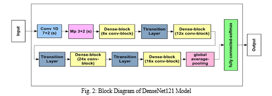
2. DenseNet121 Model
The DenseNet121 architecture, initially pre-trained on an extensive image dataset, underwent adaptation for the specialized task of breast cancer detection. This adaptation process included:
a. Fine-tuning: Adjusting the network parameters to effectively learn features specific to mammogram images, enhancing its ability to detect subtle patterns indicative of breast cancer.
b. Training Methodology: Employing binary cross-entropy loss function and the Adam optimizer, akin to the approach used for the custom CNN model, to maintain consistency and optimize training efficiency.

These model architectures were designed to maximize the detection and classification accuracy of breast cancer from mammogram images.
F. Model Training
Both the custom CNN and the DenseNet121 models were trained on the designated training dataset. Throughout the training process, we meticulously tracked changes in loss and accuracy over multiple epochs to ensure the models' performance was continuously improving.
G. Performance Evaluation
The models' performance was rigorously evaluated on both the training and validation datasets. We analyzed key metrics such as loss and accuracy, generating detailed graphs to visually represent the progression and stability of these metrics over the epochs.
H. Comparison and Analysis
A comprehensive comparison between the CNN and DenseNet121 models was conducted, focusing on their accuracy and loss metrics. Additionally, we examined the trade-offs between computational efficiency and accuracy, providing insights into the practical implications of deploying each model.
I. Application Development
To facilitate real-time breast cancer detection, we developed a user-friendly web-based application. This application, built using Flask, FastAPI, and ReactJS, integrates the trained models and enables users to upload mammogram images for immediate analysis and cancer identification.
IV. RESULTS AND DISCUSSIONS
The breast cancer detection system features a user-friendly interface that seamlessly integrates advanced Convolutional Neural Network (CNN) and DenseNet121 models. This intuitive platform accurately analyzes mammogram images, delivering real-time predictions about the presence of carcinoma. Rigorous testing and evaluation yielded the following key outcomes:
A. Model Performance Comparison
Accuracy: Both the custom CNN and DenseNet121 models achieved high accuracy levels in detecting breast cancer. While the CNN model offered a balanced approach with satisfactory accuracy and efficiency, DenseNet121 stood out in feature extraction, achieving higher accuracy but at the cost of greater computational requirements.
Loss Metrics: The loss metrics for both models were carefully tracked during training and validation. DenseNet121 consistently showed lower loss values, indicating better performance in learning from the dataset compared to the custom CNN.
Efficiency: The custom CNN model demonstrated greater computational efficiency, making it suitable for applications where resources are limited. In contrast, DenseNet121 required more computational power and memory due to its complex architecture and extensive feature extraction capabilities.
B. Real-time Inference
The web-based application developed using Flask, FastAPI, and ReactJS provides real-time breast cancer detection capabilities. Users can easily upload mammogram images through the user-friendly interface. Once an image is uploaded, the system quickly processes it using the integrated models, delivering immediate and reliable predictions. This real-time inference capability is crucial for timely diagnosis and intervention, potentially improving patient outcomes. The application is designed to handle a high volume of concurrent users without compromising performance. It leverages the power of modern web technologies to ensure fast and efficient processing. Flask and FastAPI provide a robust backend for handling requests and managing the machine learning models. ReactJS ensures a smooth and responsive user experience on the frontend. Additionally, the system includes security features to protect user data and maintain confidentiality. Overall, the application aims to assist healthcare professionals and patients by providing rapid and accurate breast cancer detection.
C. User Engagement and Empowerment
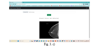
The intuitive interface of the web application is designed to encourage active user engagement. Users can easily interact with the system, upload images, and receive instant feedback on their health status. This feature empowers users with critical health insights, promoting proactive health management and early detection of potential issues. The integration of advanced machine learning models like CNN and DenseNet121 in breast cancer detection marks a significant step forward in medical diagnostics. The comparative analysis of these models highlights the strengths and trade-offs of each approach, providing valuable insights for future developments. CNN Model: The custom CNN model strikes a balance between accuracy and computational efficiency. Its relatively simple architecture makes it suitable for deployment in environments with limited computational resources. However, its performance, while commendable, is slightly lower than that of DenseNet121 in terms of accuracy and feature extraction capabilities. DenseNet121 Model: DenseNet121 excels in feature extraction due to its densely connected architecture. It achieves higher accuracy, making it a robust option for breast cancer detection. However, this comes at the cost of increased computational load, necessitating more powerful hardware for real-time applications. The real-time inference capability of the developed web application ensures that users receive an immediate analysis of their mammogram images. This feature is critical for early diagnosis and intervention, potentially improving patient outcomes. Additionally, the user-friendly interface fosters greater engagement and empowers individuals with vital health information. The following images display the output generated by the models developed in this project for breast cancer detection. These outputs demonstrate the models' capability to analyze mammogram images and provide predictions regarding the presence or absence of carcinoma. Each image showcases the model's interpretation and classification of specific cases, highlighting its potential to aid medical professionals in diagnosing breast cancer effectively.


Figure 1. a) is the user interface (UI) for uploading mammographic images for cancer classification. This image demonstrates the intuitive UI designed for the project, enabling users to easily upload mammographic images for analysis. The interface is streamlined to facilitate straightforward interaction, allowing users to select and submit images with minimal effort. Once uploaded, the images are processed by the integrated CNN and DenseNet121 models to deliver real-time predictions regarding the presence of carcinoma. This user-friendly design is intended to support both medical professionals and patients, providing accessible and immediate diagnostic insights. Figure 2. B) illustrates the detection of breast cancer in the uploaded mammographic image. The image depicts the output generated by the model, indicating the presence of carcinoma. The highlighted areas represent regions where the model has identified potential signs of breast cancer, showcasing its ability to accurately process and analyze mammographic data. This visual confirmation aids medical professionals in quickly assessing the results and making informed decisions regarding further diagnostic steps or treatments. Figure 1. c) illustrates that breast cancer was not detected in the uploaded mammographic image. The image demonstrates the model's output, indicating an absence of carcinoma in the provided mammogram. This result showcases the model's ability to accurately analyze and classify mammographic images, providing clear and reliable feedback. Such outcomes are crucial for reassuring patients and guiding medical professionals in determining the next steps in the diagnostic process.
References
[1] Ali Bou Nassif, Manar Abu Talib, Qassim Nasir, Yaman Afadar, Omar Elgendy, “Breast cancer detection using artificial intelligence techniques”in 2021. [2] F.A. Spanhol, L.S.Oliveria, C. Petitjean, L. Heutte, “Breast cancer histopathological image classification using convolutional neural networks”, in: Proceedings of the International Joint Conference on Neural Networks, 2016-October, 2016, pp. 2560. [3] Spanhol, F. A., Oliveira, L. S., Cavalin, P. R., Petitjean, C., &Heutte, L. (2017,Oct) “Deep features for breast cancer histopathological image classification”.In 2017 IEEE International Conference on Systems, Man, and Cybernetics (SMC) (pp. 1868-1873). [4] José Rouco, Paulo Aguiar, Catarina Eloy, António Polónia, “Classification of breast cancer histology images using Convolutional Neural Networks” in 2019. [5] Huang G., Liu Z., Der Maaten V., Weinberger K. Densely connected convolutional networks. 2016. [6] Moh’d Rasoul Al-hadidi, Abdulsalam Alarabeyyat, Mohannad Alhanahnah, “Breast Cancer Detection using K-nearest Neighbor Machine Learning Algorithm” in 2016. [7] Yu-Dong Zhang, Chichun Pan, Xianquing Chen, Fubin Wang, “Abnormal breast identification by nine-layer convolutional neural network with parametric rectified linear unit and rank-based stochastic pooling” in 2018. [8] Nalini Singh, Ambarish G Mahapatra, Gurukalyan Kanungo, “Breast Cancer Mass Detection in Mammograms using K-means and Fuzzy C-means Clustering” in 2011. [9] Souza, M.D., Prabhu, G.A., Kumara, V. et al. EarlyNet: a novel transfer learning approach with VGG11 and EfficientNet for early-stage breast cancer detection. Int J Syst Assur Eng Manag (2024). https://doi.org/10.1007/s13198-024-02408-6 [10] J. Padhye, V. Firoiu, and D. Towsley, “A stochastic model of TCP Reno congestion avoidance and control,” Univ. of Massachusetts, Amherst, MA, CMPSCI Tech. Rep. 99-02, 1999. [11] P. M. Manjunath, Gurucharan and M. DSouza Shwetha, \"IoT Based Agricultural Robot for Monitoring Plant Health and Environment\", Journal of Emerging Technologies and Innovative Research vol. 6, no. 2, pp. 551-554, Feb 2019 [12] A. Karnik, “Performance of TCP congestion control with rate feedback: TCP/ABR and rate adaptive TCP/IP,” M. Eng. thesis, Indian Institute of Science, Bangalore, India, Jan. 1999. [13] Melwin D\'souza, Ananth Prabhu Gurpur, Varuna Kumara, “SANAS-Net: spatial attention neural architecture search for breast cancer detection”, IAES International Journal of Artificial Intelligence (IJ-AI), Vol. 13, No. 3, September 2024, pp. 3339-3349, ISSN: 2252-8938, DOI:http://doi.org/10.11591/ijai.v13.i3.pp3339-3349 [14] J. Padhye, V. Firoiu, and D. Towsley, “A stochastic model of TCP Reno congestion avoidance and control,” Univ. of Massachusetts, Amherst, MA, CMPSCI Tech. Rep. 99-02, 1999. [15] Salins, R. D., Prabhu, G. A., Martis, J. E., & MS, S. (2023). A Novel Skin and Mole Pattern Identification Using Deep Residual Pooling Network (DRPN). International Journal of Intelligent Engineering & Systems, 16(3)
Copyright
Copyright © 2024 Mohith K P, Rahil Husain, Anzar Iqbal, Vijay Kumar Gottipati, Gella Nagendram, V Mano Priya, Sushma Shetty. This is an open access article distributed under the Creative Commons Attribution License, which permits unrestricted use, distribution, and reproduction in any medium, provided the original work is properly cited.

Download Paper
Paper Id : IJRASET63683
Publish Date : 2024-07-19
ISSN : 2321-9653
Publisher Name : IJRASET
DOI Link : Click Here
 Submit Paper Online

