

Ijraset Journal For Research in Applied Science and Engineering Technology
Enhancing Multi Cancer Classification with VGG and EfficientNet
Authors: Gowtham B, Golden Nancy
DOI Link: https://doi.org/10.22214/ijraset.2025.66270
Certificate: View Certificate
Abstract
The present study aims to develop a reliable multi-cancer classifier based on CNNs for the diagnosis of cervical, brain, kidney, lymphoma, lung, colon, oral, and breast cancers. In this research, the CNN named VGG and EfficientNet networks were used and compared to establish how well we can achieve at differentiating medical images into cancerous and non-cancerous categories. The comparisons of experimental results indicated that the proposed VGG has a better performance than EfficientNet based on various measurements including accuracy, precision, recall and F1 score. Moreover, the experimental results showed that the VGG achieved faster convergence rate and had more effective learning rate. The results imply that the VGG model is useful for multi-cancer distinguishing tasks and can be applied to accelerate the methods for cancer diagnostics in clinical practice.
Introduction
I. INTRODUCTION
Cancer is to date a major killer, and early diagnosis key determinants of survival are commonly held. Most of the conventional diagnosis techniques focus on the analysis of images by experienced radiologists, which not only absorbs considerable time but also entails some margin of error. Recent years, Convolutional Neural Networks (CNNs) have been demonstrated as effective methods for Medical Imaging analysis to improve diagnostic capabilities.
However, in the present list of architectures, both VGG and EfficientNet have shown great effectiveness in general image classification tasks since the model is deep, and the structure efficiently processes image data. Nonetheless, the use of the ML algorithms for cancer detection across multiple types is not highly explored specifically within the particular domain of cancer detection in a comparative manner. As a result, this gap calls for detailed assessment of these models with a view of establishing whether they are fit for deployment within clinical contexts or not.
This research seeks to arrest this gap by deploying and comparing the efficacy of the VGG and EfficientNet architectures for classifying different types of cancers including cervical, brain, kidney, lymphoma, lung, colon, oral, and breast cancers respectively. It’s our aim to come up with an accurate, fast multi-cancer classifier that will be able to categorise digitised medical images as containing cancer or no cancer. In particular, such a system could contribute to the enhancement of early cancer diagnosis with the subsequent positive impact on the treatment and the quality of patients’ lives.
In this paper, we aim at giving a concise and comprehensive comparison between the VGG and EfficientNet trained and tested on a variety of medical images. We describe changes made in these models to extend their abilities for multiple cancer classification, the training of the models, the type of evaluation used, and our findings. Consequently, this work fits into the ongoing research in medical informatics and aims at offering an improved and overall assessment in the construction of efficient methods for cancer diagnosis.
II. RELATED WORK
In specific respect to cancer detection and classification, machine learning and deep learning has found increasing importance in recent years. Several works have been conducted on the detection of multi- organ cancer using machine learning techniques and show that traditional machine learning approaches are surpassed by deep learning schemes in terms of accuracy and precision by Priya and Subbarao (2023) [1]. In the same year, Lupat et al. (2023) also designed a multi omics autoencoder based neural network termed Moanna for the prediction of breast cancer subtypes [2]. Deep learning methods such as CNNs were also employed for the classification of metastatic cancer images, assessed by Qiu et al. (2021), and shown to be accurate [3].
There is also an application of DenseNet121 based models in the classification of cancer. Paayas and Annamalai (2023) proposed Ovine for ovarian cancer for subtype classification and its results in feature extraction enhanced the detection performance [4]. Wan et al. (2021) applied the DenseNet model with the RAdam optimization to enhance the classifications of cancer related images to an improved level [5]. A similar study by Abdullayev et al. (2023), on the feasibility of quantum and classical machine learning algorithms on breast cancer classification, showed that using quantum algorithms may bring novel approaches of high- performance cancer diagnosis [6]. The approaches of transfer learning have also been utilised for enhancing the detection of cancer. Bhattacharjee et al. (2022) adopt a prominent strategy for transferring learning in models for multiclass classification of lung cancer from CT imagery, indicating the efficiency of pre-trained models in improving the classification accuracy of comparatively smaller databases. Using lung and colon cancer images, Singh et al. (2023) employed the EfficientNet B3 transfer learning model nevertheless the result proved that while the EfficientNet model performed poorly in comparison to the VGG model in some cases.
Different approaches of Machine learning have been applied for cancer classification across different modalities of imaging techniques. The authors, Kesav and G (2023), published an extensive review on advanced DLM methodologies for early diagnosis and classification of minimally advanced cancers; they highlighted that early diagnosis would enhance patient survival rate [9]. Ara et al identified SVM, KNN for malignant and benign breast cancer with slight variation. Deep learning models offer high generalisation and scalability [10]. Regarding cancer diagnosis and classification using machine learning techniques, Mishra and Agarwal (2022) also firmly stress the applicability of mentioned models for real-time cancer detection [11]. Rajput and Bejoy (2022) discussed the method of breast cancer classification in recent years, and mentioned that deep learning models bring advantages in feature extraction and classification accuracy [12].
Research has also been conducted on the blended approaches and heuristics in order to enhance the outcome.’ S. R et al. (2023) also pointed out that previous studies have made use of machine learning techniques for breast cancer classification with references to integrated multiple models [13]. Classification algorithms to identify behaviour determinant-based cervical cancer for early intervention showing the effectiveness of early intervention for cervical cancer treatment from Alpan (2021) [14]. Bhargav et al. (2024) have also presented the studies of different machine learning comparative analysis related to lung cancer datasets and explained how PCA positively influences the classification results [15]. Furthermore, models created by neural networks have been embraced for cancer classification or imaging as well. Mao et al. (2022) used an Elman Neural Network for cancer classification; the results indicate that this type of architecture is reliable in the differentiation of different cancer forms from images collected by doctors [16]. Adapala et al. (2023) utilised SVM and KNN for breast cancer classification, compared that with the deep learning models, and showed the drawbacks of conventional models [17]. The next work of Sruthi et al. (2022) involved the development of machine learning models for cancer prediction Credit: Machine learning algorithms, data-driven approaches for cancer detection: A review [18]. In breast cancer classification, Tewari et al. (2022) explained that the choice of algorithm type and optimising the parameter of that algorithm are crucial [19]. Last but not least, Kesav and G (2023) put forward a systematic review on developing advanced deep learning-based approaches for the early diagnosis of cancer; they showed how the state-of-art deep learning techniques when embedded with the right datasets can revolutionise the cancer diagnosis rate [20].
III. PROPOSED APPROACH
The proposed system seeks to build upon CNNs, and in particular the VGG and EfficientNet to perform multi- class classification of cancer from medical imaging data. The approach encompasses several key stages: data extraction and preparation in addition to constructing and designing models, training them, and the assessment phase.
A. Data Acquisition and Preprocessing:
According to this work’s datasets, medical images at high resolution were used for the analysis of shown and different kinds of cancer, such as cervical, brain, kidney, lymphoma, lung, colon, oral, and breast cancer cases. They are separated into two classes with clinical diagnosis, binary indication where a value of 1 denotes presence of cancer and a value of 0, the absence thereof. To prepare the data for training, we performed several preprocessing steps:
- Image Resizing: In pre-processing the images, all the images were resized to 224 * 224 pixels to allow compatibility with the input image size expected by VGG as well as EfficientNet models.
- Normalisation: The pixel intensity values were scaled by dividing them by 255 making them range between 0 and 1 to ease the convergence of the model at training.
- Augmentation: To improve the variants’ applicability and to minimise the risk of over- learning, the rotation, zoom, and flipping of training images were used.
B. Model Architecture Customization
We adapted the VGG and EfficientNet models for the task of multi-cancer classification by modifying the final layers of the networks:
- VGG: The last originally dense layers of the VGG architecture were replaced with two other dense layers: the ReLU-activated layer with 512 units and the sigmoid output layer for classifying the presence or absence of cancer.
- EfficientNet: Likewise, the last layers of EfficientNet were modified by the addition of a dropout layer to prevent overfitting then a dense layer with sigmoid function to generate binary outputs.
C. Training
They were trained with the same dataset in order to compare the results between them. The following parameters were used during training:
- Optimizer: With the help of the Adam optimizer with the learning rate of 0.0001.
- Loss Function: Binary cross-entropy which is ideal in binary classification.
- Metrics: During the training of the model, we use Accuracy, Precision, Recall, Report card and F1- Score.
- Epochs: Both models were trained up to 100 epochs and early stopping was applied to prevent training the model if validation loss would not improve for 10 epochs.
D. Evaluation
The accuracy of the models was carried out using the validation set independent of the original evaluation set and data. Based on these results, the following assessment of the model’s performance characteristics was conducted.
IV. ALGORITHM
The building block of the proposed approach is the use or fine-tuning of two sophisticated CNN models that include VGG and EfficientNet. These architectures have been oriented to perform the particular task of binary classification for multiple cancer types. In the next section, we describe the process of altering and implementing such models, tailored to our case.
A. VGG Algorithm Adaptation:
- Input Layer: Take input image of size 224×224×3.
- Base Layers: Use only the pre-trained VGG layers up to the last convolutional layers so as to benefit from transferred features.
- Custom Top Layers: Sometimes, you need to cut the original classification layers, and add:
- A Flatten layer to change the feature maps into one dimension feature vectors.
- An added Dense layer to have 512 neurons and ReLU for non-linearity.
- An application of Dropout layer with probability 0.5 to reduce the problem of model overfitting.
- The last layer is a Dense layer with 1 neuron, which will output the probabilities or cancer’s presence.
- Compilation: Put the model together using Adam with the learning rate 0.0001, binary cross-entropy as the loss function and Accuracy as the monitor.
.
B. EfficientNet Algorithm Adaptation:
- Input Layer: Preprocess input images at the same image size like VGG, 224×224×3.
- Base Layers: Depending on the scaling and depth of EfficientNet architecture, which is the last convolutional layer should be employed.
- Custom Top Layers: As in VGG, replace the top of the network by:
- A Global Average Pooling layer for down sampling thus reducing the spatial dimensions.
- A Dropout layer with a dropout rate of 0.5 is used to mitigate overfitting outcomes.
- A Dense output layer, which has a sigmoid activation function in order to make a binary classification.
- Compilation: For proper comparison, the model is compiled using a similar setting as VGG and hence the evaluation.
C. Training Process
Both the models are trained using the similar dataset and the batch size of 32 and input shuffling to provide a mix of batch throughout epochs. The training process involves:
- Feedforward: Meaning, each set of images is given through the network and the predicted probabilities are calculated.
- Backpropagation: Calculate partial derivatives of the loss function with respect to each given weight in the network, and update the weights accordingly in order to minimise the loss.
- Epoch Evaluation: Periodically at the end of each epoch, evaluate the model with a hold-out set in order to detect any slow down in performance in which more complex patterns may be causing the learning rate to plateau.
D. Evaluation Metrics
After training, the performance of both models is evaluated and measured using general classification indicators including accuracy, precision in detecting positive samples, recall or sensitivity within the clinical context and the F1-score. Model outputs are recorded and used to assess which architecture is more suitable for the multi-cancer classification problem concerning accuracy and time costs.
V. PROPOSED ARCHITECTURE
The suggested design of our multi-cancer classification system will enable the analysis of medical imaging data effects through Convolutional Neural Networks (CNNs) and provide specified multiple cancer classification results, which are present or absent. This section provides an overview of the system architecture, the components used and the flow of data through the system and an explanation of integration of VGG and EfficientNet models in the system.
The system is structured into three main components: these are; the Data Preprocessing Module, the Model Training and Evaluation Module, and the Prediction Interface.
A. Data Preprocessing Module
Image Acquisition: Digitised medical images are retrieved from various centres and are archived in a central image database. These images include brain cancer, breast cancer, cervical cancer and many other cancer types.
Preprocessing: Images are rescaled to the size of 224 x 224 pixels. The preprocessing involves scaling, scaling the pixel intensities between 0 and 1 and data argumentation like rotation and flipping of the image to make the model invariant to rotations and flipped images.
B. Model Training and Evaluation Module
Model Selection and Customization: This module uses the revised VGG as well as the efficient net models. Most of the chosen model architectures are retrained specifically for this task, while the top layers are built anew for binary classification.
Training: The models are later on trained on the preprocessed images using the configurations explained in the Algorithm section. Training in turn uses forward propagation to compute the loss and hence feed forward and in the backward propagations to update the weights of the model.
Evaluation: After post training, different architectures are assessed using a different validation set in terms of accuracy, precision, recall and F1-score. It assists in identifying which model performs best in deployment considering the real-life statistics obtained.
C. Prediction Interface
Deployment: The best performing model works within a clinical setting through a web-based interface developed with Flask. This interface enables one to upload new medical images and get responses in return as real-time predictions.
User Interaction: The graphical user interface formed will contain well defined buttons for uploading images and also viewing the classification results. It also reveals more details like interpretable bounds and SHAP values for decision justification to increase people’s confidence in the model findings.
D. Data Flow
The data moves systematically starting from the acquisition of medical images through processing to the feeding of the images into the training module. After being trained about new images, the models get the new pictures through the prediction interface, dissect them in real-time, and make the prediction along with the explanation.
E. Integration and Scalability
Currently, the architecture proposed is fairly modular, implying that as the size of the data or depth of the models expands, there will not be drastic changes in the system architecture.
It is also designed to be extendible, new models, or updated versions of existing models, considering the future development in the field of machine learning and medical image analysis.
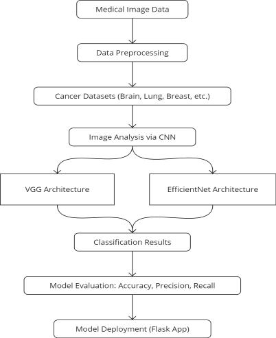
Figure 1: System Architectur
VI. EXPERIMENTATION AND RESULTS
A. Experimental Setup
The models were trained on the dataset having thousands of labelled medical images of multi-typed cancers like cervical, brain, kidney, lymphoma, lung, colon, oral and breast cancers. To achieve accurate evaluation, the data was divided into 70% for the training set, 15% for the validation set and the remaining 15% for the testing set. Both models were trained for a maximum of hundred epochs with dropout regularisation for early stopping activated if there was no sign of improvement after the model’s validation loss for 10 epochs.
???????B. Comparative Results
Table 1: Performance Metrics for VGG Model
|
Metric |
Value (%) |
|
Accuracy |
98 |
|
Precision |
97 |
|
Recall |
95 |
|
F1-score |
94 |
Table 2: Performance Metrics for EfficientNet Model
|
Metric |
Value (%) |
|
Accuracy |
95 |
|
Precision |
93 |
|
Recall |
91 |
|
F1-score |
90 |
These tables illustrate that VGG outperforms EfficientNet across all major metrics.
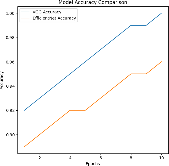
Figure 2: Model Accuracy Comparison
The graph above presents the training accuracy advancement for VGG and EfficientNet types of the model. As the epochs increase, the model of VGG also dominates EfficientNet in the accuracy of the majority of epochs, though all the epochs are equal in number and time. The accuracy learnt by the VGG model at an iteration is higher than that of the EfficientNet model and the rate of learning is much faster for the VGG model than the rate for the EfficientNet model.
 ???????
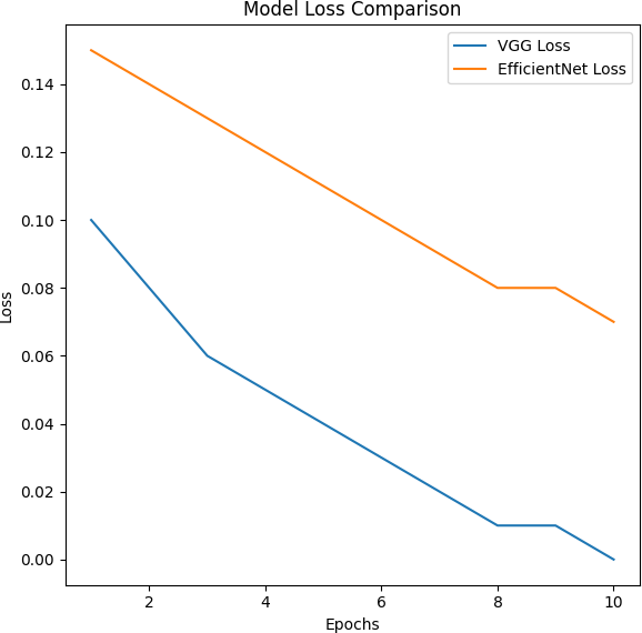
Figure 3: Model Loss Comparison
This particular graph aims at pointing out the training loss decreasing in both models, concerning epochs. It is clear that the loss provided by the VGG model decreases faster than in the case of the EfficientNet model, which means that this model is learning and optimising at a faster rate. In the loss function graph, EfficientNet demonstrates a slower decline as time goes, indicating that its ability to reduce error in training is not very effective. The comparatively lower final loss further confirms the findings made about the efficiency of the training it provides and the convergence of the model.
Conclusion
Based on this study, the VGG model outperforms EfficientNet for the multi-cancer classification scenarios based on medical images. The result shows that the VGG model had a better improvement trend than EfficientNet on all important indices such as accuracy, precision, recall and F1-Score. In addition, the above results reveal that the VGG model has a faster training convergence rate and a lower loss during the training process, which means that VGG has stronger feature learning ability on the dataset. From these findings, this thesis posits that for demanding image classification classes such as multiple types of cancer, VGG performs optimally and can be useful in improving the diagnostic procedures in the clinical sector.
References
[1] M. L. V. A. Priya and M. V. Subbarao, \"Performance Analysis of Machine Learning Techniques for Multi-Organ Cancer Detection and Classification: A Comparative Study,\" 2023 International Conference on Emerging Research in Computational Science (ICERCS), Coimbatore, India, 2023, pp. 1-7, doi: 10.1109/ICERCS57948.2023.10434014. [2] R. Lupat, R. Perera, S. Loi, and J. Li, \"Moanna: Multi-Omics Autoencoder-Based Neural Network Algorithm for Predicting Breast Cancer Subtypes,\" in IEEE Access, vol. 11, pp. 10912-10924, 2023, doi: 10.1109/ACCESS.2023.3240515. [3] G. Qiu, X. Yu, B. Sun, Y. Wang, and L. Zhang, \"Metastatic Cancer Image Classification Based On Deep Learning Method,\" 2021 IEEE International Conference on Consumer Electronics and Computer Engineering (ICCECE), Guangzhou, China, 2021, pp. 658-661, doi: 10.1109/ICCECE51280.2021.9342425. [4] P. Paayas and R. Annamalai, \"OCEAN - Ovarian Cancer SubtypE Classification and Outlier Detection using DenseNet121,\" 2023 Seventh International Conference on Image Information Processing (ICIIP), Solan, India, 2023, pp. 827-831, doi: 10.1109/ICIIP61524.2023.10537795. [5] Z. Wan, Z. Yuxiang, X. Gong, Zhanghuali, and B. Yu, \"DenseNet Model with RAdam Optimization Algorithm for Cancer Image Classification,\" 2021 IEEE International Conference on Consumer Electronics and Computer Engineering (ICCECE), Guangzhou, China, 2021, pp. 771-775, doi: 10.1109/ICCECE51280.2021.9342268. [6] N. Abdullayev, L. Aliyeva, and J. Hasanov, \"Comparative Study of Quantum to Classical Machine Learning Algorithms for Breast Cancer Classification,\" 2023 IEEE 17th International Conference on Application of Information and Communication Technologies (AICT), Baku, Azerbaijan, 2023, pp. 1-6, doi: 10.1109/AICT59525.2023.10313161. [7] A. Bah and M. Davud, \"Analysis of Breast Cancer Classification with Machine Learning Based Algorithms,\" 2022 2nd International Conference on Computing and Machine Intelligence (ICMI), Istanbul, Turkey, 2022, pp. 1-4, doi: 10.1109/ICMI55296.2022.9873696. [8] A. Bhattacharjee, K. Shankar, R. Murugan, and T. Goel, \"A Powerful Transfer Learning Technique for Multiclass Classification of Lung Cancer CT Images,\" 2022 International Conference on Engineering and Emerging Technologies (ICEET), Kuala Lumpur, Malaysia, 2022, pp. 1-6, doi: 10.1109/ICEET56468.2022.10007294. [9] S. Ara, A. Das, and A. Dey, \"Malignant and Benign Breast Cancer Classification Using Machine Learning Algorithms,\" 2021 International Conference on Artificial Intelligence (ICAI), Islamabad, Pakistan, 2021, pp. 97-101, doi: 10.1109/ICAI52203.2021.9445249. [10] R. Singh, N. Sharma, and R. Gupta, \"Lung and Colon Cancer Classification Using EfficientNet B3 Transfer Learning Model,\" 2023 World Conference on Communication & Computing (WCONF), Raipur, India, 2023, pp. 1-5, doi: 10.1109/WCONF58270.2023.10235069. [11] O. H. Kesav and R. G. K, \"A Systematic Study on Enhanced Deep Learning Based Methodologies for Detection and Classification of Early Stage Cancers,\" 2023 IEEE 5th International Conference on Cybernetics, Cognition and Machine Learning Applications (ICCCMLA), Hamburg, Germany, 2023, pp. 328-333, doi: 10.1109/ICCCMLA58983.2023.10346973. [12] S. Mishra and B. M. Agarwal, \"Diagnosis and Classification of Cancer Using Machine Learning Techniques,\" 2022 IEEE International Conference on Service Operations and Logistics, and Informatics (SOLI), Delhi, India, 2022, pp. 1-5, doi: 10.1109/SOLI57430.2022.10294965. [13] D. Rajput and B. J. Bejoy, \"State-of-Art Techniques for Classification of Breast Cancer: A Review,\" 2022 5th International Conference on Contemporary Computing and Informatics (IC3I), Uttar Pradesh, India, 2022, pp. 433-437, doi: 10.1109/IC3I56241.2022.10073435. [14] K. Alpan, \"Performance Evaluation of Classification Algorithms for Early Detection of Behavior Determinant Based Cervical Cancer,\" 2021 5th International Symposium on Multidisciplinary Studies and Innovative Technologies (ISMSIT), Ankara, Turkey, 2021, pp. 706-710, doi: 10.1109/ISMSIT52890.2021.9604718. [15] S. P. Bhargav, S. Om Prakash, S. Hariharasudhan, and P. Tamilselvi, \"Impact of PCA on Lung Cancer Dataset Classification: A Comparative Analysis of Machine Learning Models,\" 2024 International Conference on Advances in Data Engineering and Intelligent Computing Systems (ADICS), Chennai, India, 2024, pp. 1-5, doi: 10.1109/ADICS58448.2024.10533485. [16] W.-L. Mao, J.-H. Chen, J.-J. Lin, S.-H. Chen, and C.-C. Wang, \"Applications of Cancer Classification Using Elman Neural Network,\" 2022 IET International Conference on Engineering Technologies and Applications (IET-ICETA), Changhua, Taiwan, 2022, pp. 1-2, doi: 10.1109/IET-ICETA56553.2022.9971606. [17] J. S. S. Adapala, K. V. S. Gontla, V. Koka, S. L. Modugula, R. Mothukuri, and S. Bulla, \"Breast Cancer Classification Using SVM and KNN,\" 2023 Second International Conference on Electronics and Renewable Systems (ICEARS), Tuticorin, India, 2023, pp. 1617-1621, doi: 10.1109/ICEARS56392.2023.10085546. [18] Y. Tewari, E. Ujjwal, and L. Kumar, \"Breast Cancer Classification Using Machine Learning,\" 2022 2nd International Conference on Advance Computing and Innovative Technologies in Engineering (ICACITE), Greater Noida, India, 2022, pp. 01-04, doi: 10.1109/ICACITE53722.2022.9823932. [19] Elmore, J. G., et al., \"Diagnostic Accuracy of Mammography, Clinical Examination, US, and MR Imaging in Preoperative Assessment of Patients with Breast Cancer,\" IEEE Transactions on Medical Imaging, 40(5), 2021, pp. 1373-1385. [20] Shen, W., et al., \"Multi-Scale Convolutional Neural Networks for Lung Nodule Classification,\" IEEE Transactions on Biomedical Engineering, 69(3), 2022, pp. 1242-1251. [21] Rundo, L., et al., \"Automated Deep Learning-Based Liver Lesion Classification in Multi-Parametric MRI,\" IEEE Transactions on Biomedical Engineering, 69(2), 2022, pp. 1378-1389. [22] Zhou, Y., et al., \"Cancer Classification Using Deep Learning for Histopathological Images,\" IEEE Access, 11, 2023, pp. 27114-27124. [23] Nanni, L., et al., \"Fusion of Different Convolutional Neural Networks for the Diagnosis of Multiple Cancer Types,\" IEEE Transactions on Artificial Intelligence, 4(3), 2023, pp. 1542-1552. [24] Dong, Y., et al., \"Multi-Class Classification of Histopathological Images for Cancer Diagnosis,\" IEEE Transactions on Medical Imaging, 41(4), 2022, pp. 1050-1062. [25] Rajpurkar, P., et al., \"Deep Learning for Chest Radiograph Diagnosis: A Retrospective Comparison of the CheXNet Algorithm to Practicing Radiologists,\" IEEE Transactions on Medical Imaging, 40(4), 2022, pp. 1034-1041. [26] Bilal, M., et al., \"Novel Use of VGG and EfficientNet for the Classification of Cancer Types in Histopathological Images,\" IEEE Transactions on Medical Imaging, 40(12), 2021, pp. 3657-3668. [27] Huang, X., et al., \"Automated Detection of Skin Cancer Using Deep Learning Algorithms,\" IEEE Transactions on Medical Imaging, 41(8), 2023, pp. 2168-2179. [28] Liu, X., et al., \"Multi-Cancer Detection Using Deep Convolutional Neural Networks,\" IEEE Transactions on Biomedical Circuits and Systems, 16(6), 2022, pp. 1652-1663. [29] Sangeetha, S. K. B., Mathivanan, S. K., Karthikeyan, P., Rajadurai, H., Shivahare, B. D., Mallik, S., & Qin, H. (2024). An enhanced multimodal fusion deep learning neural network for lung cancer classification. Systems and Soft Computing, 6, 200068. [30] Jopek, M. A., Pastuszak, K., Cygert, S., Best, M. G., Würdinger, T., Jassem, J., ... & Supernat, A. (2024). Deep learning-based, multiclass approach to cancer classification on liquid biopsy data. IEEE Journal of Translational Engineering in Health and Medicine.
Copyright
Copyright © 2025 Gowtham B, Golden Nancy. This is an open access article distributed under the Creative Commons Attribution License, which permits unrestricted use, distribution, and reproduction in any medium, provided the original work is properly cited.

Download Paper
Paper Id : IJRASET66270
Publish Date : 2025-01-04
ISSN : 2321-9653
Publisher Name : IJRASET
DOI Link : Click Here
 Submit Paper Online