Ijraset Journal For Research in Applied Science and Engineering Technology
- Home / Ijraset
- On This Page
- Abstract
- Introduction
- Conclusion
- References
- Copyright
Green Synthesis of Copper Nanoparticles Using Leaf Extract of Phyllanthus niruri Lin. and Study of Its Antimicrobial Activity
Authors: S. Hemashree, Dr. M.L. Pruthvi, Dr. M.K. Mahesh
DOI Link: https://doi.org/10.22214/ijraset.2023.57353
Certificate: View Certificate
Abstract
Nanoscience and Nanotechnology are the study and application of extremely small things and can be used across all the other science fields such as chemistry, biology, physics and engineering. Green synthesis of nanoparticles using plant extract is a promising and ecofriendly approach, often yielding particles with unique properties. The present study deals with biosynthesis, characterization and antimicrobial activity. Of copper sulphate nanoparticles using the leaf extract of Phyllanthus niruri. In this report, aqueous phase green synthesis of specifically copper sulphate nanoparticles using leaf extract (both fresh and dry leaf) aqueous leaf extract of leaf was used for the synthesis of nanoparticles. The synthesised nanoparticles of copper sulphate were confirmed by color change when 25ml of 1% aqueous copper sulphate solution was added to 5ml og plant extract. The synthesised nanoparticles were charecterised by UV-Visible spectroscopy, SEM (Scanning Electron Microscopy), and XRD (X-Ray Diffraction). The UV-VIS spectroscopy showed the absorption peak at 378nm for fresh leaf and 376nm for dry leaf. SEM was used to study the morphological features that was predominately spherical and aggregated into large structure with not well-defined morphology and the size347nm,358nm,400nm,457nm, for fresh leaf 1.13µm,1.14µm,771nm,935nm for dry leaf. XRD using this the particle size and nature of nanoparticle was found the average diameter of copper nanoparticles is 12.8nm for dry leaf and 6.10nm for fresh leaf. antimicrobial activity was done using different bacterial and fungal species, in antibacterial activity St. aureus show maximum inhibition that is 75% in fresh leaf and 63%in dry leaf while compare to others, S. typhi showed 0% inhibition it was not resistant towards P.niruri. In antifungal activity F.oxysporum has shown maximum inhibition that is 77% for both fresh and dry leaf, penicillium atramentosum showed 0% inhibition it was not resistant towards P.niruri
Introduction
I. INTRODUCTION
A nanoparticle or ultrafine particle is usually defined as a particle of matter that is between 1-100 nanometer. And different shapes provide unique chemicals, physical and optical properties.[1] Nanoscience and Nanotechnology are the study and application of extremely small things and can be used across all the other science fields such as chemistry, biology, physics and engineering.[2]
The American physicist and Nobel prize laureate Richard Feynman introduced the concept of Nanotechnology in 1959, Prof. C.N.R. Rao is the father of nanotechnology in India. During the annual meeting of the American physical society, Feynman presented a lecture entitled “There’s Plenty of Room at the Bottom’’ at the California institute of technology. Professor Noria Taniguchi Japanese scientist coined the term Nanotechnology in 1947. K. Eric Drexler popularized the word nanotechnology. [3]
Nanotechnology can increase the surface area of material. This allows more atoms to interact with other materials. An increased surface area is one of the chief reasons where nanometer scale materials can be stronger, more durable and more conductive than their larger scale (called bulk) counterparts. [4] Nanotechnology mainly consists of processing of separation, consolidation and deformation of material by one atom or one molecule. [5]
Metallic nanoparticles are multifunctional in nature and they have been extensively used in variety of sectors like industries, medicine, drug delivery, cancer treatment [6] The green synthesis metallic Cu nanoparticles are noticeable materials primarily due to their unique properties and cost- effective compare to other metal like gold and silver. A copper nanoparticle is a copper-based particle 1 to 100 nm in size. copper nanoparticles can be prepared by natural processes or through chemical synthesis. These nanoparticles are of a particular interest due to their historical application as coloring agents and biomedical as well as the antimicrobial ones. There are various methods described to chemically synthesize Cu NPs. An older method involves the reduction of copper hydrazine carboxylate in an aqueous solution by heating through ultrasound under inert argon atmosphere as a result a combination of copper oxide and pure copper nanoparticles clusters are formed.
Copper nanoparticles can also be synthesized using green chemistry to reduce environmental impact. Copper nanoparticles display unique characteristics including catalytic and antifungal/ antimicrobial activities. [7]
As the demand for plant extracts continues to surge, their widespread utilization has gained significant momentum. we have used the leaves of Phyllanthus niruri in this project for the green synthesis of nanoparticles its antimicrobial activity. Phyllanthus niruri is a widespread tropical plant commonly found in coastal areas, known by the common name gale of wind, stonebreaker and it belongs to family Phyllanthaceae. Phyllanthus niruri has been used in Ayurvedic medicine for over 2000 years and has a wide number of traditional uses including internal use for jaundice, gonorrhea, frequent menstruation, and diabetes, sores, swelling and itchiness. Widely used for liver disease in traditional medicine. Traditional medicine systems, such as ayurvedic and Unani medicine have utilized the leaves and fruit to treat gallstones and jaundice. [8]
Nanotechnology plays an essential role in material science capable of diverse novel application in biomedical science, pharmaceutical science, medicine, nutrition [9] Medicine and Healthcare: Drug Delivery: Nanoparticles can be designed to deliver drugs to specific target sites in the body, improving treatment efficacy and reducing side effects. Cancer Treatment: nanoparticles can selectively target cancer cells and deliver therapies directly to tumors, minimizing damage to healthy tissue. Medical Imaging: nanoparticles are used as contrast agents in imaging techniques like MRI and CT scans, enhancing the visualization of tissue and organs. Diagnostic Tools: nanoscales sensors and assays allow for sensitive and rapid detection of disease and pathogens. Automotive and Aerospace: nanocomposites contribute to lighter and stronger vehicle components. nanotechnology enhances fuel efficiency through improved engine materials and coating.[10]
Objectives:
- Synthesis and characterization of copper nanoparticles from Phyllanthus niruri L. fresh leaf extract
- Synthesis and characterization of copper nanoparticles from Phyllanthus niruri L. dry leaf extract
- Study of antimicrobial activity of copper nanoparticles from Phyllanthus niruri L. fresh leaf extract
- Study of antimicrobial activity of copper nanoparticles from Phyllanthus niruri L. dry leaf extract
II. MATERIAL AND METHODS
A. Chemicals
Analytical grade copper sulphate pentahydrate (CuSO4.5H2O) was used in this study.
B. Collection of Leaves
Leaves of Phyllanthus niruri were collected from Yuvaraja’s College campus, University of Mysore, Mysuru.
C. Preparation of Fresh Leaves Extract
20g of fresh leaves of Phyllanthus niruri was weighed and washed thoroughly thrice in distilled water for few minutes. The washed leaves were dried, chopped into fine pieces and was boiled in 100ml of distilled water for 15 mins in 500ml borosil beaker. The extract obtained was filtered through muslin cloth and again filtered with Whatman no: 1 filter paper and then it is immediately used for the biosynthesis of copper nanoparticles.
D. Preparation of Dry Leaves Extract
Cleaned leaves of Phyllanthus niruri is shade dried for few days and it is finely powered. 5g of fine powder was taken and boiled in 100ml of distilled water for 15 mins in 500ml borosil beaker. The extract obtained was filtered using muslin cloth and again with Whatman no: 1 filter paper, and extract was used for the biosynthesis of copper nanoparticles.
E. Green Synthesis of Copper Nanoparticles
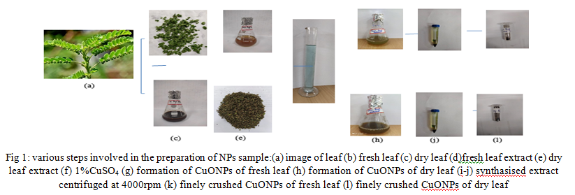
Copper sulphate was provided by the department of botany, YCM. For the synthesis of copper nanoparticles, 5ml of fresh leaves extract was added to 25ml of 1% aqueous copper sulphate in 250 ml borosil conical flask at room temperature. The same procedure is fallowed for dry leaf extract.
The color changes from orange to green as an indication of presence of copper nanoparticles in fresh leaves. From dark brown to green in dry leaves extract. Reactions are carried out in shade inside BOD incubator for 24 hours.
F. Extraction of Nanoparticle Samples
Nanoparticle samples were collected by centrifuging the reaction mixture twice at 4000rpm for 10 mins. The residue is used, obtained greenish brown color sample of copper sulphate (from using both fresh and dry leaves extract) dried at room temperature to obtain nanoparticles powder.
G. Characterization Techniques
Ultraviolet-visible spectrum All ultraviolet- visible (UV-vis) spectra were recorded on the Beckman coulter DU730 UV-vis spectrophotometer. The absorption spectra of the prepared NPs were recorded by taking the aqueous dispersion of the NPs and scanned in the range of 200- 550 nm operated at resolution 1nm at IOE, Mysore. Distilled water was taken to adjust the baseline.
(SEM)Scanning electron microscope (SEM)was used to study the morphological features of synthesized nanoparticles from fresh and dry leaf extract of Phyllanthus niruri. SEM images were recorded using Carl Zeiss Germany, model: EVO MA 15 SEM instrument at IOE, Mysore.
X-ray diffraction the particle size and nature of the copper NPs were determined using Bruker Eco D8 advance X-pert PRO operating at a voltage of 40kV, a current of 20mA with copper Kα radiation at 2θ angle ranging from 10 to 800. A thin film of the copper nanoparticles was made by dipping a glass plate in a solution and carried out for X-ray diffraction studies. The crystalline copper nanoparticle was calculated from the width of the XRD peaks and the average size of the nanoparticles can be estimated using the Debye Scherrer D= kλ/βcosθ [11]
H. Antibacterial Assay
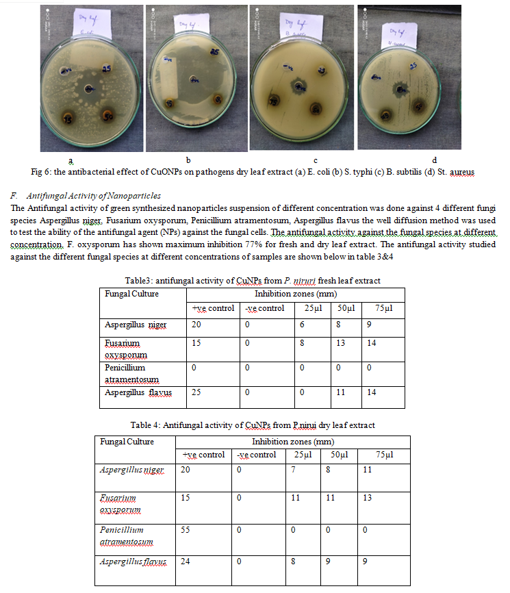
The selected bacteria are Escherichia coli, Salmonella typhi, Bacillus subtilis, Staphylococcus aureus, which were sub cultured from the pure culture in an inoculation tube containing nutrient agar media for antibacterial study. The pure culture was provided by the P.G department of Microbiology, manasagangothri campus, Mysore. The nutrient agar high medium was prepared as per the requirement according to the number of plates. A small amount of agar is added to solidify the prepared media and the pH was adjusted to 7. Then the media was homogenized for 15mins and then kept for sterilization. After sterilization the NA media was poured into sterile Petri plates under aseptic condition and allowed for solidification. Well diffusion method by agar plates was used for calculating the zone of inhibition. These 4 bacterial pathogens were then coated over a agar plate with the help of sterile swab of cotton. Then these plates were permitted to dry. After those 5 wells were bored by sterile cork borer in each agar plate. Subsequently, 25µl,50µl,75µl, of CNPs solution, positive control and negative control was taken. The antibiotic ampicillin was taken as positive control and distilled water as negative control. Then the plates were kept for complete diffusion followed by incubation at 37 ?C for 24 hrs. and measured the diameter of inhibitory zones in mm.
I. Antifungal Assay
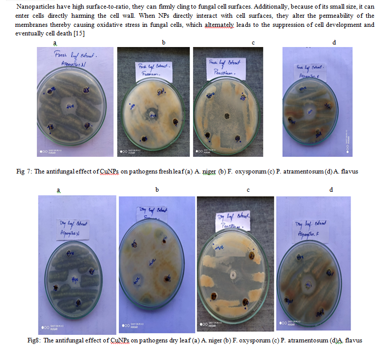
The selected fungi are Aspergillus niger, Fusarium oxysporum, Penicillium atramentosum, Aspergillus flavus which were sub cultured from the pure culture for further study. The pure culture was provided by the P.G department of Biotechnology manasagangothri campus, Mysore. PDA high medium was prepared as per the requirement according to the number of plates. A small amount of agar is added as solidifying agent. Then the media was homogenized for 15mins and then kept for sterilization. After sterilization the PDA media was poured into sterile Petri plates under aseptic condition and allowed for solidification. The antifungal activity of the NPs was determined by well diffusion method. The fungal inoculums prepared were used to test the antifungal potential. The PDA media was poured into sterile Petri plates in aseptic condition then plates were allowed to solidify in laminar air flow chamber. The 4 fungal pathogens were then coated over a media containing plates with the help of sterile swab of cotton. Then these plates were permitted to dry. After those 5 wells were bored by sterile cork borer in each agar plate. Subsequently, 25µl,50µl,75µl, of CNPs solution, positive control and negative control was taken. The antibiotic Bavistin was taken as positive control and distilled water as negative control. Then the plates were sealed and incubated at room temperature for 2-3 days. And finally antifungal activity was calculated by measuring the diameter of inhibitory zones in mm.

III. RESULT AND DISCUSSION
A. Synthesis of Copper Nanoparticles
Copper sulphate was used as precursor, 5ml of fresh and dry leaf extract was added to 25ml of 1% aqueous copper sulphate in 250 ml borosil conical flask at room temperature. The reaction is carried out in shade inside a BOD incubator for 48 hours. The color change was observed for fresh leaf extract from orange to green and for dry leaf extract brown to green. The color change suggests the synthesis of copper nanoparticles
B. UV-VIS Spectroscopy
UV-Vis analysis is one of the most important characterization methods to study nanoparticles. The surface plasmon resonances (SPR) of synthesized Sulphate nanoparticles have been studied by UV-Vis Spectrophotometer. The absorption of visible radiations due to the excitation of SPR, imparts various colors to nanoparticles. As the nanoparticles size changes, color of the solution is also supposed to change. So, UV-Vis absorption spectrum is quite sensitive to the formation of nanoparticles. All the nanoparticle samples are subjected to UV-Vis study. Fig. 2(a &b) shows the UV-Vis spectrum of the 2samples. The nanoparticles synthesized showed maximum absorptions between 350 nm to400 nm. The sharp bands of copper oxide nanoparticles were around 378nm for fresh leaf and 376nm for dry leaf, as multiple literature reviews indicate that the SRP peak for CuNPs was around 550to 600nm. This confirms that P.niruri leaf extract both dry and fresh leaf has the potential to reduce copper ions to CuONPs

C. SEM (Scanning Electron Microscopy)
Scanning Electron Microscopy provided further insight into the morphology and size details of the synthesized nanoparticles. The typical SEM image shown individual copper nanoparticles as well as number of aggregates. The morphology of the copper nanoparticles was predominately spherical and aggregated into large irregular structure with not well-defined morphology was observed. The nanoparticles were measured from the SEM image with the help of Image J software.
SEM images of copper nanoparticles of fresh leaf extract were of 347nm, 358nm, 400nm, 457nm, and dry leaf extract were 1.13µm, 1.14µm, 771nm, 935nm. Fig 3. Small nuclear particles are self -aggregated and orient themselves to form larger particles [12]

D. X- Ray Diffraction (XRD) Analysis
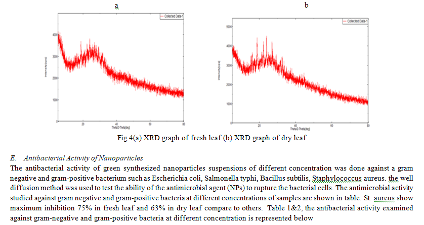
Fresh leaf X-Ray diffraction pattern of synthesized CuNPs showed 5 distinct peaks with 2θ values. 18.79, 22.30, 23.97, 27.00, 31.72.out of these 5 values 3.25,6.43,7.82, 6.48, and 6.54.can be assigned to the sets of planes (002), (011), (101), (110) and (112) respectively. These sets of planes may be indexed to the face centered cubic (FCC) lattice structure of the copper nanoparticles. The mean size of copper nanoparticles calculated using Debye-Scherrer’s equation D= kλ/βcosθ where D is the mean grain size, λ is the wavelength of copper target, β is the FWHM of the diffraction peak and θ is the diffraction angle the average diameter of the CuNPs is calculated that is 6.10nm by Scherrer’s formula using FWHM obtained from the diffraction peaks.
Dry leaf X-Ray diffraction pattern of synthesized CuNPs showed 7 distinct peaks with 2θ values. 16.15, 18.76, 22.50, 23.98, 27.09, 31.85, and 32.61.out of these 7 values 1.17, 39.10, 3.04, 42.55, 1.70, 0.92, and 1.73.can be assigned to the sets of planes (002), (002), (011), (101), (110), (112), and (103) respectively. These sets of planes may be indexed to the face centered cubic (FCC) lattice structure of the copper nanoparticles. The mean size of copper nanoparticles calculated using Debye-Scherrer’s equation D= kλ/βcosθ where D is the mean grain size, λ is the wavelength of copper target, β is the FWHM of the diffraction peak and θ is the diffraction angle the average diameter of the CuNPs is calculated that is 12.8 nm by Scherrer’s formula using FWHM obtained from the diffraction peaks. Fig 4. Graph of XRD


IV. ACKNOWLEDGMENT
The authors gratefully acknowledge faculty of Post Graduate department of Botany, Yuvaraja’s College Mysore, University of Mysore, for providing their support and laboratory facility to conduct this research work.
Conclusion
The green synthesis of copper nanoparticles using the leaf extract (both fresh and dry leaves) of Phyllanthus niruri offers a sustainable and environmentally friendly approach to nanoparticle production. This method harnesses the reducing and stabilizing properties of natural compounds or plant extracts, reducing their need for harmful chemicals. UV-visible spectroscopy analysis was used to principally monitor the reduction of copper ions into copper oxide nanoparticles. Using SEM (scanning electron microscopy) technique, particle shape, were identified, XRD (X-ray diffraction) technique is used for analyzing the crystal structure of materials. Furthermore, the study of antimicrobial activity has shown promising results, highlighting the potential of copper nanoparticles as effective agents against various pathogens. Their ability to inhibit the growth of bacteria and fungi makes them valuable for the applications in medicine and biotechnology.
References
[1] Garibo, D., Borbon-Nunez, H. A., de Leon, J. N. D., Garcia Mendoza, E., Estrada, I., Tolendano-Magana, Y.,… and Susarrey-Arce, A. (2000). Green synthesis of silver nanoparticles using Lysiloma acapulcensis using high- antimicrobial activity. Scientific reports, 10(1), 12805 [2] Nanoparticles2022. [online]. Available: https://www.merriamwebster.com/dictionay/nanoparticle. [Accessed:30-sep-2022], [3] Khan, I., Saeed, K., and Khan, I.(2019). Nanoparticles: Properties, applications and toxicities. Arabian journal of chemistry, 12(7), 908-931 [4] Ali, I. A. M., Ahmed, A. B., and AI-Ahmed, H.I. (2023). Green synthesis and characterization of silver nanoparticles for reducing the damage to sperm parameters in diabetic compared to metformin. Scientific Reports, 13(1), 2256 [5] V. Singh, S. Yadav, V. Chouhan, S. Shukla and K. Vaishnolia, Applications of Nanoparticles in various fields, pp.221-23, 2020. Available:10.4018/978-1-7998-6527-8.ch011. [6] Jain, S., and Mehata, M. S. (2017). Medicinal plant leaf extract and pure flavonoid mediated green synthesis of silver nanoparticles and their enhanced antimicrobial property. Scientific reports, 7(1), 15867. [7] Z. Guo, X. Liang, T. Pereira, R. Scaffaro and H. Thomas Hahn, ‘CuO nanoparticles filled vinyl-easter resin nanocomposite: Fabrication, characterization and property analysis’, Composites Science and Technology, vol. 67, no, pp.2036-2044,2007. Available: 10.1016/j.compscitech.2006.11.017. [8] Bagalkotkar, G., Sagineedu, s. R., Saad, M.S., and Stanslas, J. (2006) Phytochemicals from Phyllanthus niruri Linn. And their pharmacological properties: a review. journal of pharmacy and pharmacology, 58(12), 1559-1570 [9] Chauhan, A., Verma, R., Kumari, S., Sharma, A., Shandilya, P., Li, X., …and Kumar, R. (2020). Photocatalytic dye degradation and antimicrobial activities of pure and Ag- droped ZnO using Cannabis sativa leaf extract. Scientific reports, 10(1), 7881. [10] Singh. M., Manikandan. S., Kumaraguru. A. k, (2011) conducted Nanoparticles: A new technology with wide Applications. Research journal of nanoscience and nanotechnology. 1(1):1-11 [11] Acharyulu, N. P.S., Dubey, R. S., Swaminadham, V., Kollu, P., Kalyani, R. L., and Pammi, S.V.N. (2014). Green synthesis of CuONPs using Phyllanthus amarus leaf extract and their antimicrobial activity against multidrug resistance bacteria. Int.j Eng Res Technol, 3(4), pp. [12] Noorafsha, Kashyap, A. K., Kashyap, A., Deshmuk, L., and Vishwakarma, D. (2022). Biosynthesis and biophysical elucidation of CuO nanoparticles from Nycatanthes arbor-tristis Linn. Leaf. Applied Microbiology and Biotechnology, 106(17),5823-5832. [13] Caroling, G., Vinodhini, E., Ranjitham, A.M., and Shanthi, P. (2015). Biosynthesis of copper nanoparticles using aqueous Phyllanthus emblica (Gooseberry) extract - characterization and study of antimicrobial effects. Int. J. Nano. Chem, 1(2),53-63. [14] Z. Luo, Y. Qin and Q. Ye, ‘Effect of nano-TiO2-ldpe packing on ‘Microbiological an Physicochemical Quality of pacific white shrimp during chilled storage’, International Journal of Food Science & Technology, vol.50 no.7, pp. 11567-1573, 2015.Available10.1111/ijfs.12807. [15] Y. Xie, Y. He, P. Irwin, T. Jin and X. Shi, ‘Antibacterial Activity and Mechanism of Action of Zinc Oxide Nanoparticles against Campylobacter jejuni’, pplied and Environmental Microbiology, vol,77, no 67, pp 2325, 2011.Available:10.11 11/j.1365-2672.2009.04303.x.
Copyright
Copyright © 2023 S. Hemashree, Dr. M.L. Pruthvi, Dr. M.K. Mahesh . This is an open access article distributed under the Creative Commons Attribution License, which permits unrestricted use, distribution, and reproduction in any medium, provided the original work is properly cited.

Download Paper
Paper Id : IJRASET57353
Publish Date : 2023-12-05
ISSN : 2321-9653
Publisher Name : IJRASET
DOI Link : Click Here
 Submit Paper Online

