Ijraset Journal For Research in Applied Science and Engineering Technology
- Home / Ijraset
- On This Page
- Abstract
- Introduction
- Conclusion
- References
- Copyright
Identification of Brain Tumors Using SVM Classifier
Authors: Sudanala Gopala Krishna , B. Sarvesh , M. Narendhar Reddy, R. Ajay Kumar , Venkatapathi Pallam
DOI Link: https://doi.org/10.22214/ijraset.2024.65680
Certificate: View Certificate
Abstract
A vital non-invasive method that is frequently employed in the medical sector for the examination, diagnosis, and management of anomalies in brain tissue is magnetic resonance imaging, or MRI. Effective treatment of brain cancers depends on early identification, which also greatly enhances patient outcomes. However, it is a difficult and time-consuming task to accurately detect and segment tumors from MRI slices. The suggested system uses cutting-edge methods to automate the tumor classification and segmentation procedure in order to overcome this difficulty. The technique finds aberrant tissue in MRI slices by using a Support Vector Machine (SVM) classifier for tumor localization. The tumor\'s borders are then drawn using segmentation techniques, which enables an accurate tumor size measurement. In order to train an artificial neural network (ANN) to identify the type of tumor present, these segmented regions undergo additional processing to extract important properties.
Introduction
I. INTRODUCTION
Because they are difficult to diagnose and treat, brain tumors—which can be primary or secondary, benign or malignant—present a serious medical challenge. While secondary or metastatic brain tumors spread from other parts of the body, primary brain tumors start in the brain. Malignant tumors are those that have aberrant growth patterns that have the ability to spread to neighboring tissues, while benign tumors are those that are not cancerous. Additionally, brain tumor cell growth rate and appearance are assessed on a scale of 1 to 4, where higher grades correspond to more aggressive growth. Effective treatment depends on an accurate and timely diagnosis, and imaging methods such as magnetic resonance imaging (MRI) have emerged as the gold standard for identifying brain cancers. MRI makes it possible to see abnormal areas like tumors, edema, or cysts, which is vital information for diagnosis.
Tumor detection and segmentation from MRI images, however, can be a time-consuming and human error-prone procedure. Determining the borders of aberrant areas requires image segmentation, and methods like thresholding, edge detection, and clustering are frequently used to do this. Furthermore, based on the features that are retrieved from segmented MRI images, machine learning classifiers like Random Forests, Support Vector Machines (SVM), and Artificial Neural Networks (ANN) are frequently used to categorize and predict tumor types.The program called Classification Learner helps with using tools like scattering plots and confusion matrices to evaluate and compare various models in order to train classifiers.
Additionally, preprocessing techniques like edge detection and noise reduction improve image quality and guarantee that the features of the tumor are precisely recorded for analysis and classification. Combining these methods improves the efficiency of brain tumor diagnosis, lessens the need for manual intervention, and allows for accurate and quick treatment decisions.
II. LITERATUREREVIEW
1) Saptalakar. B. K, Rajeshwari. H, “Segmenttion Based Detection of Brain Tumor,” International Journal of computer and Electronics Research, Vol. 2, pp.20-23, February 2013.
The paper by Saptalakar and Rajeshwari (2013), titled "Segmentation Based Detection of Brain Tumor," addresses the critical challenge of accurately detecting brain tumors using medical imaging techniques. Early and precise detection of brain tumors is essential for effective treatment and patient survival. MRI (Magnetic Resonance Imaging) is a widely used tool for brain tumor diagnosis due to its ability to provide high-resolution images of brain tissues. However, the process of tumor detection from MRI scans is complex and requires automated techniques for accurate and efficient segmentation of tumor regions. The authors focus on the use of image segmentation as a method to isolate and identify abnormal regions in brain MRI images, which is a crucial first step for diagnosing brain tumors. The paper discusses various segmentation techniques and highlights their application in detecting and localizing tumors within the brain.
By utilizing segmentation algorithms, the tumor region can be effectively separated from the surrounding healthy tissue, making it easier to analyze and determine the nature and size of the tumor. This segmentation process is an essential component of the brain tumor detection pipeline, as it reduces the need for manual intervention and allows for faster diagnosis. The authors propose a framework that incorporates segmentation techniques to enhance the accuracy of tumor detection, which can assist doctors in making informed treatment decisions. The research is significant for advancing brain tumor detection methods and emphasizes the importance of automated image processing in the medical field for improving diagnosis efficiency and reducing human error.
2) Jayashree. M. J, Charutha. S, “An Efficient Brain Tumor Detection By Integrating Modified Texture Based Region Growing And Cellular Automata Edge Detection”
The paper by Jayashree and Charutha, titled "An Efficient Brain Tumor Detection By Integrating Modified Texture-Based Region Growing and Cellular Automata Edge Detection," explores an innovative approach for brain tumor detection using advanced image processing techniques. Accurate and early detection of brain tumors is crucial for patient prognosis and treatment planning. In this study, the authors propose a method that combines modified texture-based region growing and cellular automata edge detection techniques to enhance the accuracy and efficiency of brain tumor detection in MRI scans. The region- growing method, known for its ability to segment and grow tumor regions based on texture features, is modified to improve its ability to distinguish tumor tissues from surrounding healthy brain tissue. The texture features are carefully selected to reflect the distinct characteristics of tumor regions, which aids in the precise localization of the tumor. On the other hand, the cellular automata edge detection technique is employed to refine the boundaries of the detected tumor region. This approach helps improve the accuracy of edge detection, leading to clearer and more precise tumor delineation. By integrating these two techniques, the proposed method not only improves segmentation accuracy but also reduces computational complexity, making it suitable for real-time applications in clinical settings. The results of the study demonstrate that the proposed approach outperforms traditional methods in terms of detection accuracy, efficiency, and robustness. This integrated method has significant potential in medical imaging, particularly in brain tumor diagnosis, where precise tumor localization and size estimation are essential for treatment planning.
3) AlbertSingh.N, Amsaveni.V,“ Detection of Brain Tumor using Neural Network”, in ICCCNT, pg,1- 5, July 2013.
The paper "Detection of Brain Tumor using Neural Network" by Albert Singh and Amsaveni, presented at the ICCCNT conference in July2013,addresses the critical issue of brain tumor detection through advanced computational methods. Brain tumors, particularly in the early stages, are challenging to detect and diagnose using traditional methods. This study focuses on employing neural network techniques to improve the accuracy and reliability of brain tumor detection in medical images, particularly MRI scans. The authors highlight the importance of leveraging machine learning, specifically neural networks, to enhance the precision of brain tumor identification. Neural networks, known for their ability to recognize patterns and classify complex data, are trained on a dataset of MRI images to distinguish between normal and tumor- affected brain regions. The process involves preprocessing of the images to extract relevant features, followed by the application of a neural network model to classify these features into tumor or non-tumor categories. The network is trained using a range of image features, ensuring that the model can generalize well and accurately detect tumors in unseen MRI scans. The results of the study show that neural networks provide an effective solution for brain tumor detection, with high accuracy in identifying the presence of tumors and helping in early diagnosis. This approach has the potential to significantly assist medical professionals by automating the detection process, reducing diagnostic time, and minimizing human error. The study concludes that neural network-based methods are a promising tool for improving the speed and accuracy of brain tumor detection in clinical settings.
4) Nguyen Thanh Thuy, Tran Son Hai, Le Hoang Thai, “Image Classification using Support Vector Machine and Artificial Neural Network”, Vietnam.
The paper "Image Classification using Support Vector Machine and Artificial Neural Network" by Nguyen Thanh Thuy, Tran Son Hai, and Le Hoang Thai explores the application of machine learning techniques, specifically Support Vector Machines (SVM) and Artificial Neural Networks (ANN), for the task of image classification. Image classification is a fundamental problem in computer vision, where the goal is to assign a label to an image based on its content. The authors investigate the performance of SVM and ANN, two powerful and widely-used classification algorithms, in classifying images into different categories. SVM is known for its effectiveness in handling high-dimensional data and finding optimal hyperplanes for classification tasks, while ANN is renowned for its ability to model complex relationships and perform non- linear classification tasks. The paper compares these two approaches in terms of classification accuracy, efficiency, and robustness in handling various types of image datasets. The authors utilize a set of labeled images, applying feature extraction techniques to reduce the dimensionality of the data before feeding it in to both classifiers. Through extensive experiments, the paper demonstrates that both SVM and ANN can be highly effective for image classification, with each technique having its strengths depending on the specific application. SVM excels in scenarios where a clear margin of separation between classes exists, while ANN performs better with more complex, non-linear data patterns. The paper concludes that combining both techniques, or selecting the appropriate one based on the image dataset and application, can lead to high- performance image classification systems suitable for a range of practical uses, including medical imaging, facial recognition, and remote sensing.
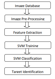
III. BLOCKDIAGRAM

Fig2. Overview of SVM Implementations
IV. SVM CLASSIFICATION
The data points that are closest to the decision surface are known as support vectors. They are the hardest to categorize. They directly affect where the decision surface should be placed. We can demonstrate that the function class with the lowest capacity (VC dimension) is the source of the ideal hyper plane. A selection of training samples, the support vectors, and the quadratic programming problem fully specify the decision function of support vector machines, which maximize the margin around the separation hyper plane.
V. RESULTS

Fig3.Results display
Conclusion
The application of the Support Vector Machine (SVM) classifier to identify brain tumors from MRI images is the main goal of this paper, \"Identification of Brain Tumors Using SVM Classifier.\" The study\'s conclusion emphasizes SVM\'s efficacy and precision as a reliable machine learning technique for automated brain tumor detection. The study shows that SVM can effectively classify brain tissue regions into normal and tumor regions when combined with suitable feature extraction techniques like texture, shape, and intensity-based features. The study highlights how SVM\'s capacity to establish an ideal decision boundary between classes (tumor vs. non-tumor) enables high accuracy in identifying even minute abnormalities in brain tissue that manual diagnoses might miss. SVM is a promising tool for medical imaging applications, especially in the field of brain tumor detection, as the results show that it performs better than many conventional classification techniques in terms of classification accuracy, specificity, and sensitivity. The study also emphasizes how crucial preprocessing techniques like noise reduction and image normalization are to enhancing the SVM classifier\'s performance. The authors come to the conclusion that although SVM classifiers have a lot of promise for automated tumor detection, more sophisticated approaches like feature selection, deep learning, and hybrid models can be used to increase the model\'s efficiency, accuracy, and robustness. This work helps medical professionals and reduces human error in diagnosis by paving the way for faster and more dependable diagnostic systems for brain tumor detection. For even greater accuracy and generalization across various medical imaging datasets, it also creates opportunities for future research into combining SVM with other machine learning models.
References
[1] H. Sahbi, D. Geman, and N. Boujemaa, “Face Detection Using Coarse-to-Fine Support Vector Classifiers,” Proc. Int’l Conf. Image Processing, pp. 925-928, 2002. [2] E. Osuna, R. Freund, and F. Girosi, “Training Support Vector Machines: An Application to Face Detection,” Proc. IEEE Conf. Computer Vision and Pattern Recognition, pp. 130-136, 1997. [3] Saptalakar. B. K, Rajeshwari. H, “Segmenttion Based Detection of Brain Tumor,” International Journal of computer and Electronics Research, Vol. 2, pp.20-23, February 2013. [4] Jayashree. M. J, Charutha. S, “An Efficient Brain Tumor Detection By Integrating Modified Texture Based Region Growing And Cellular Automata Edge Detection” [5] Venkatapathi, Pallam, Habibulla Khan, S. Srinivasa Rao, and Govardhani Immadi. \"Cooperative spectrum sensing performance assessment using machine learning in cognitive radio sensor networks.\" Engineering, Technology & Applied Science Research 14, no. 1 (2024): 12875-12879. [6] C. Cortes and V. Vapnik, “Support-Vector Networks,” Machine Learning, vol. 20, no. 3, pp. 273-297, 1995. [7] Ek Tsoon Tan, James V Miller, Anthony Bianchi, Albert Montillo- “Brain Tumor Segmentation With Symmetric TextureAnd symmetric IntensityBased Decision Forests”GE Global Research, Niskayuna, NY, USA, University of California Riverside, Riverside, CA USA. [8] MARKING, N.V.W., 2014. MULTI-WAVELET BASED ON NON-VISIBLE WATER MARKING. [9] Sudhakar Alluri, Komireddy Shreyas, Lingampally Ganesh, Mangali Vamshi, Venkatapath Pallam “A System Based in Virtual Reality to Manage Flood Damage” International Journal for Research in Applied Science & Engineering Technology (IJRASET) ISSN: 2321-9653; Volume 11 Issue XI Nov 2023 [10] Venkatapathi Pallam, Vasudev Biyyala, Chandra Shekar Jadapally, Ramsai Nalla, Dr. Sudhakar Alluri “Doctors Assistive System Using Augmented Reality Glass Critical Analysis” International Journal for Research in Applied Science & Engineering Technology (IJRASET) ISSN: 2321-9653; Volume 11 Issue X Oct 2023 [11] Subhashini. J, Vijay. J, “An Efficient Brain Tumor Detection Methodology using k- means Clustering Algorithm”, International conference on communication and Signal Processing, pg. 653– 657, April 2013. [12] Sudhakar Alluri, Karnati Mahidhar, Kalluru Kavya, Dulam Srija, P.Venkatapathi “High Performance Of Smartcard With Iris Recognition For High Security Access Environment In Python Tool” Industrial Engineering Journal ISSN: 0970-2555; Volume : 52, Issue 10, No. 2, October : 2023 [13] Albert Singh. N, Amsaveni. V, “Detection ofBrain Tumorusing Neural Network”, in ICCCNT, pg, 1-5, July 2013. [14] Gudipelly Mamatha, B.Manjula and P.Venkatapathi “Intend Innovative Technology For Recognition Of Seat Vacancy In Bus” International Journal of Research and Analytical Reviews, Volume 6, Issue 02, April-June.-2019, ISSN: 2349-5138 [15] Nguyen Thanh Thuy, Tran Son Hai, Le Hoang Thai, “Image Classification using Support Vector Machine and Artificial Neural Network”, Vietnam. [16] Chinnaiah, M. C., Sanjay Dubey, N. Janardhan, Venkata Pathi, K. Nandan, and M. Anusha. \"Analysis of pitta imbalance in young indian adult using machine learning algorithm.\" In 2022 2nd International conference on intelligent technologies (CONIT), pp. 1-5. IEEE, 2022.
Copyright
Copyright © 2024 Sudanala Gopala Krishna , B. Sarvesh , M. Narendhar Reddy, R. Ajay Kumar , Venkatapathi Pallam. This is an open access article distributed under the Creative Commons Attribution License, which permits unrestricted use, distribution, and reproduction in any medium, provided the original work is properly cited.

Download Paper
Paper Id : IJRASET65680
Publish Date : 2024-11-30
ISSN : 2321-9653
Publisher Name : IJRASET
DOI Link : Click Here
 Submit Paper Online


