Ijraset Journal For Research in Applied Science and Engineering Technology
- Home / Ijraset
- On This Page
- Abstract
- Introduction
- Conclusion
- References
- Copyright
Image Transform and Hybrid Optimization Based Feature Reduction for Leaf Diseases Classification
Authors: Dr. J. L. Divya Shivani, Dr. Veera Babu A
DOI Link: https://doi.org/10.22214/ijraset.2024.58694
Certificate: View Certificate
Abstract
In recent years, there has been amazing progress in technology innovation across many sectors, including medical care, homeland security, the arts, and even agriculture. The ability to run one\'s business well is highly regarded in every sector. Because to recent advancements in technology, such as smart phones and the Internet of Things, this is now a distinct possibility. In this research, we offer a smart management system for horticulture that makes use of machine learning methods. This system is based on the smart management scheme that was previously described. As a component of horticulture management, many academics have advocated for the use of machine learning to analyse plant growth based on the features of the leaf. We used the grayscale image, which employed numerous features for classification while simultaneously normalising the pixel values. As a consequence of this, the processing of damaged leaves in their early stages takes more time and results in less accurate classification. As a result, for the purpose of this work, we do a colour space conversion in order to investigate the image in terms of hue, saturation, and value. Following that, it extracted features from the Value channel by making advantage of the histogram\'s attributes. After that, the best characteristics were chosen by a process that included both particle swarm and cuckoo search optimisation. Following this, a regression neural network was trained to classify data based on the features that had been selected in the previous step. In order to evaluate how effective the proposed strategy is, classification performance criteria such as accuracy, sensitivity, and specificity were utilised. The suggested approach was tested in a simulated Windows 10 environment using a real-time dataset and the MATLAB software. It was compared to other methods that are already in use. While evaluating performance, comparisons to the current state of the art are conducted with regard to accuracy, sensitivity, and specificity.
Introduction
I. INTRODUCTION
India is famous for its agriculture, cultural and heritage structures. Among these, agriculture plays a major role in the Indian economy's growth. But in today’s technological development, agricultural growth is in a diminishing stage by converting the agricultural land into industrial or commercial buildings.
The main reasons behind the diminishing agricultural growth are technological development and the younger generation's lack of interest in agriculture. This can be modified by introducing technology advancement in agriculture. This will help the existing old farmer and welcome the young budding farmer into agriculture. For example, introducing smart irrigation systems and crop monitoring using the internet of things and computer-aided technologies.
Based on this smart management technique in agriculture, in this paper, an automated leaf disease classification algorithm was proposed using machine learning algorithms. The proposed algorithms were selected based on analysing various techniques that are used for leaf disease classification. The techniques that are used in identifying the diseased region are discussed below.
Utilized satellite images like multispectral (MS), hyperspectral (HS), and visible images (VI) to identify the diseased beetroot leaves. In combined the features of MS and VI images using a fusion technique. Then, they performed classification using machine learning algorithms called Naive Bayes (NB) and the K-nearest neighbour algorithm (K-NN). While in they utilized a single HS image and a decision tree technique for classification. A multi-level based citrus leaf disease classification was proposed by using the ensemble algorithm Adaboost technique.
Mammography is the most trustworthy technique that is currently available and can save lives by detecting breast cancer in its early stages. Digital mammography is now the most accurate screening tool currently available for breast cancer. In this piece of research, the authors present a strategy that makes use of a multi-SVM classifier for the purpose of finding and classifying breast cancer masses in digital mammograms[2].
An automated method is used to segment the damaged mammogram by first splitting it into smaller parts, and then merging those smaller parts back together again using a region-based segmentation technique.
The starting point of this technique is the selection of a seed location [3]. The proposed algorithm employs a process known as area split and merge to isolate the affected area of a picture from its surrounding pixels and morphological procedures to digitally reduce noise. The image undergoes both of these processes.
The image types and procedures utilized for classification were outlined in the paper [4]. It emphasized the many sorts of image capture ranges, such as visual, thermal, and satellite imaging, in image types. [5] suggested a feature selection method based on the detection of illnesses in rice leaves. The best features from the standard feature set were chosen using a rough set method. For cherry leaves, [6] developed a chemical-oriented virus-affected leaf disease classification. The pathogen characteristics of twisted and rusty leaf varieties were investigated using in vitro and categorized.
The visual qualities of hyperspectral images are described by a number of bands. Only a few of the many bands had access to the crucial information. In such situations, band selection is critical. A sequential projection was used in [4] to suggest a band selection method. The suggested method was tested using HS tomato images, which were categorised using an extreme machine learning algorithm. The suggested pre-processing and feature extraction strategies in the majority of the publications were for leaf disease classification. They suggested a beet leaf categorization based on post-processing in [10].
A study of alternative methodologies for classifying Alfalfa leaf diseases was conducted. For lesion segmentation, a combination of K-median clustering and linear discriminate analysis works well [11]. The production of tan in leaves is investigated using sorghum pigment analysis, which is based on gene missing and translated components [12].
For automated leaf disease classification, a histogram-based feature extraction using colour-transformed leaf images was proposed in [13]. Similarly, utilizing ensemble voting and histogram matching approaches, the histogram properties of soybean leaves were utilized to diagnose the leaf disease in [14]. The methodologies utilized to identify leaf diseases using visible range-based images were primarily detailed in the paper [15].
A multi-level processing of cucumber leaf diseases classification was proposed in [16]. Here, they segmented the leaves using K-means, then sparse classification was performed on the texture features from the leaves. Similarly, in [17] also performed the lesion segmentation and SVM-based diseases identification in Cucumber leaves.
The articles [18–19] highlighted the techniques used for identifying the diseases in wheat leaves. In [18], the HS image was processed using spectral indices for classification. In [19], deep learning algorithms were analysed in wheat leaf disease classification. The NB algorithm was used to classify the okra and bitter gourd leaf in [20]. The other recent techniques that were utilized for leaf disease classification are highlighted in the following section.
II. RECENT WORKS
This section is to describe the recent methods that are used for the classification were discussed.
G. R. Jothilakshmi et al., (2017) approach for extracting the region of interest from sono-mammogram pictures based on the detection of unique blocks was proposed. Here, a region expanding technique was used to isolate the afflicted area, and then features were extracted from that area. Then, a radial basis neural network was used for classification after bacterial optimisation was used to pick relevant characteristics. This method's weakness is that it only looked at fungal illnesses.
Iqbal et al. (2018) investigated the detection of citrus leaf diseases. The colour and shape were found to be the best for feature extraction during the analysis. Fuzzy logic was utilized to reduce the number of features. Traditional machine learning techniques were utilized for classification, and classification measures were used to analyse the results. Because there are so many different reduction approaches, the analysis in the feature reduction section was modest.
Liu et al., (2018) proposed a deep learning algorithm for classifying the apple leaf diseases using hyperspectral images. the performance can be enhanced further by using band selection algorithm and different network instead of Alexnet.
Lu et al., (2018) In addition, we used a multispectral satellite image of tomatoes to perform the classification. In this case, principal component analysis was employed to simplify the vegetation indices used for feature extraction. Then, it was categorised using K NN, which is the method of choice when trying to determine which leaves are in good condition.
Barbedo (2018) performed a deep learning-based leaf diseases classification for twelve plant using convolution neural network. The shortcomings of this approach are that it performed the classification on diseases segmented region only.
Lin et al., (2019) also utilized the deep learning algorithm with modification in CNN layers for classifying the wheat diseases. The shortcoming of this approach is it utilized same data for training and it results in over fitting of the network.
Geetharani and pandiyan (2019) also modified the CNN layers and utilized augmentation process for classifying the thirty-nine types of diseases affected leaves. The epoch and batch normalization values were selected using manual approach.
Priyadarshini et al., (2019) modified the Le deep learning network for maize leaf diseases classification. It performed classification only on 3 types only with higher processing time and steps.
III. PAPER CONTRIBUTION
From the analysis of recent techniques and existing algorithms, the following points were observed.
- For ML algorithms, the images were transformed to grey scale domain.
- Most of the algorithms utilized segmentation-based classification.
- The feature selection was using traditional algorithms like PCA, relief algorithm and correlation properties.
- Deep learning techniques skipped the local features from the images.
Based on the shortcomings, in the existing paper, the moth flame-based Regression neural network was proposed for leaf diseases classification. In that also, the image was processed in grey scale domain. This results in limited classification. Hence to overcome this, in this paper, the following modifications were performed to enhance the diseases identification in leaves.
The modifications of the existing algorithm in this paper are as follows:
- In the pre-processing step, the RGB image is transformed into HSV image instead of grey scale image.
- Because in HSV image, the colour values of the image were preserved in value values.
- The feature extraction was performed on the Value channel of the image.
- In feature reduction part, the hybrid Particle swarm and cuckoo search-based optimization was used.
- The classification is performed through the same Generalized regression neural network.
The brief explanation of proposed Image transform and hybrid optimization technique was given below.
IV. PROPOSED METHOD
This study provided an image transform and hybrid optimization-based feature selection strategy for disease identification in leaves. The final classification was done using a regression neural network. All of the stages above were completed on nine distinct varieties of leaves. The suggested approach's procedure is depicted in Figure 1.

The steps in the figure 1 are listed below to perform the leaf diseases identification using proposed approach.
- 1 Healthy and unhealthy images of nine types of leaves were loaded.
- 2 Resize the images to [256 256]
- 3 Convert the resized RGB image to HSV image
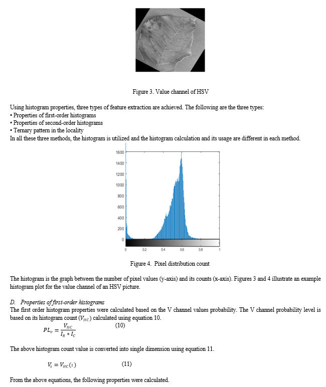
- 4 Extract the Value component from HSV image
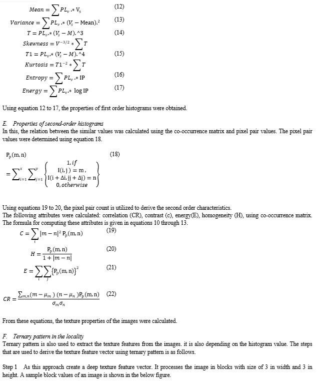
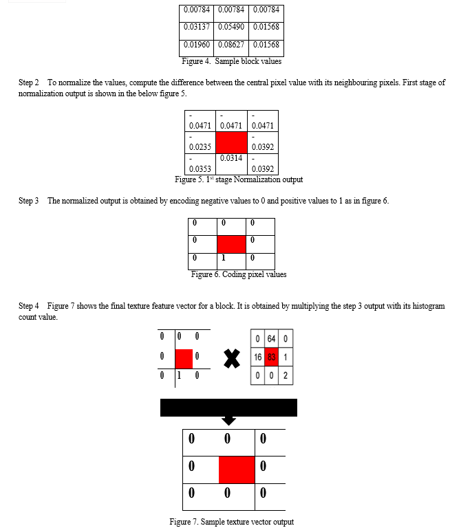
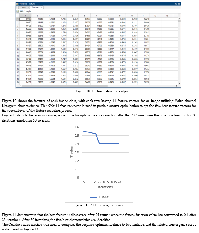
- 5 Perform feature extraction on Value component using histogram values.
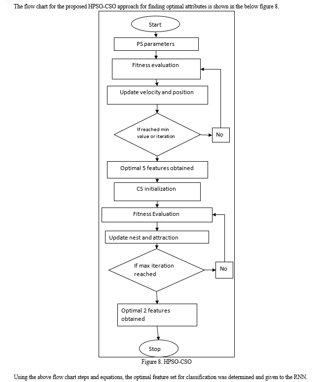
- 6 Perform first stage of feature reduction using particle swarm algorithm using following steps
- 7 Initialize swarm values, iterations and boundary values for PSO
- 8 Begin PSO by evaluating fitness function
- 9 After first iteration, update swarm position and velocity for further iterations.
- 10 Similarly, update the fitness function values.
- 11 If PSO complete all iterations or conditions reached
- 12 The top five features were obtained from PSO.
- 13 This features were reduced using Cuckoo search algorithm to select top two features
- 14 The CSO also repeat the steps 7 to 12 using its initialization and position update equations.
- 15 Once all conditions satisfied, the CSO selects top two features for classification.
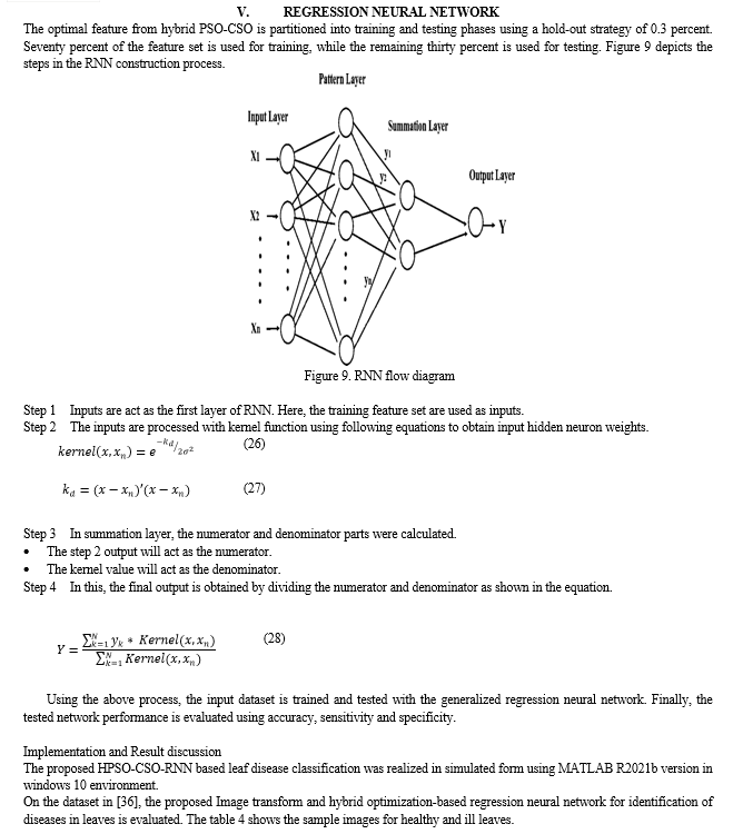
- 16 Finally, selected features were used for classification using regression neural network.
- 17 The classification was performed on 30% of selected features. The remaining features were used for training part.
- 18 The proposed method performance evaluated using classification performance metrics.
A. Kaggle Dataset
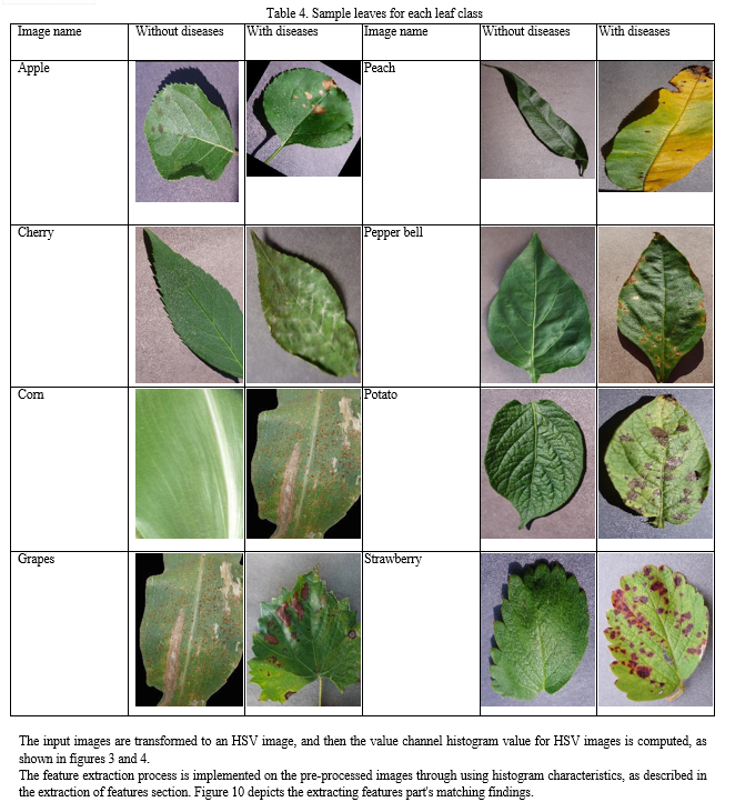
The proposed method's input is derived from the Kaggle plant disease dataset [36]. Table 1 shows the nine leaf classifications that are considered here. There are 30 healthy leaves in each leaf class, while the rest leaves are diseased. Table 1 shows the leaf classes and the number of photos in each class.
Table 1.Leaf distribution in dataset
|
Type of leaves |
Total no of images |
|
Apple |
120 |
|
Cherry |
60 |
|
Corn |
90 |
|
Grapes |
120 |
|
Peach |
60 |
|
Pepper bell |
60 |
|
Potato |
90 |
|
Strawberry |
60 |
|
Tomato |
300 |
For processing, a maximum of 990 images are taken. These images were subjected to the remaining steps of the proposed method.
B. Pre-processing
Image resizing and transformation are done on the given dataset. Because the input images are noise-free and the regression neural network can comprehend noisy data, this approach does not require any filtering.
1) Image transformation
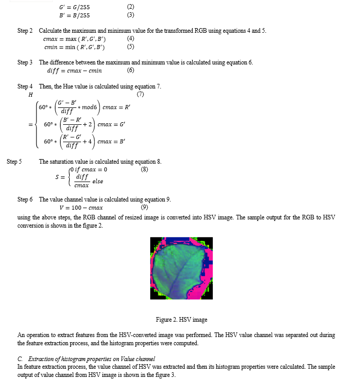
First, the image is resized to a standard size into 256 * 256 for easy processing. Then, the resized image was subjected to image transformation technique. Here, the RGB channel of image is transformed into HSV channel. Because in HSV the colour values are preserved better as compared to the grey scale transformation. The steps in RGB to hsv is shown in the below equations.


Conclusion
This study proposed a machine learning strategy for classifying various leaf diseases using a minimal statistical model. The ideal feature set is automatically obtained by solving an objective function called minimize classifier error rate using image transform and hybrid PSO-CSO optimization. This capability cut GRNN\'s training and testing times in half. The proposed approach was tested on a Kaggle dataset and found to be the best machine learning algorithm for identifying leaf diseases. In Future, the proposed method can be enhanced by processing the colour images directly for identifying the diseased region.
References
[1] G. R. Jothilakshmi, R. J. Christilda, A. Raaza, Y. SreenivasaVarma and V. Rajendran, \"Extracting region of interest using distinct block processing method in sono-mammogram images,\" 2017 International Conference on Computer, Communication and Signal Processing (ICCCSP), 2017, pp. 1-7, doi: 10.1109/ICCCSP.2017.7944091. [2] G. R. Jothilakshmi and A. Raaza, \"Effective detection of mass abnormalities and its classification using multi-SVM classifier with digital mammogram images,\" 2017 International Conference on Computer, Communication and Signal Processing (ICCCSP), 2017, pp. 1-6, doi: 10.1109/ICCCSP.2017.7944090. [3] G. R. Jothilakshmi, P. Sharmila and Arun Raaza3 “Mammogram Segmentation using Region based Method with Split and Merge Technique” Indian Journal of Science and Technology, Vol 9(40), DOI: 10.17485/ijst/2016/v9i40/99589, October 2016. [4] G.R.Jothilakshmi, ArunRaaza, V Rajendran, Y.Sreenivasa Varma and R.GuruNirmal Raj “Pattern Recognition and Size Prediction of Microcalcification Based on Physical Characteristics by Using Digital Mammogram Images” Journal of Digital Imaging, https://doi.org/10.1007/s10278-018-0075-x, 5 June 2018. [5] Veera Babu A, Dr.G.R.Jothi Lakshmi, “A Review On Detection And Classification Of Plant Diseases” Journal Of Critical Reviews, Issn- 2394-5125, Vol 7, Issue 10, 2020, Pp: 2285-2292. [6] Veera Babu A Et Al / Plant Disease Detection Methods That Integrate Picture Segmentation With Soft Computing, Neuroquantology|June2022| Volume 20|Issue 6|Page 3383-3394| Doi: 10.14704/Nq.2022.20.6.Nq22341 [7] Bauer, S. D., Kor?, F., &Förstner, W. (2011). The potential of automatic methods of classification to identify leaf diseases from multispectral images. Precision Agriculture, 12(3), 361-377. [8] Mahlein, A. K., Steiner, U., Hillnhütter, C., Dehne, H. W., &Oerke, E. C. (2012). Hyperspectral imaging for small-scale analysis of symptoms caused by different sugar beet diseases. Plant methods, 8(1), 1-13. [9] Zhang, M., & Meng, Q. (2011). Automatic citrus canker detection from leaf images captured in field. Pattern Recognition Letters, 32(15), 2036-2046. [10] Mahlein, A. K., Oerke, E. C., Steiner, U., &Dehne, H. W. (2012). Recent advances in sensing plant diseases for precision crop protection. European Journal of Plant Pathology, 133(1), 197-209. [11] Phadikar, S., Sil, J., & Das, A. K. (2013). Rice diseases classification using feature selection and rule generation techniques. Computers and electronics in agriculture, 90, 76-85. [12] Villamor, D. E. V., & Eastwell, K. C. (2013). Viruses associated with rusty mottle and twisted leaf diseases of sweet cherry are distinct species. Phytopathology, 103(12), 1287-1295. [13] Kruse, O. M. O., Prats-Montalbán, J. M., Indahl, U. G., Kvaal, K., Ferrer, A., &Futsaether, C. M. (2014). Pixel classification methods for identifying and quantifying leaf surface injury from digital images. Computers and electronics in Agriculture, 108, 155-165. [14] Omrani, E., Khoshnevisan, B., Shamshirband, S., Saboohi, H., Anuar, N. B., & Nasir, M. H. N. M. (2014). Potential of radial basis function-based support vector regression for apple disease detection. Measurement, 55, 512-519. [15] Xie, C., Shao, Y., Li, X., & He, Y. (2015). Detection of early blight and late blight diseases on tomato leaves using hyperspectral imaging. Scientific reports, 5(1), 1-11. [16] Zhou, R., Kaneko, S. I., Tanaka, F., Kayamori, M., & Shimizu, M. (2015). Image-based field monitoring of Cercospora leaf spot in sugar beet by robust template matching and pattern recognition. Computers and Electronics in Agriculture, 116, 65-79. [17] Qin, F., Liu, D., Sun, B., Ruan, L., Ma, Z., & Wang, H. (2016). Identification of alfalfa leaf diseases using image recognition technology. PLoS One, 11(12), e0168274. [18] Kawahigashi, H., Nonaka, E., Mizuno, H., Kasuga, S., &Okuizumi, H. (2016). Classification of genotypes of leaf phenotype (P/tan) and seed phenotype (Y1 and Tan1) in tan sorghum (Sorghum bicolor). Plant Breeding, 135(6), 683-690. [19] Barbedo, J. G. A., Koenigkan, L. V., & Santos, T. T. (2016). Identifying multiple plant diseases using digital image processing. Biosystems engineering, 147, 104-116. [20] Chaudhary, A., Kolhe, S., & Kamal, R. (2016). A hybrid ensemble for classification in multiclass datasets: An application to oilseed disease dataset. Computers and Electronics in Agriculture, 124, 65-72. [21] Barbedo, J. G. A. (2016). A review on the main challenges in automatic plant disease identification based on visible range images. Biosystems engineering, 144, 52-60. [22] Zhang, S., Wu, X., You, Z., & Zhang, L. (2017). Leaf image based cucumber disease recognition using sparse representation classification. Computers and electronics in agriculture, 134, 135-141. [23] Zhang, S., Zhu, Y., You, Z., & Wu, X. (2017). Fusion of superpixel, expectation maximization and PHOG for recognizing cucumber diseases. Computers and Electronics in Agriculture, 140, 338-347. [24] Shi, Y., Huang, W., Luo, J., Huang, L., & Zhou, X. (2017). Detection and discrimination of pests and diseases in winter wheat based on spectral indices and kernel discriminant analysis. Computers and Electronics in Agriculture, 141, 171-180. [25] Mondal, D., Kole, D. K., & Roy, K. (2017). Gradation of yellow mosaic virus disease of okra and bitter gourd based on entropy based binning and Naive Bayes classifier after identification of leaves. Computers and Electronics in Agriculture, 142, 485-493. [26] Lu, J., Hu, J., Zhao, G., Mei, F., & Zhang, C. (2017). An in-field automatic wheat disease diagnosis system. Computers and electronics in agriculture, 142, 369-379. [27] Chouhan, S. S., Kaul, A., Singh, U. P., & Jain, S. (2018). Bacterial foraging optimization based radial basis function neural network (BRBFNN) for identification and classification of plant leaf diseases: An automatic approach towards plant pathology. IEEE Access, 6, 8852-8863. [28] Iqbal, Z., Khan, M. A., Sharif, M., Shah, J. H., urRehman, M. H., &Javed, K. (2018). An automated detection and classification of citrus plant diseases using image processing techniques: A review. Computers and electronics in agriculture, 153, 12-32. [29] Liu, B., Zhang, Y., He, D., & Li, Y. (2018). Identification of apple leaf diseases based on deep convolutional neural networks. Symmetry, 10(1), 11. [30] Lu, J., Ehsani, R., Shi, Y., de Castro, A. I., & Wang, S. (2018). Detection of multi-tomato leaf diseases (late blight, target and bacterial spots) in different stages by using a spectral-based sensor. Scientific reports, 8(1), 1-11. [31] Barbedo, J. G. A. (2018). Impact of dataset size and variety on the effectiveness of deep learning and transfer learning for plant disease classification. Computers and electronics in agriculture, 153, 46-53. [32] Lin, Z., Mu, S., Huang, F., Mateen, K. A., Wang, M., Gao, W., & Jia, J. (2019). A unified matrix-based convolutional neural network for fine-grained image classification of wheat leaf diseases. IEEE Access, 7, 11570-11590. [33] Geetharamani, G., & Pandian, A. (2019). Identification of plant leaf diseases using a nine-layer deep convolutional neural network. Computers & Electrical Engineering, 76, 323-338. [34] Priyadharshini, R. A., Arivazhagan, S., Arun, M., &Mirnalini, A. (2019). Maize leaf disease classification using deep convolutional neural networks. Neural Computing and Applications, 31(12), 8887-8895. [35] Dhingra, G., Kumar, V., & Joshi, H. D. (2019). A novel computer vision based neutrosophic approach for leaf disease identification and classification. Measurement, 135, 782-794. [36] Singh, U. P., Chouhan, S. S., Jain, S., & Jain, S. (2019). Multilayer convolution neural network for the classification of mango leaves infected by anthracnose disease. IEEE Access, 7, 43721-43729. [37] Karlekar, A., & Seal, A. (2020). SoyNet: Soybean leaf diseases classification. Computers and Electronics in Agriculture, 172, 105342. [38] Devi, K. S., Srinivasan, P., &Bandhopadhyay, S. (2020). H2K–A robust and optimum approach for detection and classification of groundnut leaf diseases. Computers and Electronics in Agriculture, 178, 105749. [39] Adeel, A., Khan, M. A., Akram, T., Sharif, A., Yasmin, M., Saba, T., &Javed, K. (2020). Entropy?controlled deep features selection framework for grape leaf diseases recognition. Expert Systems. [40] U?uz, S., &Uysal, N. (2021). Classification of olive leaf diseases using deep convolutional neural networks. Neural Computing and Applications, 33(9), 4133-4149. [41] Chao, X., Hu, X., Feng, J., Zhang, Z., Wang, M., & He, D. (2021). Construction of Apple Leaf Diseases Identification Networks Based on Xception Fused by SE Module. Applied Sciences, 11(10), 4614. [42] New Plant Diseases Dataset. Kaggle.com. (2021). Retrieved 26 October 2021, from https://www.kaggle.com/vipoooool/new-plant-diseases-dataset. [43] SK, P. K., Sumithra, M. G., & Saranya, N. (2021). Particle Swarm Optimization (PSO) with fuzzy c means (PSO?FCM)–based segmentation and machine learning classifier for leaf diseases prediction. Concurrency and Computation: Practice and Experience, 33(3), e5312. [44] Veera babu A, Dr.G.R.Jothi Lakshmi, Dr.J.l.divya shivani, “an efficient identification and detection of plant leaf diseases using region-based thresholding algorithm”, semiconductor optoelectronics, issn-1001-5868, vol. 42, no. 1 (2023),pp: 754-764 [45] VEERA BABU A, Dr.G.R.JOTHI LAKSHMI, “Soft Computing and Image Processing for Detection and Analysis of Plant Infections”, Mathematical Statistician and Engineering Applications, ISSN- 2094-0343, Issue 3s2, Volume 71, July 2022, PP: 1173-1185. [46] VEERA BABU A, Dr.G.R.JOTHI LAKSHMI, “Plant Disease Detection Methods That Integrate Picture Segmentation With Soft Computing”, Neuro Quantology, Issn- 1303-5150, Vol 20, Issue 6, 2022, Pp: 3383-3394. [47] VEERA BABU A, Dr.G.R.JOTHI LAKSHMI, “A Review On Detection And Classification Of Plant Diseases” Journal Of Critical Reviews, Issn- 2394-5125, Vol 7, Issue 10, 2020, Pp: 2285-2292.
Copyright
Copyright © 2024 Dr. J. L. Divya Shivani, Dr. Veera Babu A. This is an open access article distributed under the Creative Commons Attribution License, which permits unrestricted use, distribution, and reproduction in any medium, provided the original work is properly cited.

Download Paper
Paper Id : IJRASET58694
Publish Date : 2024-02-29
ISSN : 2321-9653
Publisher Name : IJRASET
DOI Link : Click Here
 Submit Paper Online

