Ijraset Journal For Research in Applied Science and Engineering Technology
- Home / Ijraset
- On This Page
- Abstract
- Introduction
- Conclusion
- References
- Copyright
Improving MRI Image Clarity and Noise Reduction for Enhanced Brain Tumour Detection
Authors: Mrs. Mandeep Kaur, Dr. Rahul Thour
DOI Link: https://doi.org/10.22214/ijraset.2025.66957
Certificate: View Certificate
Abstract
Brain diseases are serious conditions that must not be ignored, as brain failure can pose a significant threat to overall health. Early detection and intervention are critical in managing various brain-related disorders. One of the primary diagnostic methods for detecting brain tumors and other neurological issues is MRI imaging. MRI is a preferred technique due to its efficiency, real-time imaging capabilities, and lack of radiation. However, challenges such as speckle noise, Gaussian noise, and other artifacts continue to compromise the quality of MRI images. As a result, enhancing image quality is crucial for accurate brain disorder diagnosis. To overcome these challenges, various imaging techniques are employed for preprocessing, noise reduction, and image enhancement. A key approach to obtaining high-quality images from noisy MRI data is image restoration and enhancement. Given the high-frequency characteristics of MRI, noise is often present in brain scans. Preprocessing plays an essential role in improving image quality by applying filters to eliminate noise. Techniques like mean, median, Wiener, and other filters are commonly used to address issues such as speckle, salt-and-pepper, and Gaussian noise. This research offers a comprehensive overview of various MRI image preprocessing and enhancement techniques, outlining their objectives and effectiveness.
Introduction
I. INTRODUCTION
MRI imaging is a non-invasive, radiation-free and real-time method for capturing internal body structures, aiding doctors in detecting diseases or abnormal tissues. MRI, known for its efficiency and speed, has become a valuable tool for various medical applications. However, MRI imaging can be impacted by signal dependence, which restricts the resolution of images and complicates human interpretation and diagnosis. Consequently, reducing speckle noise in medical MRI image processing is a crucial challenge. Explores advanced techniques in brain image analysis, focusing on improving image quality and disease classification accuracy. It begins with a foundational understanding of MRI and CT, followed by employing image processing methods to reduce noise and enhance clarity. The research then evaluates segmentation techniques and applies deep learning, specifically CNNs, to refine disease classification. Ultimately, the goal is to advance diagnostic capabilities in medical imaging[1]. Reviews various methods for brain tumor detection and segmentation from MRI images, highlighting the challenges posed by tumor tissue variability and its resemblance to normal tissues. The review examines different automated techniques aimed at improving the accuracy and efficiency of segmentation, discussing both their advantages and limitations. It also explores the application of these methods in clinical procedures, emphasizing their role in enhancing brain tumor diagnosis[2]. A novel two-module computerized method for brain tumor detection using MRI, aimed at improving speed and accuracy. The first module enhances image quality through a combination of adaptive Wiener filtering, neural networks, and independent component analysis. The second module employs Support Vector Machines for tumor segmentation and classification. Tested on various brain tumor types, the method demonstrated exceptional performance, achieving high sensitivity, specificity, and accuracy, while significantly reducing processing time compared to existing methods[3].Introduces a three-step preprocessing method and a novel Deep Convolutional Neural Network (DCNN) architecture for the effective diagnosis of brain diseases, including glioma, meningioma, and pituitary tumors, from MRI images. The model leverages batch normalization for faster training and computational efficiency, with fewer layers and training iterations. Experimental results demonstrate its outstanding performance, achieving an overall accuracy of 98.22%, with particularly high detection rates for glioma, meningioma, and pituitary tumors, underscoring the architecture’s robustness and potential for rapid, accurate diagnosis[4].
Explores a two-step Generative Adversarial Network (GAN)-based data augmentation (DA) approach to enhance brain tumor detection from MRI images, addressing the challenge of small and fragmented medical datasets. The method combines Progressive Growing GANs (PGGANs) for realistic noise-to-image generation and Multimodal UNsupervised Image-to-image Translation (MUNIT) for refining generated images to closely resemble real ones. Results demonstrate that this approach significantly improves tumor classification, boosting sensitivity from 93.67% to 97.48%, outperforming traditional DA methods in medical imaging tasks[5]. Evaluates a deep learning-based denoising approach (dDLR) for improving brain MRI image quality by comparing it with traditional methods such as DnCNN and SCNN. Experimental and clinical results show that dDLR outperforms these methods in terms of structural similarity (SSIM) and peak signal-to-noise ratio (PSNR), enhancing image clarity while preserving important details. In clinical tests, dDLR significantly improved the quality of images with lower acquisition settings, offering potential for high-quality brain MRI reconstruction with reduced noise[6].Explores the integration of edge detection and compression techniques to improve the performance of content-based image retrieval (CBIR) systems for brain tumor diagnosis. Focusing on preserving image quality post-compression, the research introduces a hybrid approach combining Canny-based edge detection, Huffman lossless compression, and Discrete Wavelet Transform (DWT). This method aims to enhance processing speed without compromising image quality, addressing previous gaps in CBIR systems that overlooked image integrity, and also incorporates noise reduction for better brain image analysis [7].
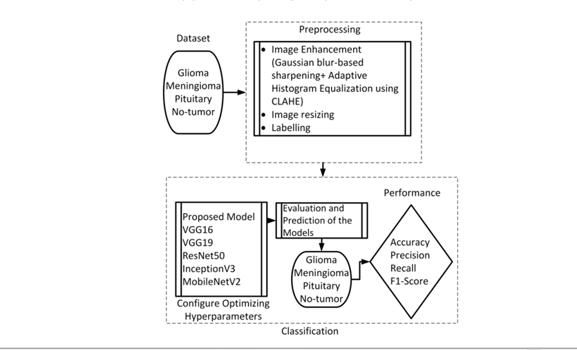
A novel deep learning-based approach for brain tumor classification using MRI images, combining image enhancement techniques such as Gaussian-blur sharpening and Adaptive Histogram Equalization (CLAHE). The model leverages Convolutional Neural Networks (CNNs) to classify glioma, meningioma, pituitary tumors, and normal cases, achieving a high classification accuracy of 97.84%. Compared to pre-trained models like VGG16 and ResNet50, the proposed method demonstrated superior performance, with notable precision, recall, and F1-score, highlighting its potential as a reliable tool for accurate brain tumor diagnosis[8]. A Automated approach for brain tumor detection using MRI images, focusing on tissue segmentation and tumor classification. The method utilizes a combination of skull stripping, Berkeley wavelet transform for segmentation, and a support vector machine (SVM) classifier to analyze tumor features. Experimental results demonstrate high accuracy (96.51%), sensitivity (97.72%), and specificity (94.2%), outperforming manual detection by radiologists. The approach offers improved speed, accuracy, and quality, making it a valuable tool for clinical decision support in brain tumor diagnosis[9]. A multi-stage method for edge detection of brain tumors in MRI images, integrating noise removal via the Balance Contrast Enhancement Technique (BCET), segmentation using Fuzzy c-Means (FCM) clustering, and edge detection with the Canny method. The approach demonstrates resilience to noise and improves tumor segmentation accuracy by 10-15% compared to expert estimates. The results, obtained using MATLAB, show promise for reliable tumor detection across varying tumor characteristics, such as location, type, and size[10]. Feature extraction and organized storage of abnormalities in brain MRI and CT scans, such as tumors and hemorrhages. The methodology integrates multiple phases, including image extraction, transformation, and progression, which involve noise reduction, skull removal, and image enhancement. By segmenting the images based on T1, T2, and PD-weighted intensities, and extracting features for classification, the system enhances diagnostic accuracy. Experiments on over 200 datasets show promising results, demonstrating the effectiveness of the approach in improving brain image analysis and detection accuracy[11].

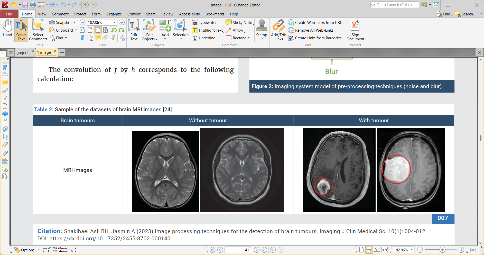
Figure1. MRI Brain Image with Tumor and without Tumor[1].
II. PRE-PROCESSING AND IMAGE QUALITY ENHANCEMENT
A. Pre-processing of MRI Images
Input MRI Image and Pre-processing: The input is an MRI image, which is pre-processed using a Gaussian filter. This filter helps in reducing noise from the image by smoothing it out, preventing blurriness. Known as a smoothing operator, it removes fine details naturally present in the image. The filter’s impulse response is based on a Gaussian function (GF), which defines the probability distribution of noise. It effectively eliminates Gaussian noise. The filter is linear and operates as a low-pass filter with a Gaussian function of a specific standard deviation.
MRI images are often affected by low contrast, Rician noise and speckle noise, which can impact image clarity. To address these issues, several methods can be applied to enhance the image quality and improve system performance.
Preprocessing is essential for removing unwanted noise from MRI images, including speckle and Gaussian noise, as well as other irregularities. This stage involves various techniques like noise suppression, image restoration, contrast enhancement, smoothing, and sharpening to improve the overall quality of the MRI images. These steps aim to reduce background noise, preserve critical information, enhance the edges of objects, and increase the contrast between the region of interest and the surrounding speckle.

Figure 2. Flow chart of the suggested scheme [8].
Key preprocessing methods include[12]:
- Image Restoration: This process aims to minimize or remove the degradation that occurs during the image acquisition phase. A level set function is used for proper image orientation.
- Smoothing and Sharpening: These techniques are applied to optimize image resolution in both the spatial and frequency domains. They are particularly effective in emphasizing object edges and fine details. Various filters, such as the Gabor filter and Gaussian function, are used to achieve this.
- Contrast Enhancement: This technique improves the visibility of MRI images. By adjusting the image’s value range, histogram equalization is utilized to boost contrast and ensure uniform intensity throughout the image.
- Noise Suppression: Noise, such as speckle noise, Rician noise, can degrade the visual quality of the image by replacing pixels with new ones that distort the original details. Speckle noise, a common issue in MRI images, is multiplicative and random in nature, obscuring important image features like edges, shapes, and intensity values. Various noise reduction techniques, including linear and nonlinear mean filters, median filters, Gaussian low-pass filters, wavelet filters, Gabor filters, and Wiener filters, are used to address this problem. A system developed by T. Rahman and M. S. Uddin, for instance, used a Gabor filter to reduce speckle noise and histogram equalization to enhance image quality. They also compared region-based segmentation and cell segmentation techniques, ultimately using region-based segmentation with MATLAB for improved results.
- Image Quality Enhancement: Techniques like histogram equalization and contrast enhancement are widely used to improve image quality. Enhancing an image involves adjusting its contrast, spatial resolution, brightness, and reducing noise. Histogram equalization helps by adjusting the distribution of gray levels in the image, providing a clearer representation of the image’s global features. Image quality is assessed using both objective and subjective fidelity metrics, which measure the accuracy and overall fidelity of the processed image.
III. PRE-PROCESSING AND IMAGE ENHANCEMENT TECHNIQUES
Table 1 presents a collection of various study techniques and methods used in medical imaging for tumor detection and classification, particularly in brain tumor analysis. Each study utilizes a distinct combination of image processing, machine learning, and deep learning strategies to address challenges such as noise reduction, segmentation, and classification accuracy. The techniques range from advanced methods like Convolutional Neural Networks (CNN), Support Vector Machines (SVM), and Generative Adversarial Networks (GANs) to more traditional image enhancement approaches like wavelet transforms and edge detection. By employing these methods, the studies aim to improve image quality and enhance diagnostic outcomes in medical imaging, especially for identifying brain tumors in MRI and CT scans.
Table 1: Description of Preprocessing techniques
|
Study |
Techniques Used |
Description |
|
[1] |
Image processing, CNN |
Focuses on noise reduction and segmentation methods to enhance image quality, followed by deep learning (CNN) for disease classification. |
|
[2] |
Automated segmentation methods |
Reviews methods for tumor detection and segmentation, emphasizing the challenges posed by tumor variability and their resemblance to normal tissues. |
|
[3] |
Adaptive Wiener filtering, Neural networks, SVM |
A two-module approach with image quality enhancement (filtering and neural networks) followed by tumor segmentation and classification using SVM. |
|
[4] |
DCNN, Batch normalization |
A three-step preprocessing method combined with a novel deep convolutional neural network for diagnosing glioma, meningioma, and pituitary tumors. |
|
[5] |
PGGAN, MUNIT |
A two-step GAN-based data augmentation approach, combining noise-to-image generation and image refinement for improved tumor detection. |
|
[6] |
DnCNN, SCNN, dDLR |
A comparison of deep learning-based denoising methods (dDLR, DnCNN, SCNN) for enhancing MRI image quality, focusing on SSIM and PSNR metrics. |
|
[7] |
Canny edge detection, Huffman compression, DWT |
A hybrid approach integrating edge detection, lossless compression, and wavelet transform to improve CBIR performance and maintain image quality. |
|
[8] |
Gaussian-blur sharpening, CLAHE, CNN |
Combines image enhancement techniques (Gaussian-blur, CLAHE) with CNNs for high-accuracy classification of brain tumors in MRI images. |
|
[9] |
Skull stripping, Berkeley wavelet transform, SVM |
Integrates skull stripping and wavelet transform for segmentation, followed by SVM classification for brain tumor detection with high accuracy. |
|
[10] |
BCET, FCM clustering, Canny edge detection |
A multi-stage approach using BCET for noise removal, FCM clustering for segmentation, and Canny edge detection for tumor boundaries. |
|
[11] |
Image extraction, T1/T2/PD segmentation, Feature extraction |
Focuses on extracting and organizing features from MRI and CT scans for enhanced diagnosis of tumors and hemorrhages, improving classification accuracy. |
Conclusion
Enhancing the quality of input images is a critical task in image processing. This paper explores commonly used techniques for preprocessing and improving the quality of MRI images. Although MRI is favored by physicians due to its lack of radiation compared to other imaging methods, it presents challenges due to the introduction of noise, which leads to various issues. Additionally, MRI images often suffer from poor contrast, speckle noise, Gaussian noise, and other imperfections. Therefore, improving image quality is essential for accurately identifying the region of interest. To address these challenges, the paper discusses various image processing methods, including preprocessing, noise reduction, and quality enhancement techniques. Several filters for noise removal are also examined, tailored to different types of noisy MRI images.
References
[1] Asli, Barmak Honarvar Shakibaei, and Anaëlle Jasmin. \"Image processing techniques for the detection of brain tumours.\" Imaging Journal of Clinical and Medical Sciences 10.1 (2023): 004-012. [2] Roy, Sudipta, et al. \"A review on automated brain tumor detection and segmentation from MRI of brain.\" arXiv preprint arXiv:1312.6150 (2013). [3] Asiri, Abdullah A., et al. \"Optimized Brain Tumor Detection: A Dual-Module Approach for MRI Image Enhancement and Tumor Classification.\" IEEE Access 12 (2024): 42868-42887. [4] Musallam, Ahmed S., Ahmed S. Sherif, and Mohamed K. Hussein. \"A new convolutional neural network architecture for automatic detection of brain tumors in magnetic resonance imaging images.\" IEEE access 10 (2022): 2775-2782. [5] Han, Changhee, et al. \"Combining noise-to-image and image-to-image GANs: Brain MR image augmentation for tumor detection.\" Ieee Access 7 (2019): 156966-156977. [6] Kidoh, Masafumi, et al. \"Deep learning based noise reduction for brain MR imaging: tests on phantoms and healthy volunteers.\" Magnetic resonance in medical sciences 19.3 (2020): 195-206. [7] Charaya, Saurabh. \"NOISE REMOVAL TO ENAHNCE IMAGE QUALITY DURING BRAIN TUMOUR DETECTION.\" NeuroQuantology 20.9 (2022): 6763. [8] Rasheed, Zahid, et al. \"Brain tumor classification from MRI using image enhancement and convolutional neural network techniques.\" Brain Sciences 13.9 (2023): 1320. [9] Bahadure, Nilesh Bhaskarrao, Arun Kumar Ray, and Har Pal Thethi. \"Image analysis for MRI based brain tumor detection and feature extraction using biologically inspired BWT and SVM.\" International journal of biomedical imaging 2017.1 (2017): 9749108. [10] Zotin, Alexander, et al. \"Edge detection in MRI brain tumor images based on fuzzy C-means clustering.\" Procedia Computer Science 126 (2018): 1261-1270. [11] Snehkunj, Rupal, Ashish N. Jani, and Nalin N. Jani. \"Brain MRI/CT images feature extraction to enhance abnormalities quantification.\" Indian Journal of Science and Technology 11.1 (2018): 1-10. [12] Kaur, Gurjeet, and Sukhwinder Singh. \"Image Quality Enhancement and Noise Reduction in Kidney Ultrasound Images.\"
Copyright
Copyright © 2025 Mrs. Mandeep Kaur, Dr. Rahul Thour. This is an open access article distributed under the Creative Commons Attribution License, which permits unrestricted use, distribution, and reproduction in any medium, provided the original work is properly cited.

Download Paper
Paper Id : IJRASET66957
Publish Date : 2025-02-14
ISSN : 2321-9653
Publisher Name : IJRASET
DOI Link : Click Here
 Submit Paper Online

