Ijraset Journal For Research in Applied Science and Engineering Technology
- Home / Ijraset
- On This Page
- Abstract
- Introduction
- Conclusion
- References
- Copyright
Kidney Stone Detection and Stone Analysis Using Image Processing
Authors: Mr. Narasimha Raju Paka, Basamma Guggalashettara , Ankitha H K, Archana K
DOI Link: https://doi.org/10.22214/ijraset.2024.62433
Certificate: View Certificate
Abstract
The primary goal of the project is to detect the kidney stone from the digital ultrasound image of the kidney by performing various image processing techniques. Due to the varied texture and existence of speckle noise, detecting regions of interest in ultrasound pictures is a difficult process. Ultrasound scanning is the most common method of examining a patient for the presence of kidney stones. We created an application that aids the medical practitioner in selecting the region to be evaluated can also referred to as renal calculi using the suggested technology. The feature extraction is done on cropped portions that may have stones in them. The KNN classifier is accustomed to categories images based on training data.
Introduction
I. INTRODUCTION
The production of crystals in the urine induced by genetic predisposition distinguishes renal calculus, identified as kidney stone formation. There are many individuals, as well as children, are impacted by kidney stones, most cases go unnoticed unless there is severe abdominal pain or an irregular urine color. Furthermore, persons with kidney stones exhibit common symptoms such as fever, discomfort, and nausea, which might be mistaken for other illnesses. Kidney stone identification is critical, especially early on, to receive adequate medical treatment. The appearance of renal calculi in the kidney reduces renal functionality with able probably cause dilatation.
Persons whomever have not known analysis with this sequence will be affected by the severity of chronic kidney disease (CKD) / chronic renal failure (CRF). By the agency of this asymptomatic character, it is frequently detected through antibiotic review further disorder such as cardiovascular disease (CVD), diabetes, and another medication issues this incline. to the urogenital apparatus. Days, computer-assisted tools like ultrasound imaging, computed tomography (CT), and X rays gives the almost all proper features diagnostic apparatus for nephrolith transmit and recognition. The aim of this project is to detect the kidney stone from a digital ultrasound image of the kidney by performing image processing techniques.
But the image produced by the ultrasound techniques is inappropriate for promote methodology due to low contrast and the occurance of speckle noise. Hence, the study also examined the effectiveness of various diagnosis techniques on the ultrasound image to improve the classification of the image. Further, enhanced ultrasound image will adapted to locate the correct location of the stone. The main motive of this project was to designed an elementary and straightforward technique to find the renal calculi. This detection can be done in any available PC’s and hence any normal being can check an ultrasound for a renal calculi and dissolve it in the stone.
II. LITERATURE SURVEY
Kidney stones are a common urological condition affecting a significant portion of the global population. The application of picture editing method for kidney finding own manifest encouraging data in enhancing the correctness and order of diagnosing kidney stones.
This is only about the key issues over the world to detect the proper location of stone throughout the kidney.In the formation of kidney stones, calcium is more common among them. This research explores the advanced technique to detect boundary, segmented area, and enhance detection of stone from the kidney with present locations left or right.
The main objective of that predict is to efficiently detect kidney stone problems among in consequence of image, and to better the detection rate in terms of rightness too sensitivity. Concerning outcome of our current life style kidney stone has become a common health issue.
An Automated kidney stone classification is implemented using Back Propagation Network and image and data processing techniques. Image enhancement is done of removing noise and brighten the image.
The initial stages of these kidney stones diseases are noticed latterly or which cannot be detected easily, in turn damages the kidney as they row to become larger.In this project the survey of different algorithms and classifications are analysed followed all recognition of stone present in the kidney.
Hospital and detached sonography figure not new to evaluate the proposed scheme and algorithm. The suggested scheme has been assessed by different performance measuring parameters. The attribute removal motion exist old to measure the precise coordinates of the stone and the overall appearance of the stones created from the picture
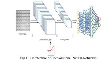
- Convolutional Layer: Convolutional layers perform a convolution on the input before forwarding the output to the next layer. The picture element within a convolution's receptive area are all converted to a single value. The convolutional layer's final output is a vector.
- Batch normalization Layer: Batch normalization is a network layer that enables each layer to learn more independently from the others. It's used to normalize the production the preceding layers. Standardizes the inputs to a layer for each mini-batch. This stabilizes the educational path and reduces the figure of instruction period required to build deep networks dramatically.
- Max Pooling is a convolution method within which Kernel collects the maximum value from the area it convolves. Max Pooling basically means that we will only forward the most relevant information
- Layer ReLu: That occur a function of activation.
- Softmax layer: Typically, this stage serves as the ultimate output Layer in a multi-class classification neural network diagram like that show in picture 2, “workflow on a few advance system”. Its primary purpose is to scale the neural network’s output to a range between zero and one .This layer is known for its complete connectivity, here each node is liked to every node in the proceding layer.
III. ALGORITHMS
In a multi-class classification neural network setup, this particular stage acts as the final layer of output as depicted in Figure 2, “Workflow regarding the planned system”. Its main function is to normalize the output values of the neural network, ensuring they fall within the range of zero to one. This layer is characterized by full connectivity, meaning each node is connected to every node within some preceding layer.
A. Alexnet's Transfer Learning
Knowledge consolidation in machine learning refers to a method point a duplicate level at length sole duty exist improve or reused for another task. Essentially, the information obtain among resolve solitary pain is leveraged to enhance performance continously a dissimilar yet connected problem. This model comprise eight layers, consisting of excellent crulicue sheet, trio totally link film, and a softmax layer.
B. Convolutional Neural Network
The diagram in figure 1 illustates the planning on a difficulty nerve grid, which is a type of deep-learning model. CNNs differ from fully connected networks in that they use specific layers such as convolutional, fully connected, and max-pooling layers. T/he complete interconnectivity of CNNs can make them prone to overfitting with certain datasets. CNNs consist of two main sections :feature extraction, which involves convolution and merging coat, in addition to classification, locus quite associate loops are used. The final part regarding the Cable news network is the SoftMax layer, which produces the networks output.
- Input Layer: The input layer of a neural network serves to receive initial data for processing. The lots of input features match untill the total neurons in this layer. These artificial neurons play the role of ingesting the features this one do exist processed within the system. The entire character of features can be obtained rest on the total

2. Output Layer: The output layer of a nerve lattice exist a crucial component that receives and processes input from preceding surface till originate the last works, typically representing predictions or classifications. This layer is designed to transform the input data into the required format for the specific task, often converting it into a set of class probabilities or values .In the context regarding greek root system models, the output layer serves as the final stage where predictions are directly generated root at length some learned patterns and features. This layer is essential across various neural network architectures, particularly those used in closed-loop control systems.

IV. PROPOSED METHODOLOGY
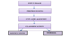
In this project, we propose utilizing both ALEXNET with complexity neuron webbing while bit regarding a apparatus before enhance performance and accuarcy. The projects process is illustrated in Figure 2.
Pre-processing enhances image quality by eliminating unwanted distortions and enhancing specific visual characteristics important for subsequent analysis as well as processing .used to improve image data by removing undesired distortions and boosting particular seeing quality this one occur relevant for further processing and analysis. An undesired distortion also be removed by testing out various activation, loss, and optimizer procedures. Further by Changing the unit, layers numbers and the batch size the undesired distortions would be reduced significantly. The visual features of performance metrics of images would be boosted by the following techniques. They are as follows: adding more number of layers, increasing the Epochs, changing the image size and also make use of move study technique.
A. Module 3
- Input Layers: The add loop of a neural network is spot deatils live initially fed into the model. These symbols nerve within this layer determines some entire digit towards trait or inputs for the network. These input neurons serve as the starting point, providing raw data for processing by subsequent layers of neurons within those nerve matrix structure.
- Hidden Layer: The data among a few load surface is passed before that masked film for processing. A specific unseen veneer get store among some store blanket and conducts computations on this data.Depending at length this copy along with a given amount of data, many unseen surface can be used. every digit about dendrite inside all masked film may differ, but typically exceeds each digit towards input features some veiled sheet about this network typically have more neurons than the input features, and the structure is known as a nonlinear network due to the computation performed at each layer. Each layers output is generated through matrix operations involving the previous layers output, incorporating learned weights and biases, and applying activation functions to introduce nonlinearity.
V. EXPERIMENTAL RESULTS AND ANALYSIS
Image processing algorithms provide a more accurate and precise procedure during locate renal rocks compared to conventional methods. This Project proposes to spot the Stone from CT scanned medical images using multi clustering model and morphological process. The segmentation refers to the process of partitioning a digital image into multiple segments. The Kidney CT is taken and its noises are removed using filters. The morphological process occur worn before smoothen the Stone region from the noisy background.
CNN algorithm gives more accuracy than the machine learning.
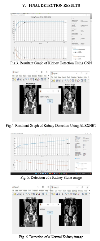
Figures 3 and 4 show those recognition towards nephrolith using Alexnet and CNN, respectively. The graph towards renal calculus sight shows that the action fall take away as the time period of 8 minutes 45 seconds with a accuracy of 69.39% using CNN whereas in ALEXNET technique, the process was completed within a time slot of 1min 17seconds with a validation accuracy of 96%.
The contrast between existing and anticipated work is seen in figure1.
TABLE 1 . Comparison Between The Proposed Work And Other Related Works
|
S.NO |
YEAR |
TITLE OF REFERENCES |
ALGORITHM |
IMPORTANCE |
DATA SET |
ACCURACY |
|
1. |
2012 |
Diagnosis of Kidney Stone Disease neural net for |
LVQ, RBF and feedforward planning Make use of perceptron algorism.n. |
Early detection of kidney stones and best model of diagnosis. |
Real set data gathered from different medical laboratories |
92% |
|
2. |
2019 |
Urinary Stone Detection in CT Images Utilize Cable news network |
Complexity Neuron webbing |
CT scan and kidney stone detection |
Random s1 ,s2 database |
95% |
|
3. |
2019 |
Chronic Kidney Disease (CKD) Prediction using CNN |
Mathematical concepts and |
XGBoost, Logistic Regression, Neural networks, Naive Bayes Classifier |
Not Specified
|
90% |
|
4. |
2020 |
urinalysis among sonography pictures by using Canny edge detection and CNN classification |
Central Neural Networks
|
Pre -processing , noise filtering and segmentation
|
collected images from Google
|
70-85% |
|
5. |
2020 |
Neural Net for kidney Stone Detection |
Cuckoo Search Algorithm |
generate ROI, using of sobel filtering and edge detection method |
Real set data collected from Hospitals inside that form of DICOM |
94.61% |
|
6. |
2021 |
Early urinalysis within Ultrasound scan Images Using Median Filter and Rank Filter |
Median filtering algorithm is used. |
median filter , rank filter |
Not Specified
|
82.2% |
|
7. |
2021 |
Improving the correctness of predicting disease
|
clustering Neural Network S |
Noise filter, SOM cluster |
Kaggle Database |
93% |

The figure 5 and 6 clearly gives some variance in the middle of a normal Kidney and the Kidney with stone. Likewise we can able to notice the stone existing within that nephro and for the further treatment.
Conclusion
This paper was examined and deployed to detect whether a kidney stone is present or not using pretrained ALEXNET and CNN. On a public dataset, the suggested technique was tested on both ALEXNET and CNN. CNN achieved a maximum accuracy of 69 percent, whereas ALEXNET achieved a maximum accuracy of 96 percent. With more time and more thorough research, the proposed system would be improved in the future.
References
[1] DC Elton , EB Turkbey , PJ Pickhardt “A Deep Learning system designed to automatically detect kidney stone s and perform volumetric segmentation using non contrast ct scans .detection and volumetric segmentation on non-contrast CT scans . February 2022. [2] S Shinde, U Kulkarni, D Mane, using machine learning for medical image analysis with deep learning: August, 2021 – Springer. [3] S Sudharson, P Kokil ,” deep neural networks for kidney ultrasound image classification” Computer Methods and Programs in Biomedicine, 2020. [4] M George, HB Anita ,”Analysis of Ultrasound Images Using Machine Learning Techniques: A Review” - Pervasive Computing and Social Networking, 2022. [5] LA Fitri, F Haryanto, H Arimura, C YunHao, K Ninomiya ,”Automated classification of urinary stones based on microcomputed tomography images using convolutional neural network” - Physical Medica, 2020. [6] H Alghamdi, G Amoudi, S Elhag, K Saeedi ,”Deep learning approaches for detecting COVID-19 from chest Scanned images: A survey” - IEEE Access, 2021. [7] Chen, Joy Iong-Zong. \"Design of Accurate Classification of COVID19 Disease in X-Ray Images Using Deep Learning Approach.\" Journal of ISMAC 3, no. 02 (2021): 132-148. [8] F Ma, T Sun, L Liu, H Jing , “Detection and diagnosis of chronic kidney disease using deep learning 2020. [9] S Sudharson, P Kokil “Computer-aided diagnosis system for the classification of multi-class kidney abnormalities in the noisy ultrasound images” -Programs in Biomedicine, 2021. [10] Shanthi K.G. Sivalakshmi P. Sesha Vidhya S. Sangeetha Lakshmi K., Smart drone with real time face recognition, Materials Today: Proceedings,2021.
Copyright
Copyright © 2024 Mr. Narasimha Raju Paka, Basamma Guggalashettara , Ankitha H K, Archana K. This is an open access article distributed under the Creative Commons Attribution License, which permits unrestricted use, distribution, and reproduction in any medium, provided the original work is properly cited.

Download Paper
Paper Id : IJRASET62433
Publish Date : 2024-05-21
ISSN : 2321-9653
Publisher Name : IJRASET
DOI Link : Click Here
 Submit Paper Online

