Ijraset Journal For Research in Applied Science and Engineering Technology
- Home / Ijraset
- On This Page
- Abstract
- Introduction
- Conclusion
- References
- Copyright
Liver Cancer Detection Using Image Processing
Authors: Dr. K Praveen Kumar , Komineni Prasad, Nukala Sunil, Adki Nikhil
DOI Link: https://doi.org/10.22214/ijraset.2024.65955
Certificate: View Certificate
Abstract
Medically termed Hepatic cancer, this comes from irregular proliferation in cells within the liver and comes in most variant known as Hepatocellular carcinoma- HCC which accounts for 75% diagnosed. Typically, it appears quite late thereby posing more risk. Precisely early diagnosis now becomes so imperative. Mainly, in the following proposal, the very key purpose in using image processing methodology shall revolve around making liver cancer identification possible quite earlier than previous. By making use of Computed Tomography images, the study seeks to identify malignant liver tumours using advanced image enhancement techniques. In particular, anisotropic diffusion filters are used for image filtering. The morphological operations will be applied in combination with a simple yet effective sequence of dilation and erosion operations. The proposed method shall accurately highlight and outline tumor areas in Computed Tomography scans, which may have a significant impact on new early cancer detection methods.
Introduction
I. INTRODUCTION
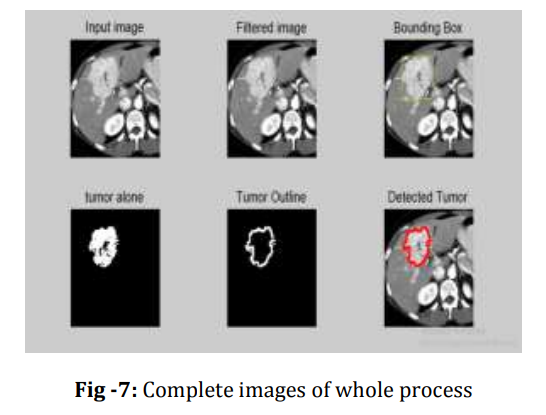
Cancer nomenclature has its origin in history dating back to 460-370 BC attributed to Hippocrates known as the "FATHER OF MEDICINE". Billions of cells of the human body divide each day to produce new cells to replace those that died. Biological organization at the most basic levels begins with cells, which unite to form tissues, and tissues unite to make organs. Sometimes, cell division is irregular, leading to uncontrolled proliferation of cells, which then form lumps or growths known as tumors. This research project presents a simple approach for cancer detection based on image processing techniques. Digital image processing is the use of computer algorithms to analyze and process visual information. The software tool used for this research is MATLAB. Computed tomography liver cancer images are used to highlight tumor regions. The liver cancer detection process encompasses three critical stages: image preprocessing, image processing, and tumor region highlighting. Image preprocessing involves enhancement using anisotropic diffusion filters to eliminate noise and image imperfections. Thresholding potentially introduces noise, making this stage crucial since minor deviations can significantly impact detection accuracy. Following image improvement, segmentation occurs to determine tumor areas during processing. Morphological operations, including dilation and erosion, facilitate image segmentation. These processes form basic techniques that have both simplicity and efficiency based on simple mathematical theory not including complex set theory mathematical calculations. In the last step, there is emphasis on a tumor in an image for enhanced visualization of that specific region. To have thorough understanding, the author uses sub-plotted images on each stage of the process: original image, the filtered image, and image that borders the tumor. And finally, the outlined or highlighted tumor region on the original input image
II. LITERATURE REVIEW
- Rong Zhu, et.al., in the paper "Improved Anisotropic Diffusion Filter in Image Processing" emphasized the dominance of anisotropic diffusion as a noise reduction mechanism. The paper explains the advanced algorithm designed to focus on the removal of salt and pepper noise in digital images.
- Ravi S, et.al., with "Morphological Operations for Image Processing: Understanding and Applications" underlined the availability of morphological operations based on set theory theories. Their research primarily focused on structural imperfection removal within image processing domains.
- Wassem Abdulrahman, et.al., in "Diagnosis of Liver Tumors Using Image Processing" concentrated on precise abdominal scanner image segmentation, introducing innovative methodologies for tumor region extraction in CT scanning.
- Amit Verma, et.al., with "A Survey on Digital Image Processing Techniques for Tumor Detection" have profoundly analyzed the existing methodologies for tumor detection along with comparative evaluations and presented better approaches for detection and classification.
- Jinshan Tang, et.al., in "An Adaptive Anisotropic Diffusion Filter for Noise Reduction in MR Images" has proposed an advanced filter based on adaptive threshold selection which, in performance, is remarkable while used on real magnetic resonance imaging datasets.
- Gabriel Ramos-Llordén, et.al., through "Anisotropic Diffusion Filter With Memory Based on Speckle Statistics for Ultrasound Images," presented a probabilistic memory-based approach toward over-filtering problems while using the selective philosophical concept of tissues.
- Alireza Mazloumi Gavgani, et.al, employed "Noise Reduction Using Anisotropic Diffusion Filter in Inverse Electrocardiology," in order to help noise reduction during measurements of the surface potential body, due to its potential of improving inverse electrocardiology problem resolution.
- Reitseng Lin, et.al., with "Morphological Operations on Images Represented by Quadtrees" presented algorithmic techniques that applied morphological operations directly on quadtree-represented images, producing dilated/eroded representations.
- Ruchika Chandel, et.al., in "Image Filtering Algorithms and Techniques" exhaustively discussed various techniques of image filtering, focusing on the smoothing of images as one of the most important and widely used image processing applications.
- N. Howard et al, in "A Novel Fully Automated Liver and Tumor Segmentation System," attempted to design an advanced Hepatocellular Carcinoma detection framework by using the images from Computed Tomography with high sensitivity, trying to mitigate the reduced specificity.
III. PROPOSED METHODOLOGY
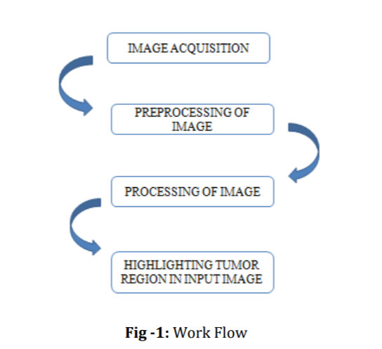
Liver cancer detection using image processing can be done with three main phases. They are,
- Preprocessing of image
- Processing of image
- Highlighting the tumor in given image
Computed tomography (CT) images are used for observation of liver tumors. MATLAB is the software used for processing the images given.
A. Work Flow of Process
B. Software Description
MATLAB is the most popular software used for Digital Image Processing. MATLAB (matrix laboratory) is multipurpose tool used for matrix manipulation, plotting of functions and data, implementation of algorithm and creating user interface. For detecting liver cancer using image processing, MATLAB software is used. It is a general usage programming language. When it is used to process images by generally writing function files, or script files to perform the necessary operations. It forms a formal record of the processing used and the final results can be tested and replicated by others. It provides many important advantages for forensic image processing.
C. Preprocessing of Image
Preprocessing of an image is the first and foremost step in image processing. The main aim of preprocessing in image processing is to improve the quality of image, suppressing unwanted distortions in image due to noise and enhance image features for further processing. Normally in performing medical image processing, preprocessing of an image plays a crucial role so that the input image does not have any impurities or imperfections, and it is done to be better for the upcoming process such as segmentation, feature extraction, etcFig3. Dot Matrix
D. Anisotropic Diffusion Filter
Diffusion filters contain two different filters called isotropic diffusion filter and anisotropic diffusion filter. Isotropic filters are linear and anisotropic filters are non-linear filters. Linear filters are homogeneous in nature and is with constant conductivity. Hence to overcome this smoothening Perona and Malik proposed the non liner method called anisotropic filters. Anisotropic filters are also called as perona-Malik equation. It is the powerful image enhancer.
The main aim of this filter is to reduce noise without removing significant parts of given image, sharp edges and significant lines. Image processing with anisotropic diffusion in MATLAB code contains some important parameters. The anisotropic diffusion is represented as,
Diff_image=anisodiff(Im, NUM_ITER, KAPPA, LAMBDA, DELTA, OPTION)
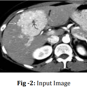
Here, Im is the input image, Num_itr is used to represent number of iterations, Kappa is the conduction coefficient, lambda is the maximum value of 0.25 for stability, delta is integration constant. There are two options first prefers smaller region over wide region whereas next prefers wide region over smaller one. In this project the input image is computed tomography image of liver as shown in the figure 2. This image is preprocessed for further processing.
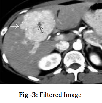
After applying anisotropic diffusion filter the given image is filtered and the noise and imperfections are removed for clear inference. It plays an important role in processing the image in next stage for clear observation of liver tumor region. The filtered image is shown in the figure 3
E. Processing
Processing of image is performing certain operations on the image to obtain certain information from the image. It is a type of signal processing where the input is an image and output is a feature extracted from the image. Digital image processing helps in manipulation of image using digital computers with help of certain algorithms. It avoids building of noise and distortion of image during processing.
F. Thresholding
It is one of the image processing method which converts the image from gray scale to binary images which is one of the segmentation method by setting up threshold value. It is most commonly used in binary images but can be applied for coloured images also. The pixel values greater than the threshold is converted into white (binary value 1) and the pixels lesser than the threshold is converted into black (binary value 0).The binary image should contain certain necessary information like position and shape of objects. The steps to be followed for thresholding are, Set the initial threshold value, specifically the 8-bit value of original image. Separating the image into two parts, Pixels less than threshold –background Pixels greater than threshold – foreground Identify the average mean value of two images. Calculate the new threshold by finding the average of two mean values.
G(x,y)=f(x)=1,if f(x,y)>T; G(x,y)=f(x)=0,if f(x,y)≤TVI.
G. Bounding Box
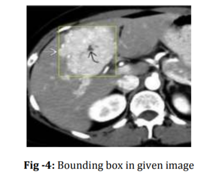
Bounding box are imaginary boxes that are created around an object. It is also one of the method of identifying the target on the image. In digital image processing bounding box is nothing but the rectangular border that covers or encloses the digital images. The coordinate in the upper left corner is x and the coordinate in the lower right corner is y.Here we extract the tumor region present in the liver by covering it with a rectangular box which is nothing but bounding the tumor region. A specified pixel range of the original liver computed tomography (CT) image will be selected in the form of box which represents the tumor region of the image. As shown in figure 4, the original image is being preprocessed for further image enhancement and filtered for reducing the noise signals present in the image and a rectangular box (bounding box) is made in the specified region. This region is the tumour region whose threshold value is greater than the normal threshold value.
H. Morphological Operations
Morphological operation is a non-linear operation which is related to shapes or morphological features of an image. This operation depends on the ordering of the pixels in the image and not their numerical value. The operation deals with structuring element producing output image of the same size.The morphological operation has two common processes which is, Dilation and Erosion
I. Dilation
The dilation adds pixels to the edges of the object in the image. The number of pixels added in an image depends on the dimensions of that image. The assess of the output image is maximum in case of dilation and in binary image the pixel is to made 1 with respect to the neighbouring pixels. The dilation expands the object and fills the holes.
J. Erosion
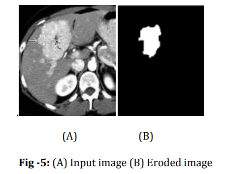
The erosion removes pixels to the edges of the object in the image. The number of pixels subtracted in an image depends on the dimensions of that image. The assess of the output image is minimum in case of erosion and in binary image the pixel is to made 0 with respect to the neighbouring pixels. The erosion contracts the object and removes small objects in the image. The eroded image is shown in the figure 5
K. Highlighting Tumor Region In Given Image
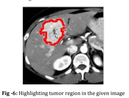
The last step is to present a clear indication of tumor region from the original image for our easy interpretation. By showing the tumor region alone is not sufficient for identification for normal humans. Hence this phase gives the obvious detection of tumor with its border in the given input image. When tumor is shown in any other image, it will not be that effective. One can find easy to observe in given input image and so this method is implemented. The final phase not only indicates the tumor normally it also border the tumor region in different color for spontaneous observation. Because the image is already given in the grey scale, so it is useless to show the tumor region in black or white. The other option is to represent it in red or green or blue. Here the tumor region will be indicated in red color as shown in the figure 6.
Finally for clear observation all the processes image in step by step is sub plotted and shown clearly. The figure 7 shows the complete processed images of whole process.
IV. RESULTS AND DISCUSSIONS
Liver cancer detection using image processing is implemented and the tumor region is found medical images used for analysis and the complete observation is shown in the table 1.
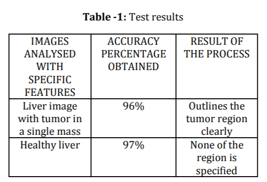
This method liver cancer detection using image processing is analyzed with different kinds of sample for complete analysis. When image of liver cancer with single mass is given as input it outlines the tumor region with red color after processing. When images of healthy liver is given the indication will be of no specific region indication. Some deviations and inaccuracy takes place when very small tumors around the single mass of tumor are present. According to the proposed algorithm, it identify only the single mass of tumor in the liver. Many suspecting regions are discussed with medical persons for getting better accuracy.
Conclusion
The new method for the detection of liver cancer based on image processing through morphological operations like dilation and erosion on abdominal computed tomography scans is introduced in the proposed research. The results of the experiment prove that the suggested methodology may be used by medical professionals for the diagnosis of hepatocellular carcinoma. Empirical evidence also proves that the morphological operations are superior, with fewer computational requirements and bypassing the need for complex mathematical modeling as in other image segmentation techniques. The existing method, though promising in nature, is recognized to have various limitations, and the most significant one is its exclusive design of the system, with single-tumor mass detection in view. Future scope is envisioned by aggregating comprehensive clinical and computed tomography image datasets that enhance accuracy of evaluations and provide robust mechanisms of validation for detecting multiple liver tumors. Additionally, the future research trajectory includes exploring magnetic resonance imaging modalities and implementing advanced classification algorithms to refine tumor categorization and diagnostic precision, thereby bridging current technological gaps in medical image analysis and potentially revolutionizing early-stage cancer detection strategies.
References
[1] Chen EL, Chung PC, Chen CL, Tsai HM, Chang CI. \"Anautomatic diagnostic system for CT liver image classification\" 1998; 45(6):78394.https://www.ncbi.nlm.nih.gov/pubme d/9609. [2] Reitseng Lin, E.K. Wong. \"Morphological operations on images represented by quadtrees\" 2002. https://ieeexplore.ieee.org/document/545858 [3] Yuki Wakida, Yoshito Mekada, Ichiro Ide \"Development of hepatocyte cancer detection method from dynamic Computed tomography images\" 2004. https://www.researchgate.net/publication/310050 161. [4] Jinshan Tang , Qingling Sun , Jun Liu , Yongyan Cao. \"An Adaptive Anisotropic Diffusion Filter for Noise Reduction in MR Images\" 2007. https://ieeexplore.ieee.org/abstract/document/43 03737. [5] Y. Masuda, A. H. Foruzan, T. Tateyama, Y. W. Chen, \"Automatic liver tumor detection using EM/MPM algorithm and shape information \", IEICE technical 2010. https://ieeexplore.ieee.org/document/5542834. [6] Häme Y, Pollari M. \"Semi -automatic liver tumor segmentation with hidden Markov measure field model and non-parametric distribution estimation. MedImage Anal\" 2011 https://www.ncbi.nlm.nih.gov/pubmed/21742543. [7] Alireza Mazloumi Gavgani, Yesim Serinagaoglu Dogrusoz. \"Noise reduction using anisotropic diffusion filter in inverse electrocardiology\" 2012. https://ieeexplore.ieee.org/document/6347341. [8] William J. Richbourg, Jianfei Liu, Jeremy M. Watt, VivekPamulapati,Shijun \"Tumor Burden Analysis on Computed Tomography byAutomated Liver and Tumor Segmentation,\"IEEETRANSACTIONS ON MEDICAL IMAGING, 2012https://www.ncbi.nlm.nih.gov/pmc/articles/P MC3924860/
Copyright
Copyright © 2024 Dr. K Praveen Kumar , Komineni Prasad, Nukala Sunil, Adki Nikhil. This is an open access article distributed under the Creative Commons Attribution License, which permits unrestricted use, distribution, and reproduction in any medium, provided the original work is properly cited.

Download Paper
Paper Id : IJRASET65955
Publish Date : 2024-12-16
ISSN : 2321-9653
Publisher Name : IJRASET
DOI Link : Click Here
 Submit Paper Online

