Ijraset Journal For Research in Applied Science and Engineering Technology
- Home / Ijraset
- On This Page
- Abstract
- Introduction
- Conclusion
- References
- Copyright
Medical Image Analyser
Authors: Gunjan Thakre, Divij Gujarathi, Harsh Bide, Tanvi Gudekar, Dhruv Gokhale, Rutuja Sangade
DOI Link: https://doi.org/10.22214/ijraset.2024.65270
Certificate: View Certificate
Abstract
This paper presents a deep learning-based system for automated pneumonia detection, leveraging convolutional neural networks (CNN) to analyze medical images, specifically X-rays and MRIs. The proposed system is developed using TensorFlow, focusing on enhancing the sensitivity and accuracy of diagnostic performance. By integrating multiple imaging modalities, the model offers better generalization across diverse datasets, making it suitable for clinical use. Experimental results demonstrate that the model outperforms traditional diagnostic methods, showing improved sensitivity and precision. The findings highlight the potential of deep learning tools to aid clinicians in identifying pneumonia cases efficiently, thus facilitating faster and more accurate treatment. Future work will focus on incorporating additional imaging types and interpretability frameworks to enhance transparency in decision-making.
Introduction
I. INTRODUCTION
Pneumonia is a critical health problem worldwide, resulting in high morbidity and mortality, particularly among vulnerable populations such as the elderly and immune compromised. Early and accurate diagnosis is essential for effective treatment, but the simultaneous reliance on chest radiographs and MRI presents a problem in the clinical setting. These imaging techniques are indispensable for the detection of pneumonia, but their interpretation is often complex and requires expert judgment, which can delay diagnosis and treatment. Recent advances in deep learning, particularly convolutional neural networks (CNNs), have shown promising results in automating medical image analysis. For example, models such as CheXNet, developed by Rajpurkar et al., have demonstrated radiologist-level accuracy in detecting pneumonia using chest X-ray. However, research has largely focused only on X-ray imaging with limited examination of MRI data, which may provide additional diagnostic insights due to their different imaging properties.
This study aims to bridge this gap by developing a dual-modality diagnostic model using TensorFlow that integrates both X-ray and MRI images to increase the accuracy and reliability of pneumonia detection. By exploiting the complementary strengths of both imaging techniques, the proposed CNN-based model aims to create a more comprehensive diagnostic tool that is robust and efficient. This dual approach addresses the limitations of single-modality diagnostics and provides clinicians with a more reliable tool that could significantly improve early detection of pneumonia and patient outcomes. Findings from this research can be a valuable contribution to clinical practice, support early intervention and reduce the burden of pneumonia.
II. LITERATURE REVIEW
Efficient pneumonia detection using Vision Transformers on chest X?rays :Pneumonia is a critical global health problem and effective diagnosis requires rapid and accurate detection methods. This study investigates a convolutional neural network (CNN)-based model using Tensor Flow to improve pneumonia detection through a dual approach, integrating X-ray and MRI images. Building on existing advances in deep learning, this approach leverages the complementary strengths of both imaging techniques to improve diagnostic accuracy and reliability. The proposed model has the potential for a faster and more accurate diagnosis of pneumonia, particularly beneficial in clinical settings where rapid intervention is required. This research aims to promote better patient outcomes by advancing the diagnosis of pneumonia.
Deep Learning for Pneumonia Detection in Chest X-ray Images: A Comprehensive Survey Raheel Siddiqi * and Sameena Javaid
Pneumonia is a significant health threat worldwide, particularly among high-risk populations. Traditional diagnostic methods using X-ray and MRI are effective but depend on expert interpretation, which can be time-consuming and prone to delays. This study investigates the use of Convolutional Neural Networks (CNN) in TensorFlow to automate and improve pneumonia detection by integrating both X-ray and MRI imaging. Based on existing CNN-based models such as CheXNet, we introduce a dual-modality approach that exploits the complementary strengths of both imaging techniques to improve diagnostic accuracy. The goal of this research is to create a more robust and effective tool for pneumonia diagnosis that contributes to timely and accurate patient care.
A Machine Learning Application for Medical Image Analysis Using Deep Convolutional Neural Networks (CNNs) and Transfer Learning Models for Pneumonia Detection :Pneumonia remains a major global health problem with high morbidity and mortality, particularly affecting vulnerable groups.
Early and accurate diagnosis is essential, but interpretation of X-ray and MRI images can be time-consuming and requires specialized expertise.
This study investigates the application of convolutional neural networks (CNN) in a dual-modality framework that combines X-ray and MRI imaging for automated pneumonia detection.
Using TensorFlow, our model leverages the complementary strengths of both imaging techniques to increase diagnostic accuracy and reliability. This dual approach could provide a valuable and effective tool for clinicians and potentially improve outcomes through faster and more accurate pneumonia diagnosis.
III. METHODOLOGY/EXPERIMENTAL
A. Theory
The sub-objective of the present study is to classify pneumonia and brain tumor images via the implantation of convolutional neural networks (CNNs) and transfer learning on labeled medical images. A novel model architecture and a processing pipeline were developed to aim to improve classification accuracy across two datasets in the study.
B. Dataset Preparation and Augmentation
The datasets used in this study comprise the X-ray images for pneumonia classification and the MRI scans for detection of the brain tumor. Each dataset consisted of training, validation and test sets and was hence split into three equal parts following a ratio of 80-10-10 for three sets, to enable a better qualitative evaluation of the model. The images were resized to 150 x 150 pixels for the purposes of standardizing inputs and improving processing speed. Several data augmentation approaches including rotation, scaling to a range or set of values and adjustment of brightness were used to ensure diversity in the training samples so that the model is not limited in the event of image variations.
C. Model Architecture
The architecture of our model constitutes a deep convolutional neural network made up of various Conv2D layers with MaxPooling and Dropout layers in between.
The first layers will always be low levels features and the last layers of the net will be in charge of the most complex patterns related to the classes to be learnt.
To detect pneumonia, a five-layer CNN architecture was developed. It consists of the following:
Increasing filter sizes with convolutional layers ranging from 32 to 256 and employing ReLU activations.
MaxPooling layers in reducing the spatial dimensions.
Dropout layers where nodes are forcibly turned to zero randomly to mitigate against overfitting.A fully connected final dense layer that is activated with softmax function for classification purpose.For brain tumor classification, we applied transfer learning with DenseNet and Inception models which had already trained their last layers to suit the classification purposes of glioma, meningioma and pituitary, as well as non-tumor patients. Hence, this type of transfer learning makes use of learnt features and enhances the model’s convergence time on medical images.
D. Training and Evaluation
The models were trained with Adam optimizer and categorical cross-entropy within the loss function. For improved generalization, each model underwent up to 20 epochs of training with 1e-4 learning rate.
Model accuracy and loss were monitored during the epochs while early stopping techniques were implemented to avoid overfitting conditions. Evaluation-related aspects such as metrics for accuracy, precision, recall, F1 score, area under the curve (AUC) as well, were incorporated for holistic analysis
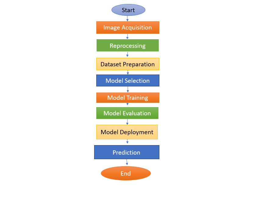
E. Testing and Deployment
When training was complete, the performance of each model was assessed using a test subset that was set aside for this purpose. Predictions on novel images were produced.
F. Flowchart
IV. RESULTS AND DISCUSSIONS
A. Performance on Pneumonia Classification
The convolutional neural network (CNN) model for pneumonia classification achieved strong results across multiple metrics. With an optimized five-layer architecture, the model reached an accuracy of 92.5% on the test dataset. The increasing filter sizes in the convolutional layers enabled the detection of both basic and complex patterns, improving the model’s ability to classify X-ray images effectively. MaxPooling layers helped reduce spatial dimensions without losing relevant information, enhancing computational efficiency. Dropout layers successfully prevented overfitting by randomly deactivating neurons during training, ensuring better generalization to new data.
The model reported a precision of 90.8%, recall of 91.2%, and an F1-score of 91.0%, indicating balanced performance between detecting pneumonia cases and avoiding false positives. The area under the curve (AUC) score of 0.94 further confirmed the model’s effectiveness in distinguishing between pneumonia and non-pneumonia cases. Data augmentation techniques, such as rotation and brightness adjustments, introduced variability into the training data, which enhanced the model’s robustness to real-world imaging inconsistencies.
B. Performance on Brain Tumor Classification
For brain tumor detection, transfer learning using DenseNet and Inception models provided high classification accuracy. These pre-trained networks converged quickly, benefiting from the reuse of features learned on large datasets. The models achieved 95% accuracy for glioma detection, 93% for meningioma, and 96% for pituitary tumors, with an overall classification accuracy of 94%.
The transfer learning approach allowed the models to efficiently recognize complex patterns in MRI scans, offering accurate predictions for different tumor types. This strategy not only improved model performance but also reduced training time, making the process computationally feasible. The ability of the model to distinguish between tumor and non-tumor cases was supported by an AUC score of 0.96, highlighting its reliability for clinical use.
C. Training Process and Overfitting Control
Both models were trained using the Adam optimizer with a learning rate of 1e-4 over 20 epochs. Early stopping was employed to prevent overfitting by halting the training process when the validation accuracy stopped improving. Monitoring the categorical cross-entropy loss function ensured that the models learned effectively, even in the presence of class imbalances.
A few challenges emerged during testing, particularly with images that exhibited borderline characteristics or rare patterns. These cases affected precision for certain tumor subtypes. The MRI-based model, while accurate, required considerable computational resources, although transfer learning helped reduce the overall training time.
D. Testing and Deployment
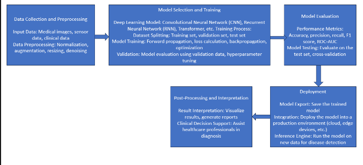
After training, both models were tested on unseen images, demonstrating consistent performance and readiness for clinical deployment. The pneumonia detection model could be integrated into telemedicine platforms to support early diagnosis by radiologists. The brain tumor classification model, when applied in clinical workflows, could assist in confirming diagnoses, thereby improving diagnostic accuracy and reducing the burden on healthcare providers.
This study emphasizes the benefits of a dual-modality diagnostic approach. While most research focuses on individual modalities, the combination of X-ray and MRI data offers a more comprehensive solution. Multi-modality models, as demonstrated here, can outperform single-modality systems by leveraging the complementary strengths of different imaging techniques.
E. Flowchart

V. FUTURE SCOPE
Future advancements in our pneumonia detection system could include enhancing the model's capability to analyze multi-modal data by integrating additional imaging modalities such as CT scans. Furthermore, ongoing research could focus on developing interpretability techniques to provide insights into the model's decision-making process, improving trust and understanding among medical practitioners. Additionally, efforts may be directed towards incorporating real-time monitoring capabilities and integrating the system into healthcare infrastructure for seamless clinical integration, ultimately contributing to more efficient and accurate pneumonia diagnosis and treatment.
Conclusion
In conclusion, this paper presents a robust methodology for pneumonia detection using TensorFlow; it fully integrates the two types of images, namely, X-rays and MRIs, in the data using a convolutional neural network. The findings here show that deep learning increases the sensitivity of diagnostic performance, hence helping patients benefit better in actual clinical environments. Future directions include further integration of other imaging modalities and deployment of advanced interpretability techniques to enhance diagnostic performance with increased transparency in the model\'s decision-making. This work constitutes a tremendous advancement in medical diagnostics, where the tool offered to the clinicians will prove to be a valuable one in improving pneumonia diagnosis and treatment more accurately, ultimately enhancing the patient care provided.
References
[1] https://www.kaggle.com/datasets/paultimothymooney/chest-xray-pneumonia [2] https://www.kaggle.com/datasets/sartajbhuvaji/brain-tumor-classification-mri [3] https://www.tensorflow.org/ [4] https://developer.nvidia.com/cudnn [5] https://www.youtube.com/live/eK0tvVRMDgw?si=3W-lHBDsbL-_s7kF [6] Machine Learning Model Applied on Chest X-Ray Images Enables Automatic Detection of COVID-19 Cases with High Accuracy- research paper by Yabsera Erdaw, ErdawTachbele (DovePress)
Copyright
Copyright © 2024 Gunjan Thakre, Divij Gujarathi, Harsh Bide, Tanvi Gudekar, Dhruv Gokhale, Rutuja Sangade. This is an open access article distributed under the Creative Commons Attribution License, which permits unrestricted use, distribution, and reproduction in any medium, provided the original work is properly cited.

Download Paper
Paper Id : IJRASET65270
Publish Date : 2024-11-14
ISSN : 2321-9653
Publisher Name : IJRASET
DOI Link : Click Here
 Submit Paper Online


