Ijraset Journal For Research in Applied Science and Engineering Technology
- Home / Ijraset
- On This Page
- Abstract
- Introduction
- Conclusion
- References
- Copyright
Deep Learning-Based Prediction of COVID-19 and Viral Pneumonia from Chest X-Ray Images
Authors: S Peruvazhuthi, K Praveen
DOI Link: https://doi.org/10.22214/ijraset.2024.63524
Certificate: View Certificate
Abstract
In recent times, the novel Coronavirus disease (COVID-19) has emerged as one of the most infectious diseases, causing significant public health crises across over 200 nations worldwide. Given the challenges associated with the time-consuming and error-prone nature of detecting COVID-19 through Reverse Transcription-Polymerase Chain Reaction (RT-PCR), there is a growing reliance on alternative methods, such as examining chest X-ray (CXR) images. Viral pneumonia symptoms include a persistent cough with mucus, fever, chills, shortness of breath, and chest pain, especially during deep breaths or coughing. These symptoms often overlap significantly with those of other respiratory infections, including COVID-19. Accurately predicting COVID-19 severity and distinguishing it from viral pneumonia is crucial for effective patient management. Deep learning models offer promise in automating this process. The chest X-ray (CXR) images undergo preprocessing through Contrast Limited Adaptive Histogram Equalization (CLAHE) to improve their quality. These enhanced images are fed into ResNet50 and EfficientNet-B0, both renowned deep learning models. Comparative evaluation demonstrates ResNet50 achieving an accuracy of 92.58%, whereas EfficientNet-B0 achieves a higher accuracy of 93.08%. This study underscores the efficacy of deep learning in COVID-19 prediction. The findings suggest EfficientNet-B0’s potential for improved diagnostic accuracy. This methodology presents a promising approach for automated, accurate COVID-19 severity prediction and differentiation from viral pneumonia, aiding timely medical interventions.
Introduction
I. INTRODUCTION
The global impact of the COVID-19 pandemic began with a significant outbreak in December 2019 in Wuhan, China, caused by the zoonotic coronavirus SARS-CoV-2. It quickly spread worldwide, resulting in approximately 70 crore confirmed cases and a significant number of deaths from Jan 2020 to May 2024, according to Worldometer data. COVID-19 is a contagious respiratory illness that affects the respiratory system, with initial symptoms such as fever, cough, and difficulty breathing typically appearing between two to fourteen days after exposure. The severity of the illness varies depending on the individual's immune response and the intensity of the disease. The virus can spread from infected individuals both before symptoms appear and for 10 to 20 days afterward. Its ability to cross international borders has had a profound impact, affecting populations across the globe.
Viral pneumonia is an infection of the lungs caused by various viruses, such as influenza, Respiratory Syncytial Virus (RSV), adenovirus, and others. It is less likely to respond to antibiotics and often requires different treatment approaches. This type of pneumonia is more common during the fall and winter months and can affect individuals of all ages, though it is particularly dangerous for young children, the elderly, and people with weakened immune systems. Symptoms includes cough, often producing mucus, fever and chills, shortness of breath. The gold standard for diagnosing COVID-19 is the Reverse Transcription Polymerase Chain Reaction (RT-PCR) test, which detects the viral ribonucleic acid (RNA) of SARS-CoV-2. A positive PCR test confirms a COVID-19 diagnosis. For the COVID-19 cases, Chest X-rays show bilateral, peripheral ground-glass opacities or consolidation. The viral pneumonias might show different patterns, such as localized opacities or diffuse infiltrates. Deep learning (DL) is a subset of machine learning known for its effectiveness across various applications [1]. DL models, which consist of multilayered neural network architectures, excel at extracting intricate features from images and have proven invaluable in tasks such as biomedical disease detection [2]. For instance, they are adept at identifying conditions like diabetes and pneumonia from CT scans, significantly enhancing computer-aided diagnosis systems [3]. Convolutional neural networks (CNNs) are particularly favoured for their robust performance in detecting and classifying COVID-19, capable of handling complex datasets and classifying patients according to disease risk. Transfer learning has emerged as a potent technique in COVID-19 detection [4-6], leveraging pretrained neural networks like DenseNet201, MobileNetV2, ResNet18, ResNet50, ResNet101, VGG16, and VGG19 on extensive datasets of X-ray images [7].
These models excel in learning comprehensive features from diverse image datasets, enabling effective generalization even when COVID-19 data is limited. This approach addresses challenges related to data scarcity, facilitating quicker and more accurate diagnoses.
II. LITERATURE SURVEY
Panwar et al. [8] introduced a CNN-based method for detecting COVID-19 using binary classification of chest X-ray images (CXR), achieving an overall accuracy of approximately 88.10%. In contrast, Hemdan et al. [9] explored various deep learning algorithms for COVID-19 image classification, highlighting DenseNet's highest accuracy at 90%. Further investigations into deep learning models for COVID-19 detection [10] demonstrated varied performances among DenseNet121, VGG16, Xception, EfficientNet-B7, and NASNet, with EfficientNet-B7 leading with an accuracy of 92.48%. Another study [11] focused on early detection of COVID-19 using chest X-rays (CXR), employing VGG16 and ResNet50 for feature extraction. These models classified images into three groups—viral pneumonia, normal, and COVID-19—achieving average accuracies of 89.34% for VGG16 and 91.39% for ResNet50 in detecting COVID-19 cases from a dataset comprising 15,153 images.
In [12], researchers employed transfer learning with well-known architectures including CheX-Net, DenseNet, VGG19, MobileNet, InceptionV3, ResNet18, ResNet101, and SqueezeNet to classify chest X-ray (CXR) images into three categories: COVID-19, viral pneumonia, and normal. Their dataset consisted of 423 COVID-19 images, 1,485 viral pneumonia images, and 1,575 normal images, achieving an impressive accuracy of 91.94% in distinguishing between these classes.In [13], the authors conducted a comprehensive evaluation of classic and specialized models using a COVID-19 dataset. Classic models such as VGG19, ResNet50, MobileNetV2, InceptionV3, Xception, and DenseNet121 were compared alongside specialized models like DarkCOVIDNet and COVID-Net. Their findings highlighted ResNet50's superior performance, achieving notable accuracies of 93.20% in binary classification (COVID-19 and No COVID-19) and 86.13% in multi-class classification (COVID-19, No COVID-19, and Pneumonia) with a simple modification. These results underscore ResNet50's effectiveness in COVID-19 detection and differentiation from pneumonia, emphasizing its potential as a valuable tool in medical diagnostics.
In their study detailed in [13], researchers extensively evaluated both traditional and specialized models using a COVID-19 dataset. They compared classic models such as VGG19, ResNet50, MobileNetV2, InceptionV3, Xception, and DenseNet121 alongside specialized models like DarkCOVIDNet and COVID-Net. Their findings demonstrated that ResNet50 consistently outperformed other models, showing superior performance. With a simple adjustment, ResNet50 achieved impressive accuracies of 93.20% in binary classification (COVID-19 vs. No COVID-19) and 86.13% in multi-class classification (COVID-19, No COVID-19, and Pneumonia). These results underscore the effectiveness of ResNet50 in detecting COVID-19 and distinguishing it from pneumonia, highlighting its potential as a valuable tool in medical diagnostics. The studies cited illustrate the promising application of deep learning techniques, particularly transfer learning, in enhancing the detection of COVID-19 from chest X-ray images. These methods often involve refining pre-trained models to enhance detection accuracy. Overall, these findings emphasize the importance of leveraging advanced deep learning approaches to address crucial healthcare challenges, especially in the context of COVID-19 detection using medical imaging.
III. METHODOLOGY
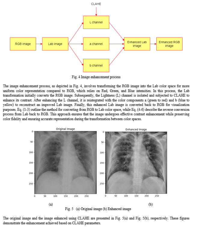
Initially, the chest X-ray (CXR) images undergo preprocessing using Contrast Limited Adaptive Histogram Equalization (CLAHE), a technique employed to enhance image contrast and detail. Subsequently, the preprocessed images are resized to a standardized dimension and normalized, ensuring consistent pixel values across all images for optimal model performance.
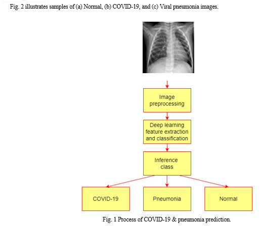
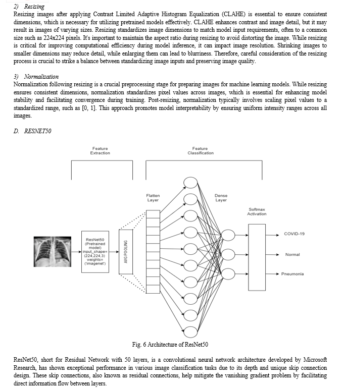
The enhanced images are then subjected to feature extraction and classification using pre-trained architectures, including ResNet50 and EfficientNetB0. These architectures leverage their learned representations from extensive datasets to extract relevant features and classify the images into respective categories, such as COVID-19, pneumonia, and normal cases. Fig. 1 depicts the process of COVID-19 & pneumonia prediction.
A. Dataset
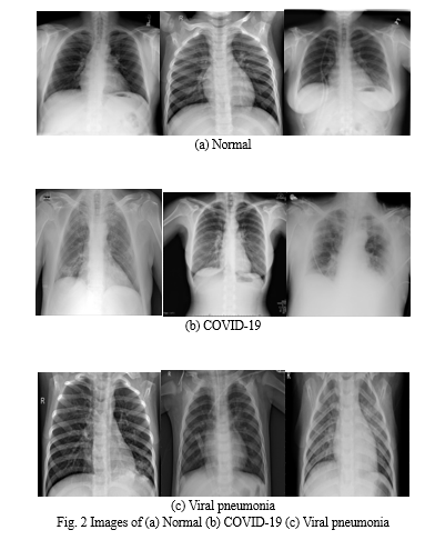
The COVID-19 Radiography Database [14,15], utilized as the main dataset, includes chest X-ray images from patients diagnosed with COVID-19, pneumonia, and those with normal lung conditions. Created in 2020 through a collaboration between Qatar University in Doha, Qatar, and the University of Dhaka in Bangladesh, alongside medical practitioners from Pakistan and Malaysia, the database was released gradually and comprises a total of 15,153 images. Within this dataset, there are 3,616 images depicting COVID-19 cases, 1,345 images showing viral pneumonia, and 10,192 images representing normal lung conditions.
B. Image Downsampling
Downsampling is a common technique used in machine learning and data analysis to mitigate class imbalance. It involves reducing the number of samples in the majority class to align with the number of samples in the minority class. This approach helps prevent bias in predictive modeling towards the majority class, thereby enhancing the accuracy of machine learning algorithms, particularly in tasks like classification where imbalanced datasets can lead to inaccurate predictions.
In the COVID-19 Radiography Database, down sampling was implemented to address an initial imbalance in the number of images across the three classes: COVID-19, viral pneumonia, and normal lung conditions. Initially, the dataset had varying numbers of images per class. By downsampling, the number of images for COVID-19 and normal lung conditions was decreased to match the number of images for viral pneumonia, which was set at 1,345 images. This adjustment ensured a more balanced representation of classes in the dataset, thereby improving the robustness and fairness of subsequent machine learning analyses and models trained on this data.
C. Image Preprocessing
Image preprocessing is crucial for improving the quality and usability of image data in deep learning algorithms. It involves applying specific techniques to raw image inputs to make them more appropriate for training deep neural networks. Three fundamental preprocessing techniques widely used in deep learning applications are CLAHE (Contrast Limited Adaptive Histogram Equalization), resizing, and normalization.
These methods prepare image data by improving contrast, standardizing dimensions, and scaling pixel values, respectively. By implementing these preprocessing steps, images are optimized for better performance and accuracy in tasks such as image classification and object detection.
1) CLAHE (Contrast Limited Adaptive Histogram Equalization)
CLAHE (Contrast Limited Adaptive Histogram Equalization) is extensively used in image processing to enhance image quality by focusing on local contrast improvement. This method effectively enhances image clarity and detail sharpness while minimizing noise amplification.
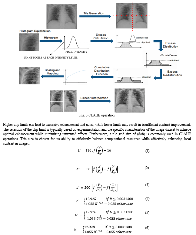
The CLAHE algorithm comprises three main stages: tile generation, histogram equalization, and bilinear interpolation. Initially, the input image is divided into non-overlapping sections known as tiles. Subsequently, histogram equalization is applied to each tile using a specified clip limit.
Histogram equalization involves several steps including histogram computation, excess calculation, excess distribution, excess redistribution, and scaling and mapping using a cumulative distribution function (CDF). Fig. 3 represents the operation of CLAHE. Initially, the histogram of each tile in the input image is computed and divided into bins. Values exceeding a specified clip limit are redistributed across other bins. Subsequently, a Cumulative Distribution Function (CDF) is computed based on these histogram values.
The CDF values for each tile are then scaled and mapped to adjust the pixel values of the input image. The resulting tiles are combined using bilinear interpolation to generate an output image with improved contrast.
The extent of contrast enhancement in CLAHE is controlled by the clip limit, which determines how the histogram is clipped to mitigate noise and enhance contrast. A clip limit of 2.0 is often chosen to strike a balance between effective contrast enhancement and the risk of introducing artifacts.
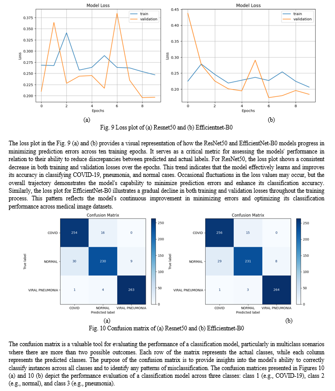
In both matrices, each row and column correspond to specific class labels, where diagonal elements indicate correct classifications and off-diagonal elements represent misclassifications. In Figure 10 (a), the model shows strong performance with 254 instances of class 1 correctly classified, 230 instances of class 2 correctly classified, and 263 instances of class 3 correctly classified. However, there are instances of misclassification, such as 16 instances of class 1 misclassified as class 2 and 30 instances of class 2 misclassified as class 1. Similarly, in Figure 10 (b), the model correctly classifies 256 instances of class 1, 231 instances of class 2, and 264 instances of class 3, with 15 instances of class 1 misclassified as class 2 and 29 instances of class 2 misclassified as class 1. These matrices provide a detailed assessment of the model's strengths in correctly classifying certain classes and areas where improvements are needed to reduce misclassifications.
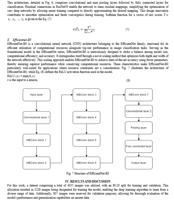
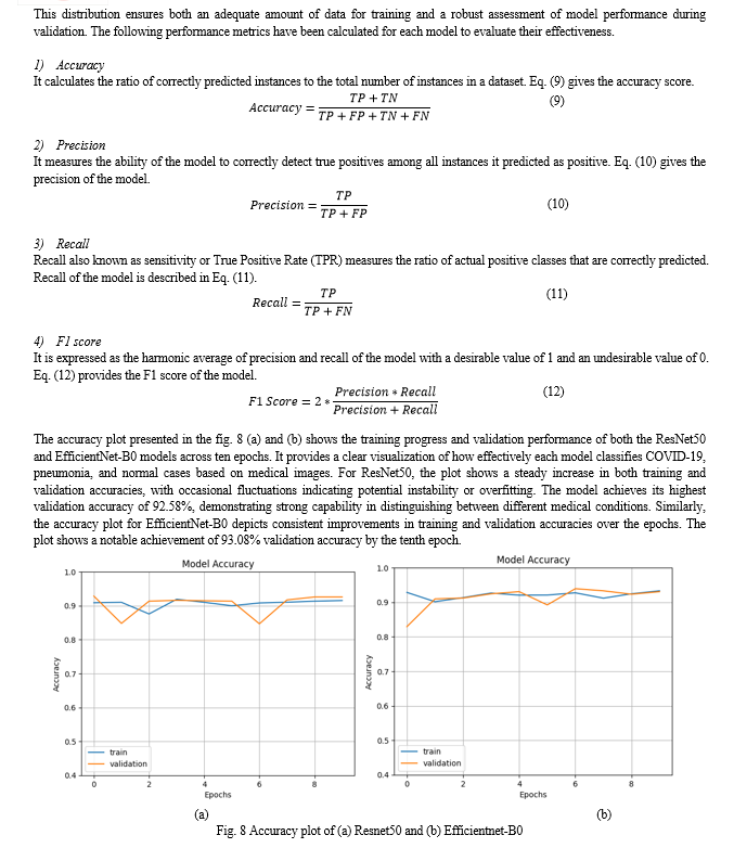
The performance comparison between the ResNet50 and EfficientNet-B0 models reveals differences in their classification capabilities. The ResNet50 model achieved accuracy, precision, recall, and F1 scores of approximately 0.925, 0.926, 0.926, and 0.925, respectively. In contrast, the EfficientNet-B0 model demonstrated slightly superior performance, with accuracy, precision, recall, and F1 scores of 0.930, 0.9307, 0.9305, and 0.9297, respectively. This suggests that EfficientNet-B0 might be more adept at capturing and generalizing features from the data, leading to more accurate predictions.
Conclusion
This work aimed to enhance medical image quality using Contrast Limited Histogram Equalization (CLAHE) and then applied two prominent deep learning architectures, ResNet50 and EfficientNet-B0, for predicting COVID-19 and pneumonia. Experimental results showed ResNet50 achieving an accuracy of 92.58%, while EfficientNet-B0 surpassed it with 93.08%. These findings underscore the capability of deep learning models in accurately categorizing COVID-19 cases from medical images. However, the performance of these models can be influenced by factors like dataset size, image quality, and variations in disease presentation. Future research could explore new image enhancement techniques or incorporate additional preprocessing steps to further enhance model performance.
References
[1] M.A. Talukder, M.M. Islam, M.A. Uddin, A. Akhter, M.A.J. Pramanik, S. Aryal, M.A.A. Almoyad, K.F. Hasan, and M.A. Moni, \"An efficient deep learning model to categorize brain tumor using reconstruction and fine-tuning,\" Expert Syst. Appl., vol. 120534, 2023. [2] M.A. Talukder, M.M. Islam, M.A. Uddin, A. Akhter, K.F. Hasan, and M.A. Moni, \"Machine learning-based lung and colon cancer detection using deep feature extraction and ensemble learning,\" Expert Syst. Appl., vol. 205, pp. 117695, 2022. [3] M. Zysman, J. Asselineau, O. Saut, E. Frison, M. Oranger, A. Maurac, J. Charriot, R. Achkir, S. Regueme, E. Klein, et al., \"Development and external validation of a prediction model for the transition from mild to moderate or severe form of COVID-19,\" Eur. Radiol., pp. 1-13, 2023. [4] N.D. Kathamuthu, S. Subramaniam, Q.H. Le, S. Muthusamy, H. Panchal, S.C.M. Sundararajan, A.J. Alrubaie, M.M.A. Zahra, \"A deep transfer learning-based convolution neural network model for COVID-19 detection using computed tomography scan images for medical applications,\" Adv. Eng. Softw., vol. 175, pp. 103317, 2023. [5] S. Agrawal, V. Honnakasturi, M. Nara, N. Patil, \"Utilizing deep learning models and transfer learning for COVID-19 detection from X-Ray images,\" SN Comput. Sci., vol. 4, no. 4, pp. 326, 2023. [6] M.M. Rana, M.M. Islam, M.A. Talukder, M.A. Uddin, S. Aryal, N. Alotaibi, S.A. Alyami, K.F. Hasan, and M.A. Moni, \"A robust and clinically applicable deep learning model for early detection of Alzheimer’s,\" IET Image Process., pp. 1–17, 2023. [7] A. Subasi, T. Keles, F. Ozyurt, S. Dogan, T. Tuncer, \"Feature extraction and fusion using deep convolutional neural networks for COVID-19 detection using CT and X-ray images,\" World J. Adv. Res. Rev., vol. 19, no. 1, pp. 914–933, 2023. [8] H. Panwar, P. Gupta, M.K. Siddiqui, R. Morales-Menendez, V. Singh, \"Application of deep learning for fast detection of COVID-19 in X-Rays using ncovnet,\" Chaos Solitons Fractals, vol. 138, pp. 109944, 2020. [9] E.E.-D. Hemdan, M.A. Shouman, M.E. Karar, \"Covidx-net: A framework of deep learning classifiers to diagnose COVID-19 in X-ray images,\" 2020, arXiv preprint arXiv:2003.11055. [10] B. Nigam, A. Nigam, R. Jain, S. Dodia, N. Arora, B. Annappa, \"COVID-19: Automatic detection from X-ray images by utilizing deep learning methods,\" Expert Syst. Appl., vol. 176, pp. 114883, 2021. [11] N. S. Kavya, T. Shilpa, N. Veeranjaneyulu, and D. D. Priya, \"Detecting Covid19 and pneumonia from chest X-ray images using deep convolutional neural networks,\" Materials Today: Proceedings, vol. 64, pt. 1, pp. 737-743, 2022. doi: 10.1016/j.matpr.2022.05.199. [12] Q. Li, X. Guan, P. Wu, et al., \"Early transmission dynamics in Wuhan, China, of novel coronavirus-infected pneumonia,\" N. Engl. J. Med., vol. 382, no. 13, pp. 1199–1207, 2020. [13] S. Agrawal, V. Honnakasturi, M. Nara, N. Patil, \"Utilizing deep learning models and transfer learning for COVID-19 detection from X-ray images,\" SN Comput. Sci., vol. 4, no. 4, pp. 326, 2023. [14] M. E. H. Chowdhury, T. Rahman, A. Khandakar, R. Mazhar, M. A. Kadir, Z. B. Mahbub, K. R. Islam, M. S. Khan, A. Iqbal, N. Al-Emadi, M. B. I. Reaz, and M. T. Islam, \"Can AI help in screening Viral and COVID-19 pneumonia?\" IEEE Access, vol. 8, pp. 132665-132676, 2020. [15] T. Rahman, A. Khandakar, Y. Qiblawey, A. Tahir, S. Kiranyaz, S. B. A. Kashem, M. T. Islam, S. A. Maadeed, S. M. Zughaier, M. S. Khan, and M. E. Chowdhury, \"Exploring the Effect of Image Enhancement Techniques on COVID-19 Detection using Chest X-ray Images,\" 2020.
Copyright
Copyright © 2024 S Peruvazhuthi, K Praveen. This is an open access article distributed under the Creative Commons Attribution License, which permits unrestricted use, distribution, and reproduction in any medium, provided the original work is properly cited.

Download Paper
Paper Id : IJRASET63524
Publish Date : 2024-07-01
ISSN : 2321-9653
Publisher Name : IJRASET
DOI Link : Click Here
 Submit Paper Online

