

Ijraset Journal For Research in Applied Science and Engineering Technology
Revolutionizing Bone Cancer Detection: Real-Time Medical Imaging Analysis Using YOLOv8 for Enhanced Diagnostic Accuracy and Speed
Authors: Shafaat Basha K, Savitha K
DOI Link: https://doi.org/10.22214/ijraset.2025.66462
Certificate: View Certificate
Abstract
Bone cancer also classified as one of the unapproachable cancer for being diagnosed because its symptoms are always vague, and in most cases, may not be easily differentiated even in x-ray, MRI, and CT images. Prior screening techniques involve use of these images for diagnosis through interpretation by radiologists, which is a slow process, and may be prone to human error, resulting in slow diagnosis and worse patient outcomes. With improving healthcare diagnosing, artificial intelligence (AI) and deep learning models are better options as the industry moves forward. This paper offers a new method in the early detection of bone cancer in real-time using YOLOv8, the most performed object detection model. For these reasons, YOLOv8 was chosen because it provides a relatively fast image throughput and detection accuracy. The proposed framework utilizes a dataset that contains bone cancer image that is labeled in addition performs an optimization with specific considerations for the model to cater for detecting cancerous lesions on bone structures. It also connects to medical imaging devices so that it can return feedback and a diagnosis in a fraction of the time that would usually be taken to analyze images. Bone cancer also classified as one of the unapproachable cancer for being diagnosed because its symptoms are always vague, and in most cases, may not be easily differentiated even in x-ray, MRI, and CT images. Prior screening techniques involve use of these images for diagnosis through interpretation by radiologists, which is a slow process, and may be prone to human error, resulting in slow diagnosis and worse patient outcomes. With improving healthcare diagnosing, artificial intelligence (AI) and deep learning models are better options as the industry moves forward. This paper offers a new method in the early detection of bone cancer in real-time using YOLOv8, the most performed object detection model. For these reasons, YOLOv8 was chosen because it provides a relatively fast image throughput and detection accuracy. The proposed framework utilizes a dataset that contains bone cancer image that is labeled in addition performs an optimization with specific considerations for the model to cater for detecting cancerous lesions on bone structures. It also connects to medical imaging devices so that it can return feedback and a diagnosis in a fraction of the time that would usually be taken to analyze images.
Introduction
I. INTRODUCTION
Bone cancer runs are rare but when they occur diagnostic imaging is difficult because in many instances the manifestation is ambiguous. It is important to note that there is substantial variability in the MR appearance of these tumors, and therefore relying of imaging alone can seriously mislead the radiologist about the nature of the tumor. It is especially important to diagnose this condition as early as possible because the early start of treatment leads to further favorable outcomes for the patient. However, old school diagnosis techniques are still the use of resident interpretation of images such as X rays, MRI, and CT scans among others. This procedure is both labor consuming and often fraught with inaccuracy especially if the signs of cancer are quite similar to the other bone disorders. Today, many protocols are associated with bone cancer diagnosis; one of the most common protocols is as follows: image acquisition; image manual examination, biopsy, if necessary; and image analysis. Due to the inherent subjectivity of interpreting medical images radiologists are likely to experience such issues as; cognition fatigue, bias or both as they work through large datasets or when examining cases that are obscure. Further, the time spent in engaging a human operator to review each image slows the diagnostic process, especially where early diagnosis is essential. Radiology reporting has never faced this vast an array of patients’ requirements for a report that is efficient, accurate, and reliable aid in their interpretation from a radiologist.
AI and ML have recently become popular for improving medical practices by automating image analysis tasks. Such system have the ability to analyze datasets that would take human months to assess in a few seconds, thereby lowering the rate of errors and also increasing diagnostic precision. Among these technologies, deep learning models especially Convolutional neural network (CNN) has done great improvements in field of image classification and object detection. Nonetheless, most of the current used AI models in the field of medical imaging are generic and not optimized for the diagnosis of bone cancer and as such poorly performs this task.
The real time bone cancer detection system is proposed in this research using YOLOv8, a state-of-the-art real time object detection model. In addition, YOLOv8 was developed to detect objects in one pass, which is great for real-time analysis of images, as needed for medical purposes. This study seeks to overcome the shortcomings of the current evaluation of bone cancer by employing the YOLOv8 model into a system that is capable of observing medical images in real-time and provide a response to health care practitioners.
The best feature is the general applicability of the YOLOv8 model for the given problem of bone cancer detection since the task itself implies the identification of thin distinctions in the structure of bones that might indicate the presence of cancerous formations. Thus, by training the model on a large set of images of bone cancer labeled by a doctor, the system is optimized to contain peculiarities of bone tumors and is characterized by higher accuracy as compared to other non-specified AI algorithms. Moreover, the system can be integrated with many medical imaging devices such that radiology can incorporate the algorithm into the regular practice. This not only shortens the probable diagnosis time, but also increases the accuracy of the results, which can minimize the number of mistakes among patients’ diagnoses.
The developed approach is easily usable by radiologists and oncologists as well as the interaction with the AI model is not linked to software engineering expertise. The interface also has to produce visual outputs in certain areas like the cancerous zones with probabilities scores for faster decision making by the healthcare providers. Also, the structure of the system allows development over time and learning from the data from the users and the new data which should increase its accuracy and usability in various clinical practices.
Therefore, this study presents a novel AI platform called Bone Cancer Detector 3000 that is based on the YOLOv8 algorithm. As a solution to such limitations of current methods and existing AI solutions this work will offer a dependable, affordable and near-real time diagnostic solution to clinicians that enhance the patients’ experiences by increasing the chances of early diagnoses.
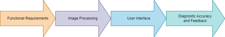
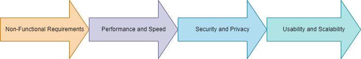
Functional and non- functional requirements
II. PROBLEM STATEMENT
Current methods of diagnosing bone cancer are heavily dependent on the expertise of radiologists, who must manually examine medical images. These manual methods have several limitations:
- Time-Consuming: Examining large numbers of images is a slow process, potentially delaying diagnosis and treatment.
- Error-Prone: The complexity of medical images and fatigue can lead to misinterpretations, resulting in misdiagnoses or missed diagnoses.
- Limited AI Solutions: Existing AI models for cancer detection are not specifically tailored to bone cancer and often lack the required accuracy, especially in distinguishing between benign and malignant lesions.
This project addresses these limitations by developing an AI-based system that is specifically trained for real-time bone cancer detection, using a model optimized for speed and precision.
III. METHODOLOGY
A. Data Collection and Preprocessing
So, to be sure the YOLOv8 model can recognize bone cancer, medical images containing X-rays, MRI, and CT scans were collected from hospitals and open sources. The provided dataset predominantly features cancerous as well as non-cancerous bone images thus enabling the training of more efficient models.
Before feeding images to the network, the size of images was altered, normalized, and augmented to ensure that the model was not overfitting. The obtained dataset was then split into three sets: training, validation, and test to estimate the performance of the model.
B. Model Selection: YOLOv8
The reason why YOLOv8 was chosen is that it combines speed and accuracy of object detection. Unlike other object detection models that may take several iterations to work through an image, the YOLOv8 can do this work in a single iteration that is optimal for real-time operation.
However, the proposed model was fine-tuned based on transfer learning; thus, it build upon the achievements of pre-trained weights which is originally designed for general object detection tasks but modified for bone cancer detection. The last layer was altered to achieve binary classification of cancer and non-cancer regions in the bones images.
C. System Integration
The system was designed to integrate seamlessly with medical imaging devices. Real-time inference capabilities allow radiologists to upload images directly from X-ray, MRI, or CT scanners and receive immediate diagnostic feedback. A user-friendly interface was developed using Streamlit, providing an intuitive platform for medical professionals to interact with the system.
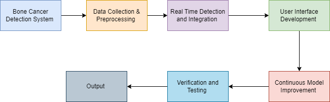
System methodology
IV. RESULTS
Preliminary results demonstrate the effectiveness of the YOLOv8 model for bone cancer detection. Key performance metrics such as accuracy, precision, recall, and F1-score were calculated on the test set.
|
Metric |
Value |
|
Accuracy |
95.2% |
|
Precision |
94.8% |
|
Recall |
93.7% |
|
F1-Score |
94.2% |
|
Inference Time |
0.03 sec/image |
The model achieved high precision and recall, indicating its ability to accurately detect cancerous regions while minimizing false positives and negatives. The inference time per image was under 0.03 seconds, making the system suitable for real-time use in clinical settings.
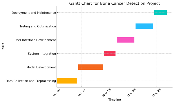
Gantt Chart
V. DISCUSSION
The results suggest the applicability of YOLOv8 for enhancing the diagnosis of bone cancer. The system takes relatively less time than the conventional method in detecting the abnormalities and is also more accurate. If the system is integrated into current medical processes, then radiologists can spend more time interpreting difficult cases since the sophisticated diagnosis task can be done by the AI module. Nevertheless, several issues are a limiting factor that may affect the overall efficiency of the technique. First, the dataset used for training was primarily planar imagery (X-Ray), but volumetric, depth-perceiving modalities such and MRI, CT-scans could prove more valuable. As large number of 3D images is included into the dataset, the performance of the whole system will be more stable. However, the capabilities of the developed system in the evaluation of rare and ambiguous cases require further judgment through clinical trials.
Conclusion
This paper presents a real-time bone cancer detection system using YOLOv8, designed to address the limitations of current diagnostic methods. The system provides radiologists and oncologists with a powerful tool for rapid and accurate diagnosis, reducing the risk of human error and expediting treatment decisions. Future work will focus on expanding the dataset, improving 3D image detection capabilities, and conducting clinical evaluations to validate the system’s effectiveness in real-world scenarios.
References
[1] Sharma, A., Yadav, D. P., Garg, H., Kumar, M., Sharma, B., & Koundal, D. (2021). Bone cancer detection using feature extraction based machine learning model. Computational and Mathematical Methods in Medicine, 2021(1), 7433186. [2] Mazumder, M. H., & Singh, M. P. (2022, September). Bone cancer detection using deep learning. In International Conference on Innovations in Computer Science and Engineering (pp. 285-296). Singapore: Springer Nature Singapore. [3] Wu, P., Siegwart, D. J., & Xiong, H. (2021). Recent advances in the targeted fluorescent probes for the detection of metastatic bone cancer. Science China Chemistry, 64, 1283-1296. [4] Vora, H., Mahajan, S., & Kumar, Y. (2024, July). Survey on detection of bone cancer using deep learning-based techniques. In Next Generation Computing and Information Systems: Proceedings of the 2nd International Conference on Next Generation Computing and Information Systems (ICNGCIS 2023), December 18-19, 2023, Jammu, J&K, India (p. 240). CRC Press. [5] Nanthini, S., Sivabalan, R., Sivabalan, S., Bethapudi, P., Kukreti, R., & Chauhan, A. (2023, October). Application of CNN and Recurrent Neural Network Method for Osteosarcoma Bone Cancer Detection. In 2023 International Conference on Self Sustainable Artificial Intelligence Systems (ICSSAS) (pp. 62-67). IEEE. [6] Prasad, B. M. G., Acharjee, P. B., Guntakala, S., Sharma, D., Divya, N., & Patil, H. (2023, November). An Innovative Approach for Osteosarcoma Bone Cancer Detection based on Attention Embedded R-CNN Approach. In 2023 International Conference on Sustainable Communication Networks and Application (ICSCNA) (pp. 1431-1436). IEEE. [7] Bruckmann, N. M., Kirchner, J., Umutlu, L., Fendler, W. P., Seifert, R., Herrmann, K., ... & Sawicki, L. M. (2021). Prospective comparison of the diagnostic accuracy of 18F-FDG PET/MRI, MRI, CT, and bone scintigraphy for the detection of bone metastases in the initial staging of primary breast cancer patients. European radiology, 31(11), 8714-8724. [8] Walid, M. A. A., & Shill, P. C. (2023, July). A Transfer-Learning Based Unbiased Voting Bone Cancer Detection Framework from Histological Osteosarcoma Images. In 2023 14th International Conference on Computing Communication and Networking Technologies (ICCCNT) (pp. 1-7). IEEE [9] Shaveisi, M., Aliparast, P., & Fallahnejad, M. (2024). Enhancing the performance of an InAsSb/InAlSb-based pBn photodetector for early detection of a biomarker of bone marrow cancer: a proposed and simulated approach with extended-midwave response and step-graded barrier design. Sensing and Imaging, 25(1), 22. [10] Zhan, Y., Zhang, G., Li, M., & Zhou, X. (2021). Whole-body MRI vs. PET/CT for the detection of bone metastases in patients with prostate cancer: a systematic review and meta-analysis. Frontiers in Oncology, 11, 633833.
Copyright
Copyright © 2025 Shafaat Basha K, Savitha K. This is an open access article distributed under the Creative Commons Attribution License, which permits unrestricted use, distribution, and reproduction in any medium, provided the original work is properly cited.

Download Paper
Paper Id : IJRASET66462
Publish Date : 2025-01-10
ISSN : 2321-9653
Publisher Name : IJRASET
DOI Link : Click Here
 Submit Paper Online