Ijraset Journal For Research in Applied Science and Engineering Technology
- Home / Ijraset
- On This Page
- Abstract
- Introduction
- Conclusion
- References
- Copyright
Skin Disease Detection Model
Authors: Ms. R Soundharya, Akanksha Shettigar, Ananya Prasad, Ashwith R Poojary, Deepa Naik
DOI Link: https://doi.org/10.22214/ijraset.2024.62566
Certificate: View Certificate
Abstract
The human skin is a remarkable organ susceptible to a myriad of know and unknown diseases. Many of these ailment are widespread, with some ranking among common worldwide. The complexity of diagnosing these diseases is compounded by challenges such as variations in skin texture, the presence of hair, and diverse skin colors. In some areas have limited access to medical facilities, individuals often neglect early symptoms, leading to exacerbated conditions over time. Furthermore, traditional diagnostic methods for skin diseases are time consuming. To address these challenges, there is a critical need to develop advanced diagnostic methods utilizing machine learning techniques to enhance accuracy cross various skin diseases. Machine learning algorithms have proven valuable in medical applications, leveraging image feature values to facilitate decision making. The diagnostic process involves three key stages: feature extraction, training, and testing. By employing machine learning technology, these algorithms learn from a diverse set of skin images o enhance their diagnostic capabilities. The primary goal is to significantly improved the accuracy of skin disease detection. This study focuses on utilizing color and texture features for the classification of skin diseases. The distinctive color of healthy skin differs from that affected by disease, while texture features effectively discern smoothness, coarseness, and regularity in images. Key features such as texture, color, and shape phyla pivotal role in image classification. The incorporation of convolution neural networks (CNN) further augments the capabilities of image classification in the realm of skin disease diagnosis.
Introduction
I. INTRODUCTION
In light of the increasing role of Artificial Intelligence in diverse applications, including healthcare, skin disorders, attributed to factors such as fungal infections, bacteria, allergies, and viruses, are prevalent. The majority of these conditions stem from unprotected exposure to UV rays, potentially altering skin texture or color and, in some cases, evolving into skin cancer. Early diagnosis imperative to mitigate progression and transmission, yet the existing time-consuming and costly diagnostic and treatment procedures pose challenges for patients. Public awareness of skin disease types and stages is often lacking, leading to delayed recognition and exacerbation.
Even dermatologists may encounter difficulties, resorting to expensive laboratory tests for accurate identification. While advanced technologies like lasers and phonetics have enhanced diagnostic speed and precision, their cost remains a limiting factor. To address this, we propose a costeffective image processing approach for skin disease diagnosis, utilizing digital images of affected skin areas. Skin disease awareness is imperative in today's society, given the rising prevalence of dermatological conditions worldwide. Skin disorders not only affect physical health but also have significant psychological and emotional impacts on individuals. However, traditional diagnostic approaches often present challenges in terms of accessibility,accuracy, and timeliness, leading to delays in diagnosis and treatment initiation. This underscores the urgent need for innovative solutions that can revolutionize dermatological diagnostics and care.
Skin disease detection model addresses this pressing need by harnessing the potential of advanced AI technology to transform skin health management. By leveraging deep learning algorithms, our platform offers rapid and precise identification of various skin conditions, enabling early detection and intervention.
Through seamless integration with user-friendly interfaces, this enhances accessibility to dermatological diagnostics, bridging the gap between individuals and quality healthcare services.
By promoting skin disease awareness and offering efficient diagnostic solutions, this model aims to empower individuals to proactively manage their skin health. Through education, early detection, and personalized treatment recommendations, our project seeks to improve health outcomes and enhance overall well-being. Together, we can revolutionize dermatological care, paving the way for a future where everyone has access to reliable and efficient skincare solutions.
II. LITERATURE REVIEW
- Paper 1
In the system designed for disease identification through image processing, the process is divided into two main stages: feature extraction and classification. Initially, feature extraction focuses on analyzing color and texture attributes of the images. Advanced algorithms are employed to meticulously assess and quantify these features, ensuring a comprehensive capture of the image's essential characteristics. In the second stage, the extracted features are fed into an artificial neural network, which acts as the classifier. The neural network is meticulously trained and tested using a robust dataset to accurately identify potential diseases. This training phase involves adjusting the network's parameters to minimize error and enhance prediction accuracy. Once trained, the classifier can effectively process new images, leveraging its learned patterns to diagnose diseases based on the visual cues captured during feature extraction. This dual-stage system ensures precise and reliable disease identification through a seamless integration of image processing and neural network capabilities .
2. Paper 2
To diagnose skin diseases such as Melanoma, Basal Cell Carcinoma (BCC), Nevus, and Seborrheic Keratosis (SK), Support Vector Machine (SVM) is utilized due to its superior accuracy compared to other methods. SVM works by analyzing the features of skin lesion images, effectively distinguishing between different types of skin conditions. This high level of accuracy is essential for early and precise diagnosis, which is crucial for effective treatment. The spread of chronic skin diseases in various regions can lead to serious health consequences if not managed promptly. Timely identification and intervention are key to preventing severe outcomes. By employing SVM, healthcare systems can enhance their diagnostic capabilities, ensuring that patients receive accurate diagnoses and appropriate care. This can help mitigate the adverse effects of chronic skin diseases, improving overall patient health and reducing the burden on healthcare resources.
3. Paper 3
Combination of CNN and SVM leverages the strengths of deep learning and traditional machine learning, respectively, to enhance performance. This approach has demonstrated A novel method for image clustering and classification has been proposed, utilizing the NAVI framework. Initially, the Scale-Invariant Feature Transform (SIFT) method is employed to detect key points within the images, ensuring robust feature extraction. Following this, Convolutional Neural Networks (CNN) and Support Vector Machines (SVM) are integrated for the tasks of classification and segmentation. The notable effectiveness, achieving an accuracy of 84% and a precision of 82%. These metrics reflect the method's ability to reliably identify and segment different image features, underscoring its potential in various applications. The integration of SIFT, CNN, and SVM thus provides a comprehensive and accurate solution for image analysis, contributing to advancements in the field of computer vision and image processing.
4. Paper 4
This paper presents a project focused on "Dermatological Disease Detection using Image Processing and Artificial Neural Networks." The approach involves utilizing a range of image processing algorithms to extract features and employing an artificial neural network for model training and testing. The system operates in two distinct stages. Initially, feature extraction is performed, targeting color and texture attributes of the skin images to capture critical visual information. In the subsequent stage, these extracted features are fed into a neural network classifier, which is trained to identify potential dermatological diseases. This dual-stage system ensures that the neural network can effectively learn from the features and accurately classify skin conditions. By integrating sophisticated image processing techniques with the powerful capabilities of artificial neural networks, the system aims to provide reliable and precise detection of various skin diseases, enhancing diagnostic accuracy and potentially improving patient outcomes.
5. Paper 5
This paper explores various segmentation techniques applied to melanoma detection through image processing. The segmentation process is crucial as it delineates the boundaries of the infected spots, enabling the extraction of more detailed features. By accurately identifying these boundaries, the segmentation techniques enhance the subsequent feature extraction phase, allowing for a more precise analysis of the affected skin areas. This detailed feature extraction is essential for effective melanoma detection, as it captures critical attributes of the lesions that may indicate malignancy. The study evaluates different segmentation methods to determine their effectiveness in accurately identifying the contours of melanoma, thereby improving the reliability of the detection process. The ultimate goal is to refine the segmentation techniques to support more accurate and early diagnosis of melanoma, which is vital for timely treatment and better patient outcomes.
6. Paper 6
Deep residual networks, known as ResNets, were introduced by the Microsoft Research Group and made a significant impact during the 2015 ImageNet and COCO competitions. ResNets achieved first place in several categories, including ImageNet detection, ImageNet localization, COCO detection, and COCO segmentation. The key innovation of ResNets is their ability to train very deep networks by using residual learning, which helps to address the vanishing gradient problem. This architecture allows the network to learn residual functions with reference to the input layers, significantly improving accuracy and performance in image recognition tasks. ResNets' success in these prestigious competitions highlights their effectiveness and has since made them a standard in the field of computer vision, influencing many subsequent developments in deep learning and image processing.
III. METHODOLOGY
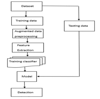
In the methodology section, we elucidate the designed system for the detection, extraction, and classification of skin disease images, specifically targeting melanoma, Eczema, and Psoriasis. The comprehensive architecture is compartmentalized into distinct modules, encompassing preprocessing, feature extraction, and classification. This section provides a detailed overview of the entire methodology employed in our research.

Expanding on The Methodological Phases :
A. Data Collection
Sources: The dataset was collected from publicly available sources including online dermatology databases and research repositories. These sources were chosen to ensure a diverse and representative collection of dermatology images spanning various skin conditions.
Acquisition Methods: The dataset was acquired through web scraping and manual collection. Web scraping involved extracting images from reputable dermatology websites and forums, while manual collection included gathering images from academic publications and platforms such as IEEE Dataport.
Data Curation: Prior to training, the dataset underwent manual curation to remove duplicates, low-quality images, and irrelevant content.Images were annotated with corresponding labels indicating the dermatological condition depicted, ensuring the dataset's integrity and suitability for classification tasks.
B. Data Splitting
Random Splitting: The dataset was randomly split into training and test sets using a stratified approach to preserve class distribution. This ensures that each class is represented proportionally in both training and test subsets, preventing bias in model evaluation.
Split Ratio: A standard split ratio of 80% training and 20% testing was adopted, striking a balance between model training performance and robust evaluation. This ratio provides sufficient data for model learning while retaining a sizable test set for unbiased performance assessment.
C. Data Augmentation
Techniques Applied: Various data augmentation techniques were applied to augment the training dataset and enhance model generalization. These techniques include random rotation, flipping, zooming, and other transformations to the training images. These transformations help introduce variations in the training data, which aids in improving the model's.
Implementation: Data augmentation was implemented using the Fastai library's built-in augmentation transforms, which seamlessly integrate with the training pipeline. Transform parameters were chosen empirically to strike a balance between augmentation effectiveness and computational efficiency.
D. Model Training
Model Architectures: ResNet50: Utilize the ResNet50 architecture, which is deeper and more complex than ResNet18, potentially offering improved performance at the cost of increased computational resources.
ResNet18: Retain the ResNet18 architecture, known for its balance between performance and computational efficiency, making it suitable for dermatology image classification tasks.
DenseNet121: Incorporate the DenseNet121 architecture, which introduces dense connectivity patterns between layers, potentially capturing more intricate features in dermatology images.
Transfer Learning: Apply transfer learning by initializing each model with pre-trained weights on the ImageNet dataset. This enables leveraging learned features from a diverse set of natural images to accelerate convergence and improve classification performance.
Fine-tuning: Fine-tune each model on the dataset by unfreezing the final few layers and training the entire network end-to-end. Fastai's inbuilt method is used to fine-tune the pre-trained ResNet18 model on the DermNet dataset. This method automatically unfreezes the final few layers of the model, applies discriminative learning rates, and trains the entire network end-toend. Fine-tuning allows the model to adapt its learned representations to the specific characteristics of dermatology images.
Training Procedure: Train each model using the same training configuration, including optimizer settings, learning rate schedule, and regularization techniques, to ensure fair comparison across architectures.
E. Model Evaluation
Performance Metrics: Evaluate the performance of each model using standard classification metrics, including accuracy, precision, recall, and F1 score. Compare the performance metrics of ResNet50, ResNet18, and DenseNet121 to assess their relative efficacy in dermatology image classification.
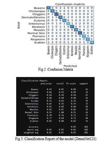
Confusion Matrices: Generate confusion matrices for each model to visualize the distribution of true positive, false positive, true negative, and false negative predictions across different classes. Compare the confusion matrices to identify common sources of misclassification and assess the models' class-wise performanc
IV. MODEL IMPROVEMENT STRATEGIES:
A. Fast AI Integration
We implemented the FastAI library into our model development pipeline, which proved to be instrumental in enhancing the performance and efficiency of our skin disease image classification system. The integration of FastAI offered several key advantages:
- High-Level API: FastAI provides a user-friendly interface with highlevel abstractions, simplifying the process of building, training,and optimizing deep learning models. This abstraction allowed us to focus on model architecture and experimentation rather than lowlevel implementation details.
- Top-Performing Models: Leveraging FastAI's pre-built models, we were able to harness the power of state-of-the-art convolutional neural network architectures without the need for extensive manual configuration. These pre-trained models served as strong starting points for our experimentation, enabling us to achieve competitive performance with minimal effort.
- Transfer Learning Support: FastAI's comprehensive support for transfer learning facilitated the seamless integration of pre-trained models into our classification task. By fine-tuning these pretrained models on our dataset, we were able to adapt them to our specific domain and achieve remarkable improvements in accuracy with just a few lines of code.
- Interpretability Tools: FastAI offers a suite of interpretability tools that enable users to gain insights into model predictions and decision-making processes. These tools, including visualization techniques and interpretability metrics, allowed us to analyze and understand the underlying factors contributing to our model's performance, enhancing our ability to interpret and trust its outputs. It helped significantly improve model accuracy from 28% to 88%. Amongst the three models, the ResNet 50 model achieved the highest accuracy of 88% with the DermNet Dataset.
B. Dataset Challenges and Solutions
- Inaccurate Dermnet Dataset: We noticed that the DermNet Dataset had disorganized images and duplicates that affected model performance. Hence we tried finding a new dataset similar to DermNet.
- New Dataset from IEEE Data Port: We obtained a curated dataset with accurate labelling from IEEE’s Dataport. The dataset had images of various skin conditions ranging from Measles, Psoriasis, Bowen, Ringworm etc. It contained a total of images of 12 types of skin conditions including normal skin.The dataset had 2 class folders Measles and No Measles .The total images in the dataset - 1,314 images.
V. DATA AUGMENTATION AND MODEL TRAINING:
A. Overfit Model
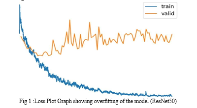
When the ResNet 50 model was retrained on the new IEEE Dataset it achieved an accuracy of 77%. Loss plot Graph was used to evaluate the trained model on the IEEE dataset . According to the loss plot, the Training loss decreased with increasing epochs but the Validation loss was increasing .This shows that the model is overfitted

B. Data Augmentation Techniques
To overcome this we decided to increase the training data using data augmentation .Rotation, width shift, height shift, shear, zoom, and horizontal flip were applied using ImageDataGenerator in Keras. This Increased dataset size from 1,314 to 6,370 images for better model generalization.
C. Model Training and Accuracy
The model was trained on the new Augmented dataset, it was observed that the Training loss decreased with an increase in epochs and so did the Validation loss indicating that the model is learning effectively and generalizing well to unseen data.
Model comparison with ResNet 18 and DenseNet 121showcased DenseNet 121 as the top-performing model with an accuracy of 99.13%.

VI. IMPLEMENTATION
The implementation phase of our project involved the practical execution of the methodologies and techniques outlined in the earlier stages. Leveraging the FastAI library and deep learning capabilities, we constructed a robust pipeline for skin disease classification. Initially, we prepared the dataset by structuring it using the DataBlock functionality, which facilitated efficient loading and transformation of the skin disease images. This involved defining the data blocks for images and categories, setting up data augmentation and normalization, and splitting the dataset into training and validation sets. Furthermore, to handle the memory constraints and computational requirements, we utilized our college's deep learning server, equippedwith powerful hardware configurations including Intel Xeon processors and NVIDIA Tesla P100 GPUs.
With the dataset prepared and the infrastructure in place, we trained several deeplearning models, including DenseNet121, ResNet18, and ResNet50, on the skin disease dataset. The training process involved fine-tuning the pre-trained models to adapt them to the specific characteristics of the skin disease dataset. Through iterative training epochs and optimization techniques, we aimed to enhance the models' performance in accurately classifying various skin diseases. The training progress was monitored through metrics such as accuracy, loss, and learning rate, and visualization tools like confusion matrices and classification reports were employed to evaluate the models' efficacy. After training, we evaluated the models' performance on a validation set to assess their ability to generalize to unseen data. This involved interpreting the model's predictions, analyzing classification metrics, and visualizing performance indicators. Additionally, implementation phase was instrumental in transforming theoretical concepts into practical solutions, culminating in the development of a sophisticated deep-learning we exported the trained models for future use and inference. Overall, the model capable of accurately diagnosing skin diseases. Skin diseases detection model:
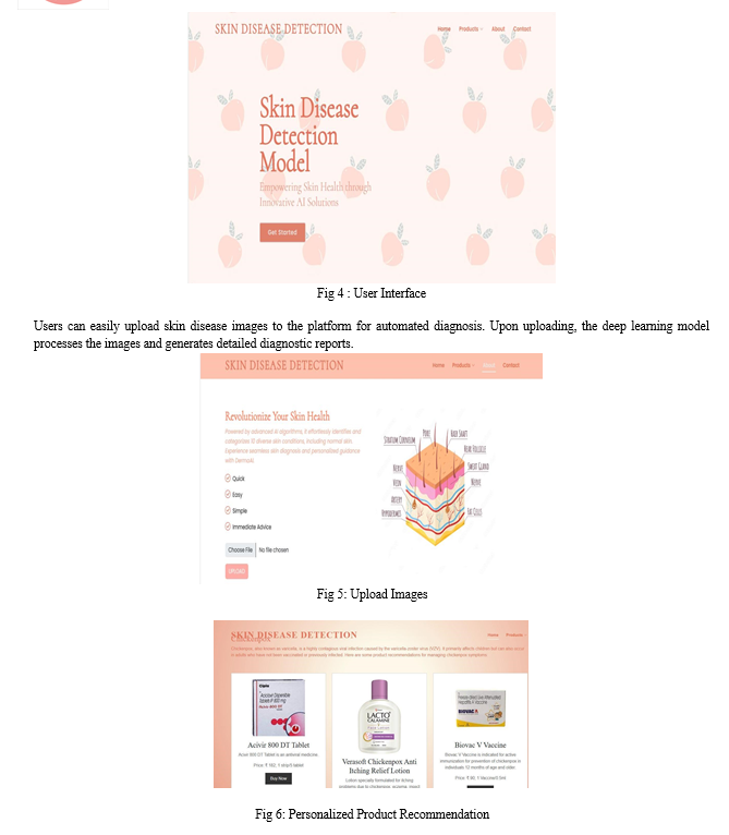
The web platform provides a user-friendly interface for users to interact with the skin disease classification system.

VII. RESULT
Following rigorous training and evaluation, the deployed model achieved a remarkable accuracy of 90.47% using DenseNet121 architecture. Through meticulous analysis of precision, recall, and F1-score metrics, the model demonstrated robustness and reliability. Visualization techniques, including confusion matrices, provided insights into classification capabilities, enhancing interpretability. This outcome signifies a significant advancement in automated skin disease diagnosis, highlighting the potential deep learning in enhancing healthcare solutions and improving patient outcomes.
VIII. FUTURE SCOPE
The project bears significant importance as it introduces a transformative approach to skin disease identification. By increasing cutting-edge deep learning and transfer learning techniques, the system expected outcomes encompass early detection and prevention of skin disease, enhanced accessibility to diagnostic tools, heightened medical awareness and the potential to serve as a valuable supplementary aid for healthcare professionals.
This innovation has the capacity to empower individual in taking proactive control of their skin health, potentially leading to better health outcomes, improved quality of life and contributing to the broader advancement of dermatological research and practices.
Conclusion
The project bears significant importance as it introduces a trans formative approach to skin disease identification. By increasing cutting-edge deep learning and transfer learning techniques, the system expected outcomes encompass early detection and prevention of skin disease, enhanced accessibility to diagnostic tools, heightened medical awareness, and the potential to serve as a valuable supplementary aid for healthcare professionals. This innovation has the capacity to empower individual in taking proactive control of their skin health, potentially leading to better health outcomes, improved quality of life and contributing to the broader advancement of dermatological research and practices.
References
[1] Ray, Arnab, Aman Gupta, and Amutha Al. \"Skin lesion classification with deep convolutional neural network: process development and validation.\" jmir dermatology 3, no. 1 (2020): e18438. [2] Hassan, Syed Rahat, Shyla Afroge, and Mehera Binte Mizan. \"Skin lesion classification using densely connected convolutional networks.\" In 2020 IEEE Region 10 Symposium (TENSYMP), pp. 750-753. IEEE, 2020. [3] Aboulmira, A., Hrimech, H. and Lachgar, M., 2022. “Comparative Study of Multiple CNN Models for Classification of 23 Skin Diseases”. International Journalof Online & Biomedical Engineering, 18(11). [4] Rathod, J., Waghmode, V., Sodha, A. and Bhavathankar, P., 2018, March. “Diagnosis of skin diseases using Convolutional Neural Networks”. In 2018 second International Conference on Electronics, communication and Aerospace Technology (ICECA) (pp. 1048-1051). IEEE. [5] Sah, A.K., Bhusal, S., Amatya, S., Mainali, M. and Shakya, S., 2019, October. “Dermatological diseases classification using image processing and deep neural network”. In 2019 International Conference on Computing, Communication, and Intelligent Systems (ICCCIS) (pp. 381-386). IEEE. [6] Yasir, R., Rahman, A., & Ahmed, N. “Dermatological Disease Detection using Image Processing and Artifi cial Neural Network.“Dhaka:International Conference on Electrical and Computer Engineering.2019 [7] Krizhevsky, A., ILYA, S., & Geoff rey, E. (2019) “ImageNet Classification with Deep Convolutional Neural Networks.” Advances in Neural Information Processing Systems. [8] Arifi n, S., Kibria, G., Firoze, A., Amini, A., & Yan, H. (2012) “Dermatological Disease Diagnosis Using Color-Skin Images. ” Xian: International Conference on Machine Learning and Cybernetics. [9] Suganya R. (2016) “An Automated Computer Aided Diagnosis of Skin Lesions Detection and Classification for Dermoscopy Images. ” International Conference on Recent Trends in Information Technology
Copyright
Copyright © 2024 Ms. R Soundharya, Akanksha Shettigar, Ananya Prasad, Ashwith R Poojary, Deepa Naik. This is an open access article distributed under the Creative Commons Attribution License, which permits unrestricted use, distribution, and reproduction in any medium, provided the original work is properly cited.

Download Paper
Paper Id : IJRASET62566
Publish Date : 2024-05-23
ISSN : 2321-9653
Publisher Name : IJRASET
DOI Link : Click Here
 Submit Paper Online

