Ijraset Journal For Research in Applied Science and Engineering Technology
- Home / Ijraset
- On This Page
- Abstract
- Introduction
- Conclusion
- References
- Copyright
Skin Lesions Classification Using CNN
Authors: Muhammed Fayez M H, Willchrist Sebastian, Dilraj S, Shabiya M I, Rotney Roy Meckamalil
DOI Link: https://doi.org/10.22214/ijraset.2024.63366
Certificate: View Certificate
Abstract
In dermatology, skin lesions present a diagnostic challenge that requires precise classification for efficient pa- tient care. Conventional categorization techniques that depend on dermatologists’ subjective visual assessment are prone to inaccuracies and inconsistencies. This work proposes a novel way to automate the classification of skin lesions using image processing algorithms, with a focus on meeting the needs of the older population, who frequently face obstacles while trying to access healthcare services. The suggested system uses state-of-the- art classification algorithms, such as machine learning and deep learning models, to accomplish accurate categorization. It also includes preprocessing approaches to improve image quality and feature extraction methods to capture pertinent lesion features. The goal of this research project is to greatly increase diagnosis accuracy and efficiency by automating the classification process. This would ultimately assist patients and medical personnel, especially in scenarios where direct contact is limited, like pandemics. Future approaches should focus on making sure that data security measures are strong and improving user interface design for senior users.
Introduction
I. INTRODUCTION
In India, skin diseases affect a significant portion of the population, but many lack access to affordable dermatological care. Our project aims to address this by developing a skin disease classifier using Convolutional Neural Networks (CNN) and MobileNetV2. Our goal is to provide a user-friendly and cost-effective solution for diagnosing skin conditions, helping those who cannot afford or lack time for in-person consultations. Through machine learning, we aim to make dermatological care more accessible and reduce the prevalence of skin diseases in India.
[2]MobileNetV2 is a convolutional neural network architecture designed specifically for mobile and edge devices. It builds on the original MobileNet, focusing on both performance and efficiency. MobileNetV2 introduces an innovative layer called the inverted residual with linear bottleneck, which significantly reduces the model’s computational complexity while maintaining high accuracy. This architecture uses depth- wise separable convolutions to minimize the number of parameters and computational load, making it lightweight and faster than traditional CNNs. Key features of MobileNetV2 include its ability to perform well on resource-constrained devices without sacrificing accuracy, and its applicability to a wide range of image classification tasks.
This paper presents a comprehensive overview of our innovative skin disease classification system, which includes a website designed to provide accessible and affordable dermatological care for individuals across India. [1] Central to our system is the provision of personalized consultancy recommendations, which are generated according to the type of the found skin lesion trained on a dataset named HAM1000 obtained from a trusted source. By considering factors such as age, gender, skin condition, medical history and skin type our system predicts the skin lesion type with an accuracy of 85 percent along with the probablity and the next most most likely lesion type to occur .
[3] In addition to personalized recommendations for Dermatologist to consult and the produces suggested according to the category of the disease, our system prioritizes user privacy and data security by ensuring that all data is kept locally on the user’s device, without being sent to any external servers. This approach leverages the capabilities of modern web browsers to store data securely within the browser itself, utilizing technologies like local storage or IndexedDB. By keeping the data local, users can rest assured that their sensitive information, including skin images and diagnostic results, remains private and under their control. This method not only reduces the risk of data breaches and privacy leaks but also ensures that users do not lose their data due to server-side issues or internet connectivity problems. Furthermore, this local storage approach aligns with data protection regulations and best practices, offering a secure and user-friendly experience while maintaining the system’s efficiency and functionality.
[7]Our system is designed with a strong focus on user expe- rience, ensuring that it is accessible and intuitive for everyone, regardless of their technical background. By providing a user- friendly interface, we aim to make skin disease diagnosis as simple as possible, allowing users to upload an image and receive a probable diagnosis with just a few clicks. The interface is clean, easy to navigate, and optimized for both desktop and mobile devices, ensuring a seamless experience across all platforms.
Our commitment to usability extends to features that guide users through each step of the process, from capturing and uploading an image to understanding the diagnostic results. Clear instructions, visual aids, and intuitive design elements help demystify the technology, making it accessible to users of all ages and technical abilities.
This user-centric approach is more than just convenience—it’s about improving public health. By empowering individuals with easy access to early skin disease detection, we aim to enhance the overall well-being of our society. Early diagnosis and treatment can prevent severe health complications, reduce healthcare costs, and ultimately contribute to a disease-free community. Our vision is to leverage technology to foster a healthier nation, ensuring that everyone has the tools they need to take control of their health, leading to a more prosperous and welfare-oriented society.
In summary, our system is designed to be user-friendly and accessible to all, regardless of technical expertise, ensuring that everyone can easily use it to diagnose skin diseases. By offering an intuitive interface and storing data locally for enhanced privacy, we aim to improve public health and contribute to a healthier, disease-free society. Our goal is to empower individuals with the tools they need for early detection and treatment, fostering a more prosperous and welfare-oriented community.
II. RELATED WORKS
A. Dermatologist-level Classification of Skin Cancer with Deep Neural Networks
Developed a deep learning algorithm that achieved dermatologist-level accuracy in classifying skin cancer using a large dataset of dermoscopic images. They utilized a CNN model pre-trained on ImageNet and fine-tuned it with over 129,000 clinical images covering more than 2,000 diseases. The model demonstrated impressive performance, achieving an area under the receiver operating characteristic curve (AUC) of 0.96, which is comparable to the diagnostic accuracy of certified dermatologists. This pioneering work highlighted the potential of deep learning in dermatology, paving the way for subsequent research in automated skin lesion classification.
B. Skin Lesion Analysis Towards Melanoma Detection
Organized a challenge to benchmark the performance of different machine learning models on the task of skin lesion classification. The ISIC 2018 challenge attracted numerous participants who applied various deep learning techniques to a standardized dataset of dermoscopic images. The best- performing models employed advanced CNN architectures like ResNet and DenseNet, achieving significant improvements in accuracy and robustness. The challenge underscored the importance of large, annotated datasets and standardized eval- uation metrics in advancing the field of automated skin lesion analysis.
???????C. Human-computer Collaboration for Skin Cancer Recogni- tion
Explored the potential of combining human expertise with deep learning models to improve the accuracy of skin cancer diagnosis. They developed a CNN model and tested its per- formance both independently and in collaboration with derma- tologists. The study found that while the CNN model alone performed well, its accuracy significantly improved when combined with human input, suggesting that a hybrid approach could be more effective than either humans or machines alone. This work emphasizes the value of integrating AI tools into clinical workflows to enhance diagnostic accuracy.
???????D. Semi-supervised Adversarial Model for Skin Lesion Seg- mentation and Classification
Proposed a semi-supervised adversarial model to address the challenges of limited labeled data in skin lesion classification. Their approach utilized a Generative Adversarial Network (GAN) to generate synthetic data and augment the training set, improving the model’s generalization capabilities. The semi-supervised model demonstrated superior performance compared to fully supervised models trained on limited data, highlighting the potential of adversarial training in medical image analysis. This study contributed valuable insights into leveraging GANs for enhancing deep learning models in the context of scarce medical data.
???????E. Automated Skin Lesion Diagnosis Using Ensemble of Con- volutional Neural Networks
Developed an ensemble approach that combines multiple CNN architectures to improve the robustness and accuracy of skin lesion classification. By integrating the predictions of several models, including VGGNet, ResNet, and Inception, the ensemble method achieved better performance than any single model. Their ensemble model was evaluated on the ISIC dataset and demonstrated high accuracy and reliability. This work highlighted the effectiveness of model ensembling in reducing variance and improving the stability of predictions in medical image classification tasks.
???????F. Diagnostic Performance of a Deep Learning CNN for Dermoscopic Melanoma Recognition in Comparison to 58 Dermatologists
Conducted a comparative study to evaluate the performance of a CNN against dermatologists in diagnosing melanoma us- ing dermoscopic images. The study involved 58 dermatologists from 17 countries who assessed a set of 100 images, while the CNN model was trained on over 100,000 dermoscopic images. The results showed that the CNN outperformed most dermatologists, achieving a higher sensitivity and specificity in detecting melanoma. This study provided strong evidence of the capabilities of deep learning models in skin cancer diagnosis and highlighted their potential as decision support tools in clinical practice.
???????G. Deep Learning outperforms Dermatologists in the Classi- fication of Skin Lesions into Seven Classes
Brinker and colleagues developed a deep learning model capable of classifying skin lesions into seven distinct categories, including melanoma, basal cell carcinoma, and benign nevi. Using a dataset of over 12,000 dermoscopic images, the team trained a CNN and compared its performance to that of experienced dermatologists. The CNN achieved higher accuracy than the dermatologists, particularly in distinguishing between benign and malignant lesions. This study underscored the effectiveness of CNNs in multi- class skin lesion classification and their potential to assist dermatologists in making more accurate and efficient diagnoses.
???????III. PROPOSED MODEL
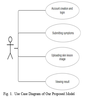
The proposed model for our skin lesion classification project not only leverages the power of Convolutional Neural Net- works (CNNs) to achieve high accuracy in diagnosing various skin conditions but also integrates seamlessly into a user- friendly web application. The model is fine-tuned on the HAM10000 dataset, where additional layers are added to specifically adapt to the nuances of dermoscopic images. Key components of the model include batch normalization to sta- bilize and accelerate training, dropout layers to prevent over- fitting, and data augmentation to enhance generalization. This model is integrated into a web application that allows users to upload their skin lesion images for analysis. Upon uploading, the model analyzes the image and provides a classification result, indicating whether the lesion is benign or malignant and identifying the specific type of lesion.Additionally, the web app offers personalized recommendations for over-the- counter medicines or advises consulting a healthcare profes- sional based on the classification results. This feature aims to provide users with immediate, actionable insights, bridging the gap between automated diagnosis and practical healthcare guidance. The proposed model aims to offer a comprehensive tool for automated skin lesion classification and medical recommendations, supporting early diagnosis and improving patient outcomes.
???????IV. SYSTEM IMPLEMENTATION
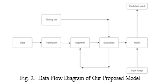
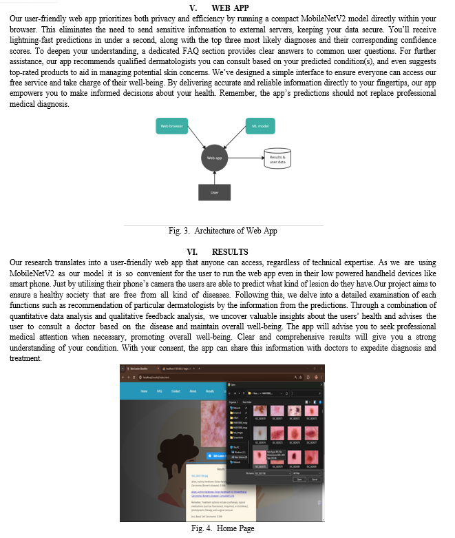
The implementation details of the project will be examined in this part, including the steps involved in data collecting, augmentation methods, preprocessing methods, suggestions, and outcomes. It will be supported by examples of the data flow diagram and use case diagram. In this case, the prediction function for skin disease detection was integrated with the MobileNetV2 model. A web application was used for the front-end design and functionality, while Python, Tensorflow.js, and MySQL were used for the back-end implementation.
???????A. Data Collection
The HAM10000 dataset, also known as the Human Against Machine with 10000 training images, is a benchmark dataset widely used in the development and evaluation of skin lesion classification algorithms. This dataset comprises 10,015 high-resolution dermoscopic images, annotated and confirmed by dermatology experts, representing seven different types of skin lesions, including benign and malignant categories such as melanomas, nevi, and basal cell carcinomas. The diversity and size of HAM10000 make it particularly valuable for training deep learning models, as it includes a wide range of skin lesion types and conditions, ensuring that models trained on this dataset can generalize well to various real-world scenarios. Additionally, the dataset includes metadata such as age, gender, and localization of the lesions, providing rich contextual information that can be leveraged for more sophisticated analysis. By providing a comprehensive and meticulously annotated collection of dermoscopic images, HAM10000 has become a cornerstone resource in the field of dermatological research, facilitating the development of more accurate and robust skin lesion classification systems.
???????B. Data Preprocessing
The process begins with data augmentation, where techniques such as rotation, scaling, flipping, and color adjustments are applied to artificially expand the dataset and improve the model’s robustness to various transformations. This helps mitigate the issue of limited training data and reduces the risk of overfitting. Next, normalization is performed to standardize the pixel values of the images, typically scaling them to a range of 0 to 1 or adjusting them to have zero mean and unit variance, which aids in faster and more stable convergence during training. Additionally, artifact removal is essential in dermoscopic images, where techniques like morphological operations or inpainting are used to eliminate noise from hair, bubbles, and other irrelevant elements. This preprocessing step enhances the clarity of the skin lesions and ensures that the CNN focuses on relevant features.
???????C. Recommendations and Results
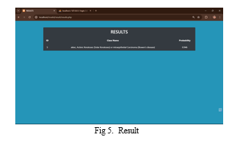
Utilizing the comprehensive user data collected from user through a Form along side other important factors such as the skin type and related medications our classifier results in an accurate prediction. Based on the result obtained it redirects them to specific dermatologists to be consulted along with their condition mentioned to the doctor. By incorporating factors such as age, gender, medical history our system calculates the top three possibilities of skin disease from the image submitted. Our system also recommends the doctors contact information and booking site to ensure that the user has no problem finding an appointment with the dermatologist. The results obtained by the user is stored in the database and could be shared with the doctor if necessary, our system makes sure that the details and information of our client does not get leaked or lost. In this subsection, we elucidate the intricacies of our recommendation algorithm and present the empirical results obtained from user interactions with the system, demonstrating its efficacy in delivering proper guidance to every users.
VII. FUTURE SCOPE
- Model Optimization: Explore techniques such as quantization, pruning, and architecture modifications to enhance TensorFlow models for improved inference speed, reduced memory usage, and increased accuracy in multi- model integration scenarios.
- Integration with Cloud Services: Investigate integration with leading cloud platforms like Google Cloud AI Platform, AWS SageMaker, or Azure Machine Learning to enable scalable deployment, training, and monitoring capabilities.
- Real-time Augmented Reality (AR) Integration: Implement AR features for users to visualize skin conditions in real-time using device cameras, enhancing user experience and facilitating more intuitive diagnosis.
- Mobile Application Development: Develop a dedicated mobile application for the skin disease classifier to pro- vide users with a seamless and optimized experience on mobile devices.
- Privacy and Ethics Considerations: Prioritize privacy and ethics by ensuring compliance with data protection reg- ulations and implementing safeguards for responsible handling of user data.
- Telemedicine Integration: Integrate the classifier into telemedicine platforms to enable remote consultations between patients and dermatologists, facilitating quicker diagnosis and treatment recommendations.
- Dataset Expansion: Expand the dataset by adding more training data, potentially resulting in a wider range of classification categories and improving the classifier’s accuracy and robustness.
- Integration with Electronic Health Records (EHR): Integrate the classifier with electronic health record systems to allow healthcare providers to access and utilize predictions directly within their workflow, streamlining patient care and diagnosis efficiency.
In summary, there is a huge future potential for classifying skin diseases using CNN models. Improvements in dataset enlargement, real-time monitoring, privacy and ethics, optimization, fine-tuning analysis, data augmentation, and interaction with cloud services will all help to improved and flexible classifiers, promoting healthier surroundings, and facilitating the maintenance of a suitable and healthy lifestyle by adopting the necessary precautions against skin disorders.
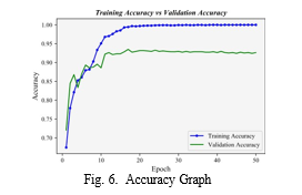 ???????
Conclusion
In this paper, we have presented an innovative skin disease classifier tailored specifically for citizens who may not have access to nearby dermatologists due to lack of money or any other factors. Through the integration of personalized skin disease classifier, simple understandable user interface and functionalities our platform aims to address the diverse amount of diseases that are present and provide proper medical care. The results of our study demonstrate the efficacy and promise of our product and ensures accurate and trustworthy results in promoting a healthy lesion free society, enhancing quality of life, and empowering people with less technical knowledge to take proactive control of their health and well-being. Looking forward, ongoing enhancements and broadening of our plat- form will reinforce its position as a valuable tool in skin disease identification, fostering the progress of comprehensive and tailored approaches to dermatological health.
References
[1] “A Model for Classification and Diagnosis of Skin Disease using Machine Learning and Image Processing Techniques”, Shaden Abdulaziz AlDera, Mohamed Tahar Ben Othman, https://dx.doi.org/10.14569/IJACSA.2022.0130531 [2] “Skin Disease Detection Using Image Processing and Neural Networks”, Sushmetha G R, Divya Shree D V, Goggi Pragna, Prathibha L, and Mary M ’Dsouza, https://journal.ijprse.com/index.php/ijprse/article/view/92 [3] ”A Smartphone-Based Skin Disease Classification Using MobileNet CNN”, Jessica Velasco, Cherry Pascion, Jean Wilmar Alberio, https://dx.doi.org/10.30534/ijatcse/2019/116852019 [4] ”AI-based localization and classification of skin disease with erythema”, Son H.M, Jeon W, Kim J, https://dx.doi.org/10.1038/s41598-021-84593- z [5] ”Skin lesion classification using deep learning and image processing”, Jibhakate A, Parnerkar P, Mondal S, Bharambe V, Mantri S.T, https://dx.doi.org/10.1109/ICISS49785.2020.9316092 [6] MobileNet Image Classification with TensorFlow’s Keras API, https://youtu.be/5JAZiue-fzY?si=AwCtIbgOFb9rhTpU [7] Dataset used for training and validating the model obtained from https://arxiv.org/abs/1803.10417
Copyright
Copyright © 2024 Muhammed Fayez M H, Willchrist Sebastian, Dilraj S, Shabiya M I, Rotney Roy Meckamalil. This is an open access article distributed under the Creative Commons Attribution License, which permits unrestricted use, distribution, and reproduction in any medium, provided the original work is properly cited.

Download Paper
Paper Id : IJRASET63366
Publish Date : 2024-06-19
ISSN : 2321-9653
Publisher Name : IJRASET
DOI Link : Click Here
 Submit Paper Online

