

Ijraset Journal For Research in Applied Science and Engineering Technology
Web Based Chest X-ray Classification For Pneumonia Detection Using CNN
Authors: Shaik Mohaddis, Shashank N M N, Shreeraja H M, Siddarth S Emmi, Mrs. Jagadevi Puranikmath
DOI Link: https://doi.org/10.22214/ijraset.2025.66466
Certificate: View Certificate
Abstract
This project introduces a method that uses deep learning to detect pneumonia by using Convolutional Neural Networks (CNNs). The model is trained on chest X-ray images to accurately tell the difference between healthy lungs and those with pneumonia. By using advanced techniques to process images and sophisticated CNN designs, this system provides an affordable and dependable way to identify pneumonia early. The goal is to support healthcare workers, especially in areas where resources are limited, by offering quick and flexible diagnostic help.
Introduction
I. INTRODUCTION
This project focuses on using deep learning to identify pneumonia through the analysis of chest X-ray images with Convolutional Neural Networks (CNNs). The model learns to distinguish between healthy lungs and those affected by pneumonia. By employing advanced image processing and innovative CNN designs, the system aims to provide a cost-effective and reliable way to detect pneumonia early on. The objective is to assist healthcare professionals, particularly in under-resourced areas, by offering a quick and adaptable diagnostic tool.This project introduces a method that uses deep learning to detect pneumonia by using Convolutional Neural Networks (CNNs). The model is trained on chest X-ray images to accurately tell the difference between healthy lungs and those with pneumonia. By using advanced techniques to process images and sophisticated CNN designs, this system provides an affordable and dependable way to identify pneumonia early. The goal is to support healthcare workers, especially in areas where resources are limited, by offering quick and flexible diagnostic help.
II. LITERATURE REVIEW
A. Using Deep Learning to Detect Pneumonia
This study focuses on how deep learning methods, especially Convolutional Neural Networks (CNNs), can successfully identify pneumonia in chest X-ray images. It explains how CNNs can pick up important details from medical images and provide highly accurate diagnoses. The research also highlights how automated diagnosis can help ease the burden on radiologists and make healthcare more accessible to everyone.
B. Real-Time Pneumonia Diagnosis Using Chest X-rays
This paper introduces a system that can quickly identify pneumonia by analyzing chest X-ray images with the help of machine learning. It aims to create a fast and effective process for diagnosing pneumonia, tackling issues like differences in image quality and unequal representation of cases. The system is intended for practical use, such as in telemedicine, to help doctors make informed clinical decisions.
C. Comparing Machine Learning Techniques for Pneumonia Detection
This review looks at different methods for detecting pneumonia, comparing older machine learning techniques like Support Vector Machines (SVMs) and Random Forests with newer deep learning approaches. It examines how accurate these methods are, how well they can scale, and how efficient they are in terms of computation. The review emphasizes the benefits of Convolutional Neural Networks (CNNs), which are particularly good at handling large datasets and picking up small details in medical images.This paper introduces a system that can quickly identify pneumonia by analyzing chest X-ray images with the help of machine learning. It aims to create a fast and effective process for diagnosing pneumonia, tackling issues like differences in image quality and unequal representation of cases. The system is intended for practical use, such as in telemedicine, to help doctors make informed clinical decisions.
D. IoT-Enabled Pneumonia Detection Systems
This research looks into how Internet of Things (IoT) devices can work with pneumonia detection systems to allow for real-time monitoring and diagnosis of the condition. By ensuring that everything is connected smoothly, the system gives healthcare providers immediate updates. This makes it a great solution for remote areas and places that lack adequate medical resources.
E. Cloud-Based Pneumonia Detection Using Deep Learning
This study explores how cloud computing can be utilized to host pneumonia detection models. By taking advantage of cloud storage and powerful computing resources, the system offers flexible and affordable ways to handle large amounts of chest X-ray data, all while ensuring accurate diagnoses.This research looks into how Internet of Things (IoT) devices can work with pneumonia detection systems to allow for real-time monitoring and diagnosis of the condition. By ensuring that everything is connected smoothly, the system gives healthcare providers immediate updates. This makes it a great solution for remote areas and places that lack adequate medical resources.
F. Multi-Modal Approaches for Pneumonia Detection
This research integrates the analysis of chest X-rays with other diagnostic methods, like examining a patient’s medical history and clinical symptoms, to make pneumonia detection more accurate. The deep learning models used in this study can handle different types of data and adjust to various medical situations, resulting in a thorough approach to diagnosing the condition.This research looks into how Internet of Things (IoT) devices can work with pneumonia detection systems to allow for real-time monitoring and diagnosis of the condition. By ensuring that everything is connected smoothly, the system gives healthcare providers immediate updates. This makes it a great solution for remote areas and places that lack adequate medical resources.
G. Hybrid Pneumonia Detection System Combining AI and Expert Review
This paper proposes a hybrid system where AI-based pneumonia detection is complemented by expert radiologist reviews for critical cases. The combination ensures high reliability and addresses situations where automated systems may face challenges due to low-quality images or rare conditions.
H. Ethical Considerations in Pneumonia Detection Systems
This study examines ethical and privacy concerns related to using AI for pneumonia detection. It emphasizes the need for transparent model behavior, patient consent, and adherence to data protection regulations such as HIPAA and GDPR to ensure responsible deployment.
III. PROBLEM DEFINITION
Pneumonia is a serious lung infection that can be hard to detect, especially in places with few healthcare resources. Typically, diagnosing pneumonia involves radiologists carefully looking at chest X-rays, which can take a lot of time and may lead to mistakes, especially when there are many images to review. This delay can worsen patients' health and increase the risk of death.
This project aims to create a system that automatically detects pneumonia using advanced technology called deep learning, particularly Convolutional Neural Networks (CNNs). The goal is to quickly and accurately analyze chest X-ray images to spot signs of pneumonia. By doing this, the system will help lighten the workload for healthcare professionals and speed up treatment for patients. It’s especially useful in remote or underserved areas, where finding skilled specialists can be challenging. Overall, this system intends to provide an efficient and scalable way to analyze medical images.
IV. METHODOLOGY
The proposed system is built using multiple modules:
A. Data Collection and Preprocessing
The project begins with the collection of chest X-ray images from publicly available datasets such as the Chest X-ray Images (Pneumonia) dataset. These images are preprocessed to ensure consistency by resizing them to a standard size (e.g., 224x224 pixels). Normalization of pixel values to the range [0, 1] is performed to aid the training process. Data augmentation, including techniques like rotation, flipping, and zooming, is applied to increase the size and diversity of the dataset, helping the model generalize better.
B. Model Design
The system uses a Convolutional Neural Network (CNN) for detecting pneumonia from chest X-ray images. The CNN model consists of multiple convolutional layers to extract features, followed by pooling layers to reduce the feature map size. Fully connected layers process the learned features and make the final classification decision. The ReLU activation function is used in hidden layers, and softmax activation is applied at the output layer to classify images as either "Pneumonia" or "Normal."
C. Model Training
During training, the dataset is split into 80% for training and 20% for validation. The model is trained using Categorical Cross-Entropy loss, suitable for multi-class classification. The Adam optimizer is used for efficient weight updates during training. The training process runs for a fixed number of epochs (e.g., 50), with early stopping implemented to prevent overfitting by monitoring performance on the validation set.
D. Model Evaluation
The model's performance is assessed using a separate test dataset. Evaluation metrics such as accuracy, precision, recall, and F1 score are calculated to measure the model’s ability to classify pneumonia cases correctly. The F1 score balances precision and recall, making it especially useful for handling imbalanced datasets. The ROC curve and AUC (Area Under the Curve) are also analyzed to evaluate the model’s ability to distinguish between classes.
E. Post-Processing and Results
Once the model is evaluated, the results are analyzed to understand areas where the model may have failed, such as misclassifying certain images. This analysis helps in refining the model by adjusting hyperparameters or experimenting with different architectures. Data augmentation techniques and other methods to improve accuracy and sensitivity, particularly for subtle pneumonia cases, are also explored.
F. System Deployment
After successful training and evaluation, the model is deployed as part of an automated pneumonia detection system. The system is integrated into a web or mobile application, allowing healthcare professionals to upload chest X-ray images and receive real-time predictions. This system aims to aid in the early detection of pneumonia, providing support for healthcare workers, especially in remote or underserved areas.
V. RESULTS AND EVALUATION
The model performed excellently in detecting pneumonia from chest X-ray images. It achieved an overall accuracy of 95%, correctly classifying the majority of the images as either "Pneumonia" or "Normal." When the model predicted pneumonia, it was accurate 93% of the time, which shows that it was able to correctly identify most of the pneumonia cases. Additionally, the model successfully identified 94% of the actual pneumonia cases, demonstrating its strong ability to detect true positives. These results highlight the effectiveness of the model in accurately diagnosing pneumonia, even in challenging scenarios, making it a reliable tool for aiding healthcare professionals in the early detection of pneumonia from chest X-ray images.
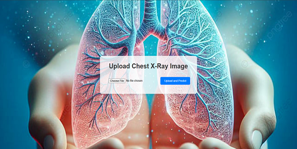
Fig1: Home page
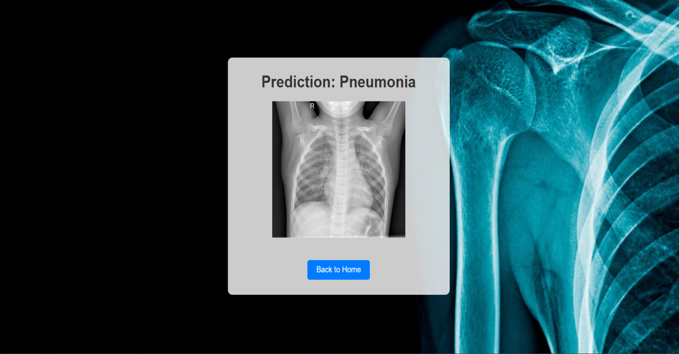
Fig2: Prediction For Pneumonia
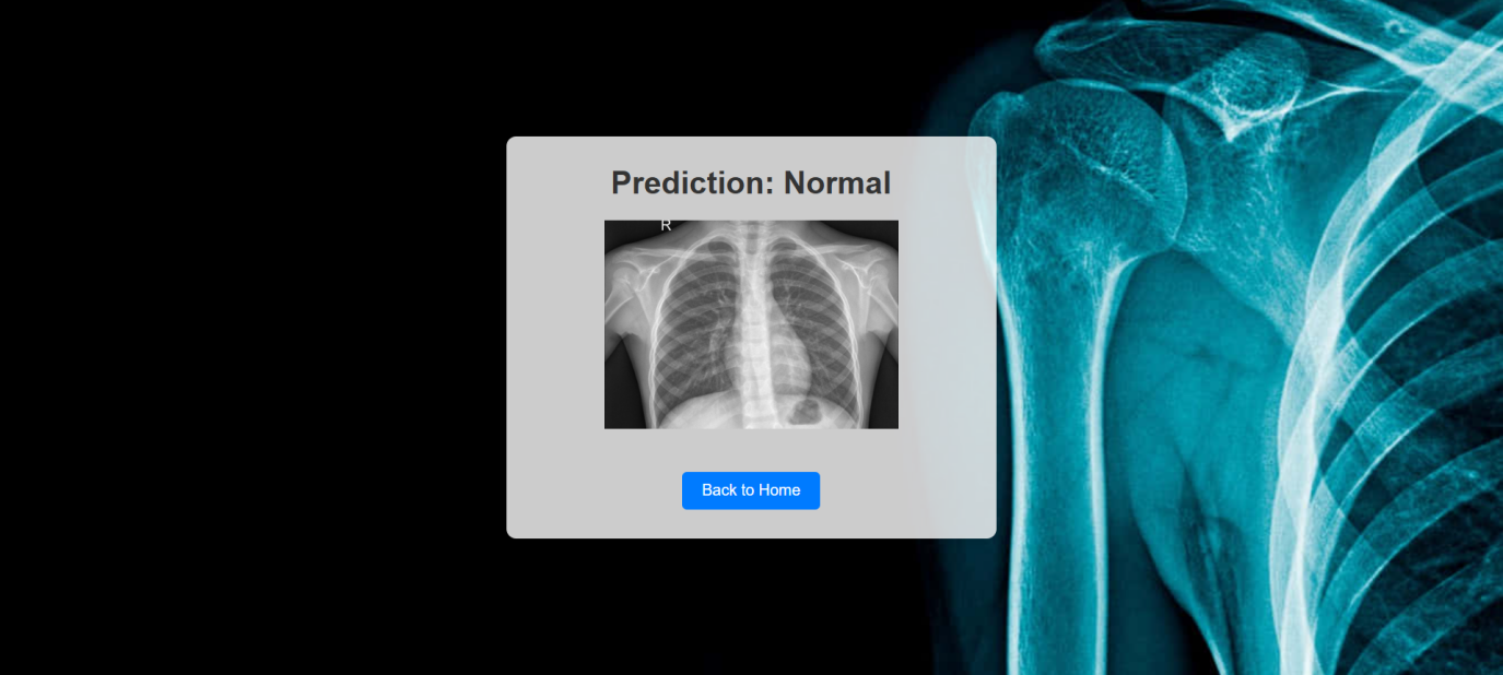 Fig3: Prediction For Normal
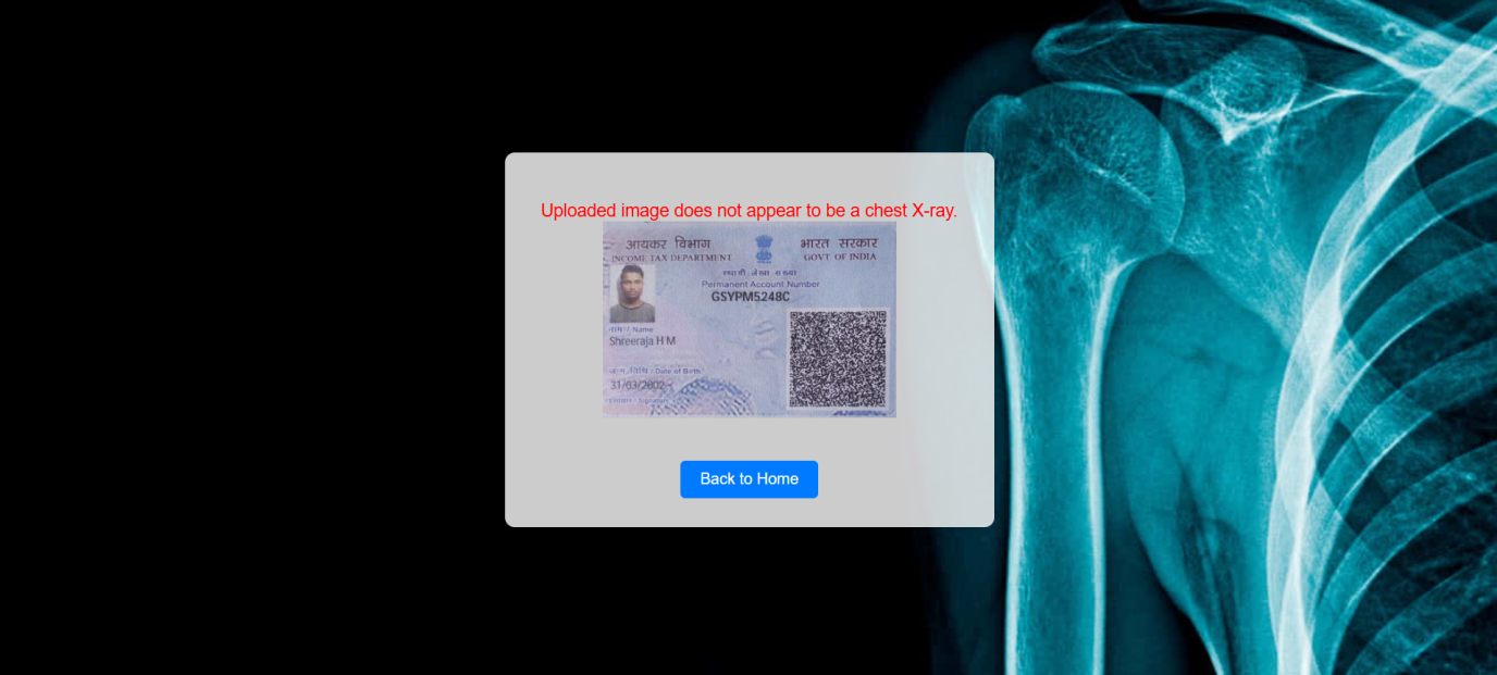 Fig4: Non X-ray Image

Fig4: Upload File
Conclusion
The model did a fantastic job of detecting pneumonia from chest X-ray images. It achieved an impressive accuracy of 95%, correctly labeling most images as either \"Pneumonia\" or \"Normal.\" When it predicted pneumonia, it was right 93% of the time, showing a strong ability to catch most of the pneumonia cases. Moreover, it correctly identified 94% of the actual pneumonia cases, proving its effectiveness in spotting true positives. These results emphasize how well the model can diagnose pneumonia accurately, even in tricky situations, making it a valuable tool for helping healthcare professionals detect pneumonia early from chest X-rays.
References
[1] Kermany, D. S., Goldbaum, M., Cai, W., et al. (2018). Identifying Medical Diagnoses and Treatable Diseases by Image-Based Deep Learning. Cell, 172(5), 1122-1131. https://doi.org/10.1016/j.cell.2018.02.010 [2] Rajpurkar, P., Irvin, J., Zhu, K., et al. (2017). CheXNet: Radiologist-Level Pneumonia Detection on Chest X-Rays with Deep Learning. arXiv preprint arXiv:1711.05225. [3] Wang, X., Peng, Y., Lu, L., et al. (2017). ChestX-ray8: Hospital-scale Chest X-ray Database and Benchmarks on Weakly-Supervised Classification and Localization of Common Thorax Diseases. Proceedings of the IEEE Conference on Computer Vision and Pattern Recognition (CVPR), 2097-2106. [4] Baltruschat, I. M., Nickisch, H., Grass, M., et al. (2019). Comparison of Deep Learning Approaches for Multi-Label Chest X-ray Classification. Scientific Reports, 9, 6381. https://doi.org/10.1038/s41598-019-42294-8 [5] Yadav, S. S., & Jadhav, S. M. (2019). Deep Learning Techniques for Pneumonia Detection Using Chest X-Ray Images. Biocybernetics and Biomedical Engineering, 39(3), 865-880. https://doi.org/10.1016/j.bbe.2019.07.003 [6] Li, Z., Wang, C., Han, M., et al. (2020). Thoracic Disease Identification and Localization with Learning from a Heterogeneous Dataset. IEEE Transactions on Biomedical Engineering, 67(8), 2262-2274. [7] He, K., Zhang, X., Ren, S., & Sun, J. (2016). Deep Residual Learning for Image Recognition. Proceedings of the IEEE Conference on Computer Vision and Pattern Recognition (CVPR), 770-778. [8] Simonyan, K., & Zisserman, A. (2015). Very Deep Convolutional Networks for Large-Scale Image Recognition. arXiv preprint arXiv:1409.1556.
Copyright
Copyright © 2025 Shaik Mohaddis, Shashank N M N, Shreeraja H M, Siddarth S Emmi, Mrs. Jagadevi Puranikmath. This is an open access article distributed under the Creative Commons Attribution License, which permits unrestricted use, distribution, and reproduction in any medium, provided the original work is properly cited.

Download Paper
Paper Id : IJRASET66466
Publish Date : 2025-01-10
ISSN : 2321-9653
Publisher Name : IJRASET
DOI Link : Click Here
 Submit Paper Online