Ijraset Journal For Research in Applied Science and Engineering Technology
- Home / Ijraset
- On This Page
- Abstract
- Introduction
- Conclusion
- References
- Copyright
Web-based Deep Learning System for Malaria Parasite Detection in Granular Blood Samples
Authors: Mr. D. Konda Babu , Togaru Naveen , Vasala Devi Pradeep , G S Sai Kowshik Varma , Kuppili Sri Charan
DOI Link: https://doi.org/10.22214/ijraset.2024.61182
Certificate: View Certificate
Abstract
Malaria is a serious health concern for modern humans, affecting people of all ages. Infected mosquitoes carry the fatal parasites responsible for malaria. Malaria can be diagnosed by examining a sample of the patient\'s blood under a microscope for parasites. The project comprises creating a web tool that employs deep learning to detect malaria parasites in blood smear photos. Convolutional neural network (CNN) models such as ResNet50, VGG19, and Customized CNN can be used to collect and categorize a set of blood smear images in order to identify patterns and characteristics. Convolutional layers, max-pooling layers, entirely linked layers, and a SoftMax layer are all utilized to create a Convolutional Neural Network (CNN) model. This technique can improve the accuracy of parasite diagnosis, increase detection rates, and reduce the disease\'s impact on global health.
Introduction
I. INTRODUCTION
Malaria is an infectious disease that kills a large number of people globally, primarily in developing nations. An accurate and fast diagnosis is critical for early treatment and effective illness management. Recent research using deep learning algorithms to analyze minuscule images of blood smears shows great promise in the detection and diagnosis of malaria. These techniques have the potential to transform malaria detection by offering a quick, accurate, and cost-effective solution, especially in resource-constrained areas. (CNNs) perform especially well in a variety of image processing applications. CNNs have been demonstrated to be effective at detecting patterns in digital blood smear images, which can be used to identify infected cells and diagnose malaria. A layered CNN model with stain normalizations was shown to be useful. According to 5-fold cross-validation results, the suggested stacked CNN model performed well and had a high accuracy rate. This paper provides an in-depth evaluation of cutting-edge deep learning algorithms for malaria diagnosis and detection. We talk about the transfer learning method and the many CNN designs for detecting malaria, such as VGGNet, Google Net, and Res Net. We also look at the obstacles and opportunities of utilizing deep learning to diagnose malaria, such as the requirement for big and diverse datasets and the possible use of deep learning methods in real-world contexts. The paper's conclusion emphasizes the advantages of applying deep learning techniques for malaria diagnosis and detection, as well as the potential implications for world health.
II. LITERATURE SURVEY
In this study, a layered C.N.N. approach for parasite detection in blood thin smear pictures was studied. The model was verified using a dataset of 27,558 cell images, and deep learning-based technology was utilized to identify malaria parasites without the need for labor-intensive engineering. To measure its performance, the model performs five-fold cross-validation across a variety of sequences. The model was evaluated on a publically available dataset and outperformed previous models. Modified Elliptic Curve Cryptography (MECC) is thus believed to be the most secure approach to store data in the cloud. [1]
Compared to ground-truth data, the suggested approach can distinguish various types of cells and parasites. Malarial parasites can be detected in all pictures. Furthermore, the method is intended to be easily applied on low-cost microcomputers, which can lower treatment costs by reducing the need for manual analysis. The system, which is based on a convolutional neural network, employs an image identification method to detect probable parasites and cell kinds. [2]
The paper suggests a mobile microscopy-deep learning method for identifying malaria. It first gives a summary of the current malaria detection techniques before discussing the need for the dl model. The suggested method uses DR networks to categorize microphotographs from stained thin blood samples as either malaria-infected or malaria-uninfected. The model may be successfully used in real-world circumstances to detect parasite infection early on, according to testing conducted on several datasets.
The study suggests a learning method that uses mobile microscopy and deep residual networks to detect parasites. Many datasets have been used to test the system's ability to discriminate between healthy and ill cells. The writers might also research the method by increasing the recognition rate. [3]
In the study's introduction, it is emphasized how dangerous malaria is, especially for mall children, and how urgent it is to have a clear diagnosis as soon as possible, especially in nations with limited resources. While certain deep learning models have shown promise in automating the malaria screening procedure, it is still difficult to reliably detect the life stages of the parasite. This work's authors use the incredibly accurate Mask RCNN object recognition and classification model to examine automated malaria screening. The segmentation masks produced by the system are immediately observable, and it has been trained on both healthy and sick red blood cells. According to the scientists, the model counting is 15 times more accurate than a manual count and has the potential to save time and money by reducing errors brought on by human counting. Lastly, it states that a distinct form of malaria screening is offered by the RConvolution Neural Network. [4]
The authors suggest detecting malaria in red blood cells by using a convolutional neural network. Many compounds associated with abnormalities in cells may be present in red blood cells; however, these have not been taken into account in prior studies that differentiate malaria parasites from other inclusions that seem to be similar. The researchers generated a new dataset using 23 blood samples that were not used for training in order to remove bias from their study. Two training datasets were used by the authors: the original training dataset and the training dataset supplemented with additional photos produced by data augmentation. A thousand pictures from every stain. [5] Researchers have developed a unique approach using a transfer learning architecture (TLA) to identify malaria parasites. Digital photos of smears from individuals with and without malaria were taken in order to compare the outcomes of this V.G.G-S.V.M model. [6]
III. SYSTEM ANALYSIS
A. Existing System
The "Deep Learning based Web App for Malaria Parasite Detection in Granular Blood Samples" technology uses a web application to recognize malaria parasites in blood smear images using deep learning techniques. Obtaining and annotating a range of blood smear image collections is the initial stage. Deep learning models for picture categorization, including ResNet50, VGG19, and a modified CNN, are constructed using this dataset. Blood smear images can be submitted for analysis by users of the web program. When uploading images, they undergo preprocessing before being fed into deep learning models. Whether or not parasites have been discovered is disclosed to users. An image depiction of the detection findings shows the zones that have been found highlighted. The solution is set up on a server, its accuracy and other metrics are evaluated, and data security and privacy measures are implemented. Constant model upgrades, user support, and maintenance are needed for this malaria detection system.
DISADVANTAGES OF THE EXISTING SYSTEM
The existing system for malaria parasite detection has several limitations:
- Limited Generalization: The system's performance is mostly determined by the quality and diversity of the training dataset. If the real-world scenarios in the training dataset are not adequately reflected, the system's performance on unseen data might be affected.
- High Resource Requirements: The high training and inference computational requirements of two deep learning models, ResNet50 and VGG19, may provide challenges in resource-constrained environments.
- False Positives and Negatives: Although deep learning models are very robust, they are not impervious to producing false positives and false negatives. These errors could lead to potentially harmful medical effects and incorrect diagnoses.
- Lack of Real-time Analysis: Patients in need of urgent care may suffer if the system is unable to provide real-time analysis or if it takes a lengthy time to detect issues.
- Dependency on Internet connectivity: The system relies on internet connectivity because it is a web application, which could be problematic in locations where having access to healthcare resources is critical but there is little to no internet connectivity.
B. Proposed System
The first stage of the screening technique entails selecting parasite candidates with the lowest grayscale intensity in order to minimize the number of feasible options. Following that, these locations are regarded to be suitable for further examination.
White blood cells (WBCs) are filtered out of the image before they can be recognized. Following that, the presence of malaria is determined by examining red blood cells.
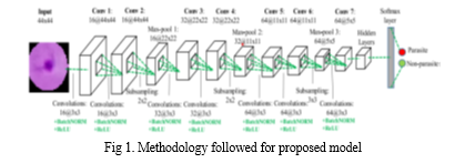
The IGMS technique can be used to find prospective parasite candidates by finding the lowest intensity regions of a grayscale image. A ring-shaped zone with a radius of 22 pixels is generated around each single pixel discovered. D. Feature Extraction: Diagnostic testing can help separate true parasites from background noise. Once the parasite candidates have been extracted, a CNN model is used. The improved CNN model includes softmax, fully connected, max pooling, and CN layers. After each convolutional layer, batch normalization layers are applied, followed by an R.E.L.U activation function. A maxpooling layer is added for every two Cn layers selected. The final feature map for CN is linked to The CNN model contains three separate fully linked layers, each with a unique label. To make the model less complex. This custom-made C.N.N. has several advantages over pre-trained networks, including a lower runtime and input size based on the normal parasite size in thick smear images. The personalized CNN model surpasses pretrained networks in accuracy, while having fewer layers and a shorter runtime. On a common Android smartphone, the system can identify parasites in 10 seconds using an input image measuring 4032 X 3024 X 3 pixels. It is designed as an Android app that connects the smartphone lens to the microscope's eyepiece. After adjusting the microscope to find the correct place in the blood smear, the user can use the app to capture photographs. The OpenCV4 Android SDK library is used to implement all image and video recognition algorithms. The Convolutional Neural Network (C.N.N.) classification model is built using convolution, max-pooling, and batch normalization. The number values above these cuboids indicate the size of the recovered features. Improving the volume and quality of training data, as well as selecting the appropriate architecture, can all aid neural network models in performing better. Feature retrieval within a CNN is possible by integrating one or more convolutional layers into the network. These layers use filters to convolve the input image, resulting in feature maps that highlight key patterns.
IV. SYSTEM DESIGN
SYSTEM ARCHITECTURE
Below diagram depicts the whole system architecture.

V. SYSTEM IMPLEMENTATION
MODULES
- The data management module: it is responsible for collecting, organizing, and maintaining a huge and diverse dataset of labels and blood smear images. includes data preprocessing to ensure that the images are of appropriate quality and format.
- Module for deep learning models: This session covers cutting-edge architectures and approaches for developing, training, and optimizing deep learning models to identify malaria parasites. Model selection, hyperparameter tweaking, and fine-tuning of pre-trained models are among the tasks it handles.
- The web application module: it is in charge of developing the program's user interface and functionalities, which include image uploading, real-time analysis, and results presentation. monitors user interactions and integrates image processing into the deep learning model.
- The offline capability module: it allows users to upload and preprocess photographs without requiring an internet connection, allowing the web application to run offline. includes synchronization algorithms for determining when to use an internet connection.
- Telemedicine Integration Module: This module includes telemedicine features that allow for remote consultation and expert participation. improves diagnostic accuracy by allowing doctors to confirm results and provide more information.
VI. RESULTS AND DISCUSSION
In this work, the performance of the modified CNN model is assessed using a five-fold cross evaluation. The tailored CNN model is resilient and effective, as evidenced by an average AUC score of 95.90% and a standard deviation of 0.18%. Our updated CNN model has average F1-score, recall, and precision values of 95.88%, 95.46%, and 96.30%, respectively.

Conclusion
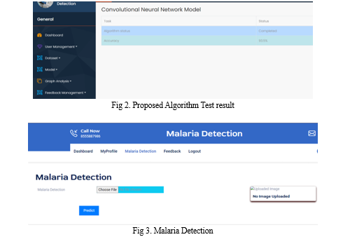
Based on the findings of the deep learning-based malaria detection investigation, it is plausible to conclude that deep learning models have showed promise in identifying malaria from tiny blood cell images. This study compares the performance of various deep learning models for recognizing malaria parasites. The results showed that the models have outstanding levels of sensitivity, specificity, and accuracy, indicating that they can reliably diagnose malaria. To make our technique more accessible, we integrated it with a web application. Users can send images of their cells, and the program will display whether or not malaria is present. The study also underlines the importance of having a large and diverse dataset for training deep learning models. Adding more layers to the model and training it on a large number of photographs can both improve performance. It also emphasizes the need for future research to improve deep learning model performance and develop more resilient models capable of handling fluctuations in image data. Overall, deep learning-based malaria detection can provide a reliable and effective diagnostic tool for early malaria diagnosis, hence slowing the disease\'s spread and improving patient outcomes.
References
[1] Yang, F., Poostchi, M., Yu, H., Zhou, Z., Silamut, K., Yu, J., ... & Antani, S. (2019). Deep learning for smartphone-based malaria parasite detect ion in thick blood smears. IEEE journal of biomedical and health informatics, 24(5), 1427-1438. [2] Umer, M., Sadiq, S., Ahmad, M., Ullah, S., Choi, G. S., & Mehmood, A. (2020). A novel stacked CNN for malarial parasite detection in thin blood smear images. IEEE Access, 8, 93782-93792. [3] Chowdhury, A. B., Roberson, J., Hukkoo, A., Bodapat i, S., & Cappelleri, D. J. (2020). Automated complete blood cell count and malaria pathogen detection using convolution neural network. IEEE Robotics and Automation Letters, 5(2), 1047-1054. [4] Pattanaik, P. A., Mittal, M., Khan, M. Z., & Panda, S. N. (2020). Malaria detection using deep residual networks with mobile microscopy. Journal of King Saud University- Computer and Information Sciences. [5] Kassim, Y. M., Palaniappan, K., Yang, F., Poostchi, M., Palaniappan, N., Maude, R. J., ... & Jaeger, S. (2020). Clustering-based dual deep learning architecture for detecting red blood cells in malaria diagnostic smears. IEEE Journal of Biomedical and Health Informatics, 25(5), 1735-1746. [6] Loh, D. R., Yong, W. X., Yapeter, J., Subburaj, K., & Chandramohanadas, R. (2021). A deep learning approach to the screening of malaria infection: Automated and rapid cell counting, object detect ion and instance segmentalion using Mask R-CNN. Computerized Medical Imaging and Graphics, 88, 101845. [7] Molina, A., Rodellar, J., Boldú, L., Acevedo, A., Alférez, S., & Merino, A. (2021). Automatic identification of malaria and other red blood cell inclusions using convolutional neural networks. Computers in Biology and Medicine, 136, 104680. [8] Militante, S. V. (2019, December). Malaria disease recognition through adaptive deep learning models of convolutional neural network. In 2019 IEEE 6th International Conference on Engineering Technologies and Applied Sciences (ICETAS) (pp. 1-6). IEEE. [9] Oyewola, D. O., Dada, E. G., Misra, S., & Damaševi?ius, R. (2022). A Novel Data Augmentation Convolutional Neural Network for Detect ing Malaria Parasite in Blood Smear Images. Applied Artificial Intelligence, 1-22. [10] Nayak, S., Kumar, S., & Jangid, M. (2019, September). Malaria detection using multiple deep learning approaches. In 2019 2nd International Conference on Intelligent Communication and Computational Techniques (ICCT) (pp. 292-297). IEEE.
Copyright
Copyright © 2024 Mr. D. Konda Babu , Togaru Naveen , Vasala Devi Pradeep , G S Sai Kowshik Varma , Kuppili Sri Charan . This is an open access article distributed under the Creative Commons Attribution License, which permits unrestricted use, distribution, and reproduction in any medium, provided the original work is properly cited.

Download Paper
Paper Id : IJRASET61182
Publish Date : 2024-04-28
ISSN : 2321-9653
Publisher Name : IJRASET
DOI Link : Click Here
 Submit Paper Online

